. 2018 Aug 20:124:149-162.
doi: 10.1016/j.freeradbiomed.2018.05.094. Epub 2018 Jun 5.

Enhanced mitochondrial DNA repair of the common disease-associated variant, Ser326Cys, of hOGG1 through small molecule intervention

Affiliations

Enhanced mitochondrial DNA repair of the common disease-associated variant, Ser326Cys, of hOGG1 through small molecule intervention

Beverly A Baptiste et al. Free Radic Biol Med. .

Abstract

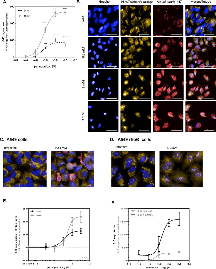
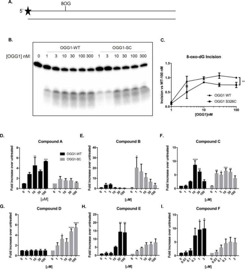
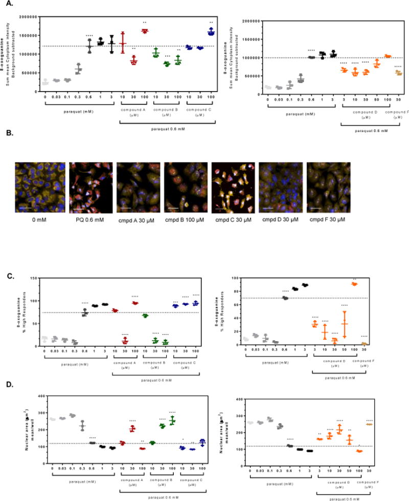
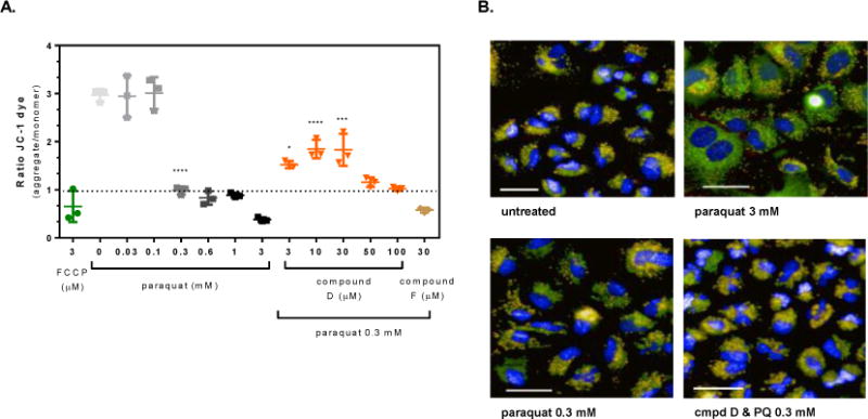
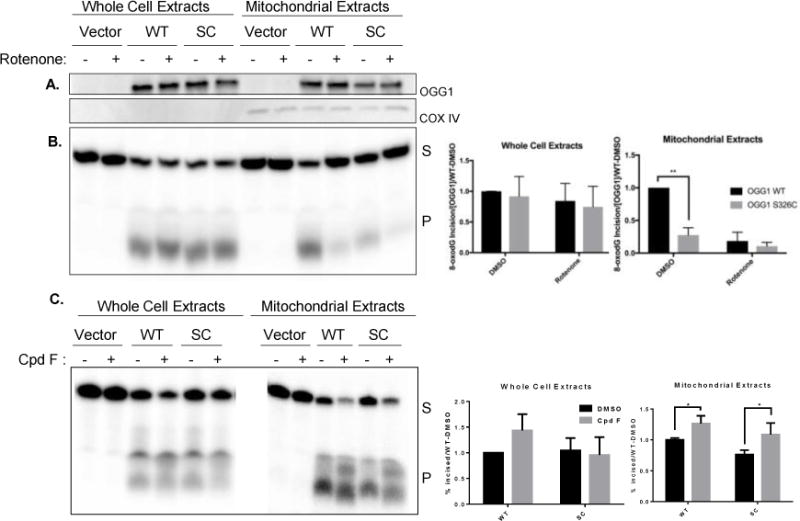
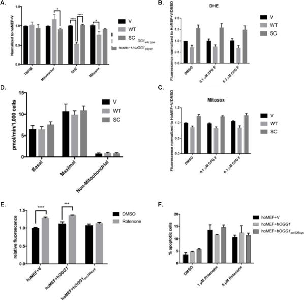
The common oxidatively generated lesion, 8-oxo-7,8-dihydroguanine (8-oxoGua), is removed from DNA by base excision repair. The glycosylase primarily charged with recognition and removal of this lesion is 8-oxoGuaDNA glycosylase 1 (OGG1). When left unrepaired, 8-oxodG alters transcription and is mutagenic. Individuals homozygous for the less active OGG1 allele, Ser326Cys, have increased risk of several cancers. Here, small molecule enhancers of OGG1 were identified and tested for their ability to stimulate DNA repair and protect cells from the environmental hazard paraquat (PQ). PQ-induced mtDNA damage was inversely proportional to the levels of OGG1 expression whereas stimulation of OGG1, in some cases, entirely abolished its cellular effects. The PQ-mediated decline of mitochondrial membrane potential or nuclear condensation were prevented by the OGG1 activators. In addition, in Ogg1-/- mouse embryonic fibroblasts complemented with hOGG1S326C, there was increased cellular and mitochondrial reactive oxygen species compared to their wild type counterparts. Mitochondrial extracts from cells expressing hOGG1S326C were deficient in mitochondrial 8-oxodG incision activity, which was rescued by the OGG1 activators. These data demonstrate that small molecules can stimulate OGG1 activity with consequent cellular protection. Thus, OGG1-activating compounds may be useful in select humans to mitigate the deleterious effects of environmental oxidants and mutagens.

Keywords: 8-Oxoguanine DNA glycosylase-1; 8-dihydroguanine; 8-oxo-7; Base excision repair; Mitochondria; OGG1(S326C); Oxidative stress.


Conflict of interest statement

CONFLICT OF INTEREST

The authors state that they have no conflicts of interest to report.

Figures

Figure 1
Figure 1. Paraquat-induced mitochondrial DNA damage is inversely related to OGG1 expression
A549 cells were treated with paraquat for 24 or 48 hr and subjected to formaldehyde fixation prior to immunofluorescence detection of 8-oxoguanine. (A) Graphical representation of the percent change in 8-oxoguanine intensity within the cytoplasm region following 24- and 48-hr treated cells with increasing concentrations of paraquat. Significance was determined using a 1-way ANOVA with a Dunnett’s posttest comparing treatment groups to an untreated control (* p<0.05, ** p<0.01, *** p<0.001, **** P<0.0001). B. Changes in 8-oxoguanine are depicted in sample images (Hoechst33342, overlaid blue; MitoTracker® orange, overlaid orange; 8-oxoG, overlaid red) from cells exposed to increasing concentrations of paraquat in panel B (line = 40 μm). (C/D) A549 cells were treated with 50 ng/mL ethidium bromide for 14 population doublings to generate A549rhoφ cells devoid of functional mitochondrial DNA. A549 and A549rhoφ cells were treated with paraquat for 24 hr, followed by staining for 8-oxoG. (C) Sample images of A549 cells from wells containing untreated cells and cells exposed to 3 mM paraquat for 24 hr (line = 40 μm). (D) Sample images like Panel C, but with A549rhoφ cells (line = 40 μm). (E) A549 cells were incubated with 5% v.v. Ogg1-BacMam for 48 hours prior to treatment with varying concentrations of paraquat for 24 hr. The cells were subjected to formaldehyde fixation for immunofluorescence detection of 8-oxoguanine and the percent change in 8-oxoguanine intensity within the mitochondria of the cells from varying concentrations of paraquat compared to control cells. (F) A549 cells were incubated with 100 nM Ogg1 siRNA containing Dharmafect® in serum-free conditions. Following 48-hr incubation, the medium was replaced to complete culture medium and the A549 cells were treated with varying concentrations of paraquat for 24 hr. The cells were stained and imaged for 8-oxoguanine. The figure represents the percent change in 8-oxoguanine intensity within the mitochondria of paraquat treated cells compared to media control.
Figure 2
Figure 2. Purified hOGG1S326C protein incises 8-oxoguanine less efficiently than purified hOGG1 protein
A. Incision of 8-oxodG was assayed using a duplex oligonucleotide containing a single 8-oxodG residue at position 11. Proteins were purified and diluted to indicated concentrations in reaction buffer. B. After 1 hour at 37°C, fragments were separated by denaturing electrophoresis and visualized using phosphoimaging. C. Results were quantitated by comparing incised bands to total radioactivity in lane. D–I. hOGG1wild type (5 nM) or hOGG1S326C (10 nM) were stimulated with compounds: A (D), B (E), C (F), D (G), E (H), or F (I) at the indicated concentrations. Black bars indicate hOGG1wild type and gray bars indicate hOGG1S326C. Asterisks represent statistical differences between untreated and indicated concentration of compound. All experiments were completed three times. Statistical analysis by 2-way ANOVA, with Tukey post-hoc multiple comparisons. (* p<0.05, ** p<0.01, *** p<0.001, **** P<0.0001)
Figure 3
Figure 3. OGG1 activators attenuate paraquat-induced oxidative damage in A549 cells
A549 cells were pre-treated for 4 hr with small molecule OGG1 activators or 0.1% DMSO prior to addition of 0.6 mM paraquat for 48 hr. The cells were stained and imaged for 8-oxoG content and the average fluorescence intensity within the cell was normalized based on the number of cells imaged. (A) Graphical representations of the change in 8-oxoG intensity within the cytoplasm of cells compared to 0.6 mM paraquat treated cells (dashed line). (B) Representative images of cells from panel A depict changes in mitochondrial 8-oxoG staining (Hoechst 33342, overlaid blue; MitoTracker® orange, overlaid orange; 8-oxoG-AleaFluor®-647, overlaid red; line = 40 μm). (C) The percent of the cell populations labeled as high responders is shown for A549 cells pre-incubated with OGG1 activators (4 hr) prior to exposure to a single concentration of paraquat (0.6 mM, dashed line) for 48 hr. (D) The images from panel A were analyzed further by examining morphology changes. The mean nuclear area (dashed line indicates no treatment, 0.6 mM PQ) for A549 cells was graphed depicting a change in overall cell health. Significance was determined using a 1-way ANOVA with a Dunnett’s posttest comparing paraquat alone treated groups to untreated control (0 mM) and OGG1 activator treated groups to 0.6 mM paraquat control (** p<0.01, *** p<0.001, **** p<0.0001). Data shown are from a representative experiment, performed three times.
Figure 4
Figure 4. OGG1 activators protect against paraquat-induced loss of mitochondrial membrane potential in A549 cells
A549 cells were pre-treated for 4 hours with the OGG1 small molecule activators or 0.1% DMSO prior to exposure to 0.3 mM paraquat for 24 hr. Mitochondrial membrane potential was measured from images captured as described in the methods. (A) Graphical representation of the change in the JC-1 ratio with increasing concentration of paraquat and cells pre-treated with the OGG1 activators prior to exposure to a single concentration of paraquat (0.3 mM) for 24 hr. (B) Representative images from untreated, 3 mM paraquat, 0.3 mM paraquat, and Compound D (30 μM) with 0.3 mM paraquat exposed cells (Hoechst 33342, overlaid blue; JC-1 monomer, overlaid green; JC-1 aggregate, overlaid orange; CellMask™ Deep Red, overlaid red; line = 40 μm). Significance was determined using a 1-way ANOVA with a Dunnett’s posttest comparing paraquat alone treated groups to untreated control (0 mM) and OGG1 activator treated groups to 0.3 mM paraquat control (* p<0.05, ** p<0.01, *** p<0.001, **** p<0.0001).
Figure 5
Figure 5. Incision of 8-oxodG is less efficient in mitochondria isolated from hOGG1S326C cells than hOGG1 cells
A. Cells were grown in the presence of DMSO (0.1%) or rotenone (5 μM) for 24 hours and then harvested. Western blot demonstrates equal expression of hOGG1 protein in hOGG1S326C and hOGG1 cells and no hOGG1 expression in vector cells. COX IV expression is shown as a mitochondrial marker. B. Cell extracts were diluted to protein equal concentrations in reaction buffer and incubated with substrate shown in Figure 1A. Fragments were separated by denaturing electrophoresis and visualized using phosphoimaging. Graphs show quantitation of incised band relative to total radioactivity in lane. The percent incised was divided by the OGG1 content in each sample and normalized to activity of hOGG1wild type extracts in each experiment. Significance was determined by 2-way ANOVA. C. Cells were grown in the presence of DMSO (0.1%) or compound F (15 μM) and harvested after 24 hours. Incision of 8-oxodG was assessed as above. All experiments included three biological repeats. Significance was determined by 2-way ANOVA.
Figure 6
Figure 6. MEFs expressing hOGG1 demonstrate marked differences in mitochondrial parameters from those expressing no OGG1 or hOGG1S326C
A. Cells were analyzed for mitochondrial parameters via flow cytometry. Stains for mitochondrial membrane potential (TMRM), mitochondrial content (Mitotracker Green), total cellular ROS (DHE) or mitochondrial ROS (Mitosox Red) were added to 90% confluent cells and fluorescence was measured in appropriate channels using a flow cytometer. All results were normalized to vector control. Results analyzed by 2-way ANOVA. B–C. Cells were grown in the presence of 0.1% DMSO, 0.1 μM, or 0.3 μM compound F for 24 hours. Measurements of mitochondrial ROS (B) and cellular ROS (C) were analyzed via flow cytometry and normalized to DMSO treated vector cells. D. Cells were analyzed via Seahorse XF24 for basal oxygen consumption, maximal oxygen consumption, and non-mitochondrial oxygen consumption, and results from three independent experiments were graphed and analyzed by 1-way ANOVA. E. Cells were grown in the presence of 0.1% DMSO or 1 μM rotenone and analyzed for mitophagy using a pH sensitive dye via flow cytometry. Results were normalized to vector DMSO control and compared for significance by 2-way ANOVA. F. Cells were grown in the presence of 0.1% DMSO, 1 μM rotenone, or 5 μM rotenone and analyzed via flow cytometry after 24 hours. Results analyzed by 2-way ANOVA. All experiments were performed repeated three times. Mean results were graphed with standard error of the mean for error bars.

References

    1. Hoeijmakers JHJ. DNA Damage, Aging, and Cancer. New England Journal of Medicine. 2009;361(15):1475–1485. - PubMed
    1. Croteau DL, Bohr VA. Repair of Oxidative Damage to Nuclear and Mitochondrial DNA in Mammalian Cells. Journal of Biological Chemistry. 1997;272(41):25409–25412. - PubMed
    1. Wallace SS. Base excision repair: A critical player in many games. DNA Repair. 2014;19:14–26. - PMC - PubMed
    1. de Souza-Pinto NC, Eide L, Hogue BA, Thybo T, Stevnsner T, Seeberg E, Klungland A, Bohr VA. Repair of 8-oxodeoxyguanosine lesions in mitochondrial dna depends on the oxoguanine dna glycosylase (OGG1) gene and 8-oxoguanine accumulates in the mitochondrial dna of OGG1-defective mice. Cancer Res. 2001;61(14):5378–81. - PubMed
    1. Shibutani S, Takeshita M, Grollman AP. Insertion of specific bases during DNA synthesis past the oxidation-damaged base 8-oxodG. Nature. 1991;349(6308):431–434. - PubMed

Publication types

MeSH terms