Multicenter Study
. 2018 Jul 27;52(1):1800766.
doi: 10.1183/13993003.00766-2018. Print 2018 Jul.

Immunological corollary of the pulmonary mycobiome in bronchiectasis: the CAMEB study

Affiliations
Multicenter Study

Immunological corollary of the pulmonary mycobiome in bronchiectasis: the CAMEB study

Micheál Mac Aogáin et al. Eur Respir J. .

Abstract

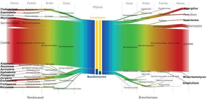
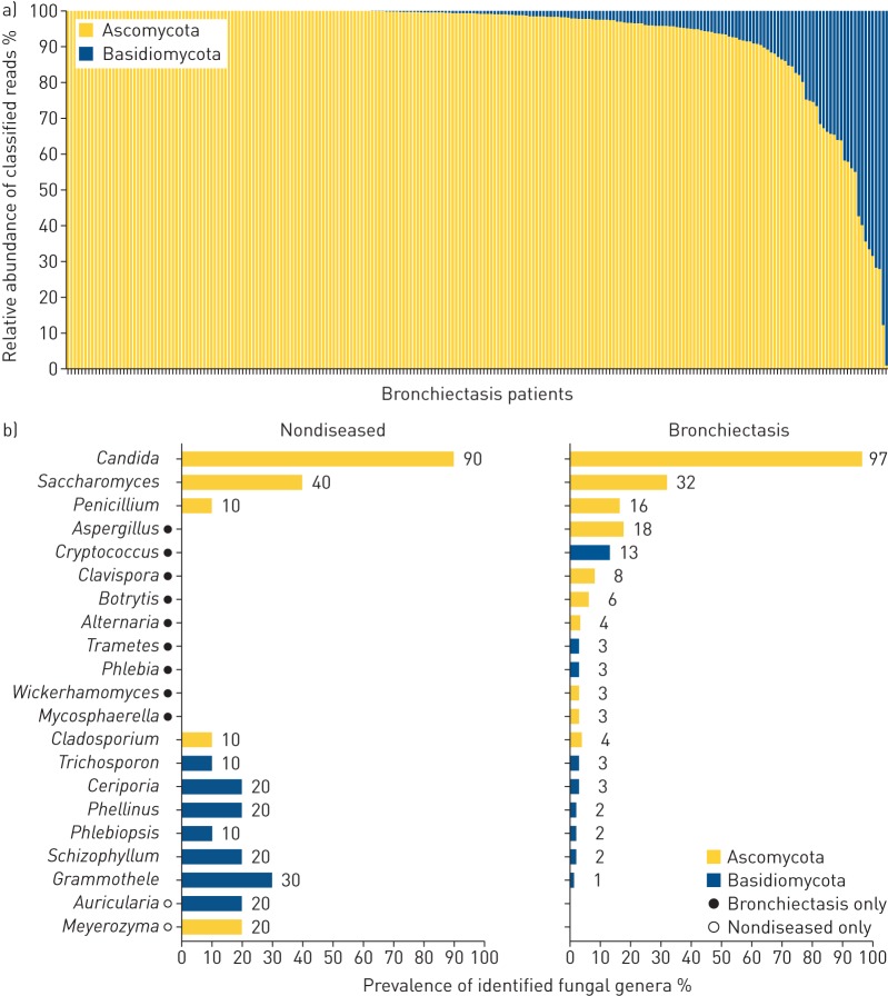
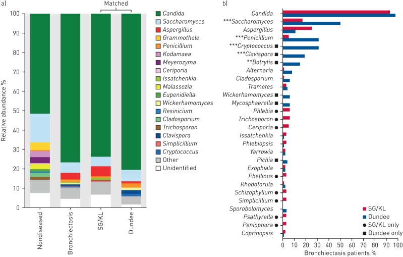
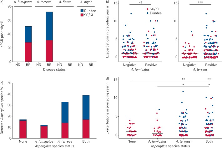
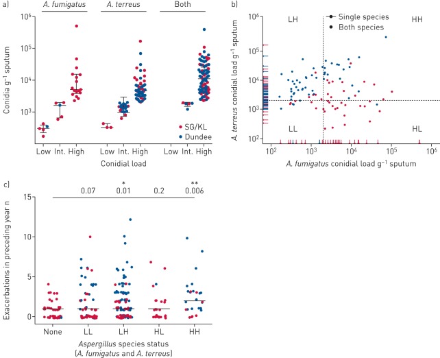
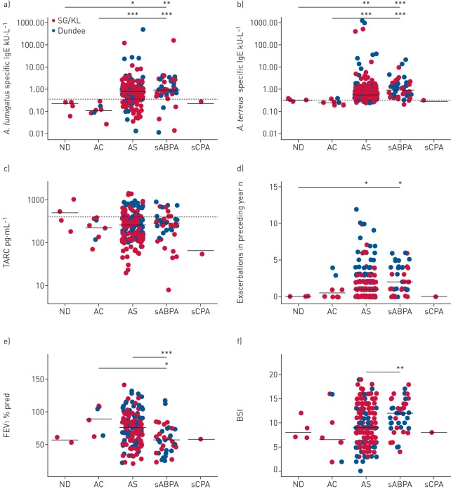
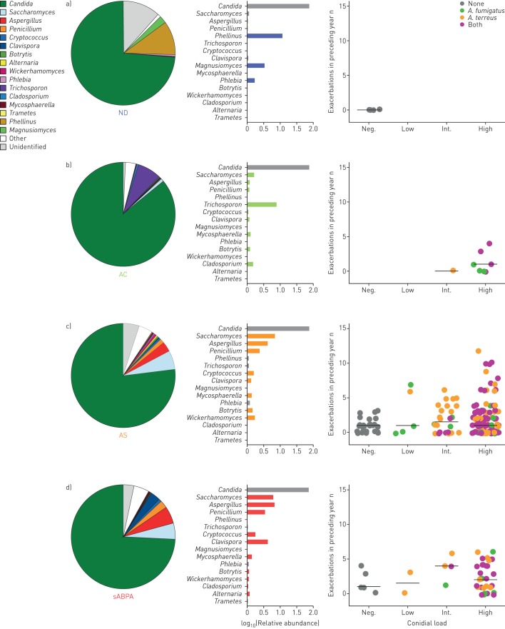
Understanding the composition and clinical importance of the fungal mycobiome was recently identified as a key topic in a "research priorities" consensus statement for bronchiectasis.Patients were recruited as part of the CAMEB study: an international multicentre cross-sectional Cohort of Asian and Matched European Bronchiectasis patients. The mycobiome was determined in 238 patients by targeted amplicon shotgun sequencing of the 18S-28S rRNA internally transcribed spacer regions ITS1 and ITS2. Specific quantitative PCR for detection of and conidial quantification for a range of airway Aspergillus species was performed. Sputum galactomannan, Aspergillus specific IgE, IgG and TARC (thymus and activation regulated chemokine) levels were measured systemically and associated to clinical outcomes.The bronchiectasis mycobiome is distinct and characterised by specific fungal genera, including Aspergillus, Cryptococcus and ClavisporaAspergillus fumigatus (in Singapore/Kuala Lumpur) and Aspergillus terreus (in Dundee) dominated profiles, the latter associating with exacerbations. High frequencies of Aspergillus-associated disease including sensitisation and allergic bronchopulmonary aspergillosis were detected. Each revealed distinct mycobiome profiles, and associated with more severe disease, poorer pulmonary function and increased exacerbations.The pulmonary mycobiome is of clinical relevance in bronchiectasis. Screening for Aspergillus-associated disease should be considered even in apparently stable patients.


Conflict of interest statement

Conflict of interest: F.T. Chew reports personal fees from Sime Darby Technology Center, Olam International and First Resources Ltd, outside the submitted work. J.D. Chalmers reports grants from AstraZeneca, grants and personal fees from GlaxoSmithKline, Boehringer Ingelheim, Pfizer, Bayer Healthcare and Grifols, and personal fees from Napp, outside the submitted work.

Figures

FIGURE 1
FIGURE 1
The pulmonary mycobiome in stable bronchiectasis: mirrored Sankey plots illustrating the relative abundance of read classification by taxonomic rank from phylum (centre; blue) to genus level (margins; red). Nondiseased (healthy) controls (n=10) are compared with patients with stable bronchiectasis (n=238). Central coloured bars demonstrate phylum-level abundance of Ascomycota (yellow) versus Basidiomycota (blue) between the cohorts. Cohort-specific fungal genera are indicated in bold.
FIGURE 2
FIGURE 2
The pulmonary mycobiome in stable bronchiectasis: a) individual patient phylum-level classification of the pulmonary mycobiome in stable bronchiectasis (n=238) illustrating the relative abundance of Ascomycota versus Basidiomycota for individual patients, and b) percent prevalence of the top fungal genera (present at >1% relative abundance) in nondiseased (healthy) controls (n=10) and patients with stable bronchiectasis (n=238).
FIGURE 3
FIGURE 3
The pulmonary mycobiome differs between Singapore/Kuala Lumpur (SG/KL) and Dundee cohorts of stable bronchiectasis. a) Genus-level classification of the pulmonary mycobiome in nondiseased (n=10), stable bronchiectasis (n=238), and matched SG/KL (n=100) and Dundee (n=100) cohorts. The relative abundance of identified taxa is colour-coded. b) Percent prevalence of observed fungal genera (present at >1% relative abundance) in matched bronchiectasis cohorts from SG/KL (n=100) and Dundee (n=100). Significant differences in prevalence between cohorts are indicated. **: p<0.01; ***: p<0.001.
FIGURE 4
FIGURE 4
Identification of specific airway Aspergillus species and their association with exacerbations in bronchiectasis. qPCR: quantitative PCR; SG/KL: Singapore/Kuala Lumpur; ND: nondiseased; BR: bronchiectasis. a) qPCR-based screening for the presence of the major specific Aspergillus species (A. fumigatus, A. terreus, A. flavus and A. niger) in ND (healthy) controls (n=10) and stable BR (n=238). b) Exacerbations for the year preceding study recruitment in patients with qPCR-detectable A. fumigatus (left) and A. terreus (right). c) Classification of qPCR-detectable Aspergillus species by percentage of the total bronchiectasis patient cohort (n=238). Patients are classified as having no detectable species (n=39), A. fumigatus only (n=26), A. terreus only (n=78) or both A. fumigatus and A. terreus (n=95). d) Exacerbations for the year preceding study recruitment in patients with no detectable species, A. fumigatus only, A. terreus only or both A. fumigatus and A. terreus. Median number of exacerbations per group is indicated. ns: nonsignificant; *: p<0.05; **: p<0.01; ***: p<0.001.
FIGURE 5
FIGURE 5
Quantification of airway Aspergillus conidial burden and its association with exacerbations in bronchiectasis (n=238). SG/KL: Singapore/Kuala Lumpur. a) Conidial burden per gram of sputum was quantified for A. fumigatus and A. terreus, respectively, and classified according to load as low (<500 conidia·g−1 sputum), intermediate (“Int.”; 500–2000 conidia·g−1 sputum) or high (>2000 conidia·g−1 sputum). Conidial load categories are illustrated according to fungal airway status as A. fumigatus only, A. terreus only or the presence of both species. b) Scatter plot of A. fumigatus (x-axis) and A. terreus (y-axis) conidial load in stable bronchiectasis. Patients with single and both species are indicated. Patients with both species are classified by their relative A. fumigatus and A. terreus burdens into low burden of both (“LL”), low burden of A. fumigatus and high burden of A. terreus (“LH”), high burden of A. fumigatus and low burden of A. terreus (“HL”), and high burden of both (“HH”). Dotted lines indicate cut-off levels for a high conidial burden (>2000 conidia·g−1 sputum). c) Exacerbations for the year preceding study recruitment in bronchiectasis patients with detectable conidial burdens of both A. fumigatus and A. terreus as classified as in (b). Median number of exacerbations per group is indicated and Benjamini–Hochberg adjusted p-values for all groups compared with “None” are shown. *: p=0.01; **: p=0.006.
FIGURE 6
FIGURE 6
Immunological classification reveals high frequencies of aspergillosis in stable bronchiectasis and an association of serological allergic bronchopulmonary aspergillosis (sABPA) with greater exacerbations, poorer pulmonary function and more severe disease. SG/KL: Singapore/Kuala Lumpur; ND: nondiseased; AC: Aspergillus-colonised; AS: Aspergillus-sensitised; sCPA: suspected chronic pulmonary aspergillosis; TARC: thymus and activation regulated chemokine; FEV1: forced expiratory volume in 1 s; BSI: Bronchiectasis Severity Index. a, b) Measured levels of specific IgE responses to a) A. fumigatus and b) A. terreus, where aspergillosis is classified immunologically as ND (n=4), AC (n=8), AS (n=182), sABPA (n=43) and sCPA (n=1) (supplementary table E4). c) TARC, a proposed ABPA marker in cystic fibrosis [24], was assessed according to the same classification. Dashed lines show respective cut-offs indicating a positive test for each marker (specific IgE 0.35 kU·L−1; TARC 386 pg·mL−1). d) Exacerbations for the year preceding study recruitment, e) pulmonary function (FEV1 % pred) and f) disease severity (BSI) were assessed according to immunological aspergillosis class. Mean values are indicated except for exacerbations and BSI, where median values are shown. *: p<0.05; **: p<0.01; ***: p<0.001.
FIGURE 7
FIGURE 7
Pulmonary mycobiome profiles illustrate specific taxa-associated patterns according to immunological classification of aspergillosis in stable bronchiectasis. ND: nondiseased; AC: Aspergillus-colonised; AS: Aspergillus-sensitised; sABPA: serological allergic bronchopulmonary aspergillosis. a) ND (n=4), b) AC (n=8), c) AS (n=182) and d) sABPA (n=43) mycobiome profiles illustrate the different composition by relative abundance of reads classified to genus level, with the b) AC, c) AS and d) sABPA states exhibiting increased exacerbations and higher conidial load. Only a single patient had sCPA and therefore data is not shown. Mycobiome profiles are represented by pie charts with colour coding according to the taxonomic legend. Adjacent colour-coded log-scaled bar charts detail the observed taxa patterns in each immunological patient class (formal statistical assessment is provided in supplementary table E5). Exacerbations in the preceding year (y-axis) are plotted against Aspergillus conidial load (x-axis) and colour-coded according to quantitative PCR detection status of Aspergillus species into A. fumigatus alone, A. terreus alone, presence of both or none. Patients are further classified according to their individual conidial load as negative (“Neg.”; no detected conidia·g−1 sputum), low (<500 conidia·g−1 sputum), intermediate (“Int.”; 500–2000 conidia·g−1 sputum) or high (>2000 conidia·g−1 sputum).

References

    1. Polverino E, Goeminne PC, McDonnell MJ, et al. . European Respiratory Society guidelines for the management of adult bronchiectasis. Eur Respir J 2017; 50: 1700629. - PubMed
    1. Dickson RP, Erb-Downward JR, Martinez FJ, et al. . The microbiome and the respiratory tract. Annu Rev Physiol 2016; 78: 481–504. - PMC - PubMed
    1. Rogers GB, Zain NM, Bruce KD, et al. . A novel microbiota stratification system predicts future exacerbations in bronchiectasis. Ann Am Thorac Soc 2014; 11: 496–503. - PubMed
    1. Cox MJ, Turek EM, Hennessy C, et al. . Longitudinal assessment of sputum microbiome by sequencing of the 16S rRNA gene in non-cystic fibrosis bronchiectasis patients. PLoS One 2017; 12: e0170622. - PMC - PubMed
    1. Huffnagle GB, Noverr MC. The emerging world of the fungal microbiome. Trends Microbiol 2013; 21: 334–341. - PMC - PubMed

Publication types

MeSH terms

LinkOut - more resources