. 2018 Jun 7;9(6):693.
doi: 10.1038/s41419-018-0716-5.

Loss of Drosha underlies dopaminergic neuron toxicity in models of Parkinson's disease

Affiliations

Loss of Drosha underlies dopaminergic neuron toxicity in models of Parkinson's disease

Ronglin Wang et al. Cell Death Dis. .

Abstract

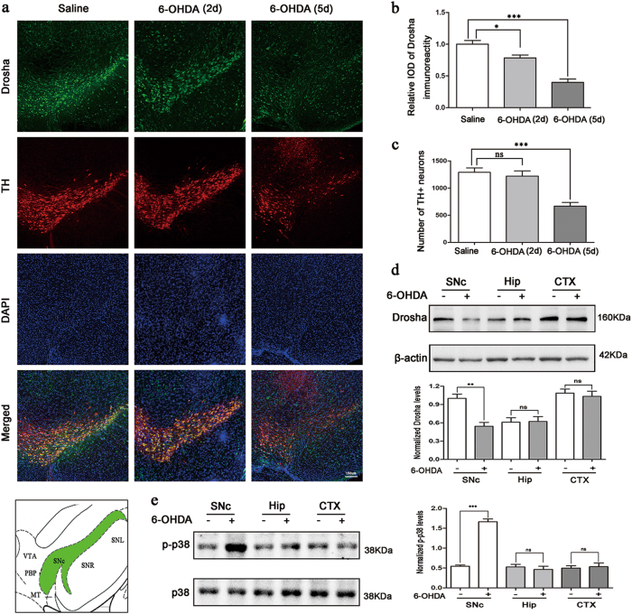
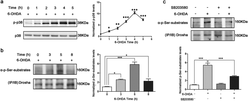
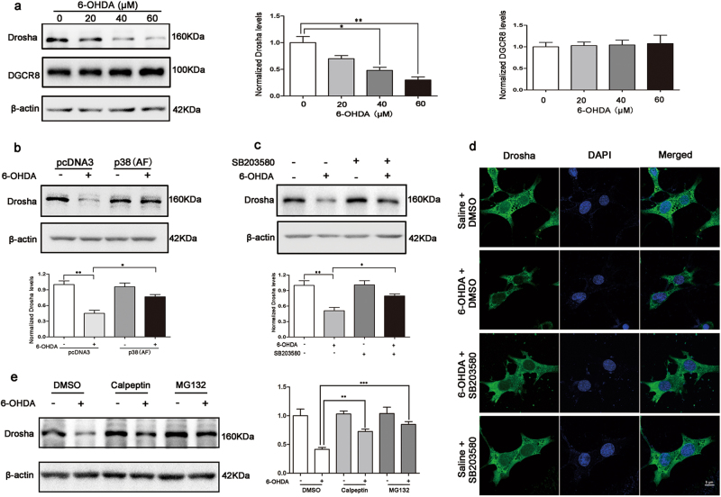
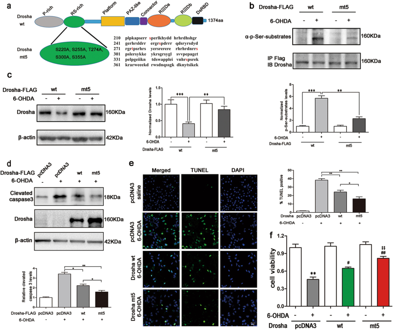
MiRNAs, a group of powerful modulator of gene expression, participate in multiple cellular processes under physiological and pathological conditions. Emerging evidence shows that Drosha, which controls the initial step in canonical miRNA biogenesis, is involved in modulating cell survival and death in models of several diseases. However, the role of Drosha in Parkinson's disease (PD) has not been well established. Here, we show that the level of Drosha decreases in 6-OHDA-induced cellular and animal models of PD. 6-OHDA induced a p38 MAPK-dependent phosphorylation of Drosha. This triggered Drosha degradation. Enhancing the level of Drosha protected the dopaminergic (DA) neurons from 6-OHDA-induced toxicity in both in vitro and in vivo models of PD and alleviated the motor deficits of PD mice. These findings reveal that Drosha plays a critical role in the survival of DA neurons and suggest that stress-induced destabilization of Drosha may be part of the pathological process in PD.


Conflict of interest statement

The authors declare that they have no conflict of interest.

Figures

Fig. 1
Fig. 1. 6-OHDA reduced the stability of Drosha in a mouse model of PD.
a High panels: Drosha levels and TH-positive DA neurons in SNc of saline control mice and 6-OHDA lesioned PD mice. Saline or 0.3 ul 6-OHDA (20 μM) was injected into the SNc of mouse brain. Five days after injection, the brains were perfused with 0.9% NaCl solution and cold 4% paraformaldehyde in phosphate buffer. Then the brains were removed for immunofluorescence. The dilution ratio of Drosha was 1:100 and TH was 1:1000 (n = 3). Lower panels: The position of SNc in the midbrain. b The quantitative value of Drosha. (ANOVA test followed by Tukey HSD, *P < 0.05, ***P < 0.001, n = 3). c The number of TH-positive neurons. (ANOVA test followed by Tukey HSD, ***P < 0.001, n = 3). d Western blot analysis of Drosha level in different brain regions. Five days after injection, the brains were perfused with 0.9% NaCl solution and removed for immunoblot. An anti-Drosha antibody was used to determine the level of Drosha at a dilution ratio of 1:500. An anti-β-actin antibody was used as a loading control. The data are expressed as mean ± S.E.M. (Student’s t-test, **P < 0.01, n = 3). e Western blot analysis of p-p38 level in different brain regions. The brain was removed for immunoblot 2 days after injection. The data are expressed as mean ± S.E.M. (Student’s t-test, **P < 0.001, n = 3)
Fig. 2
Fig. 2. 6-OHDA caused a p38 MAPK dependent phosphorylation of Drosha.
a p38 MAPK was activated with the treatment of 6-OHDA. SN4741 cells were treated with 40 μM 6-OHDA in indicated time. The cell lysate was analyzed by Western blot using an antibody against phospho p38 (9211). An anti-p38 antibody was used as a loading control. Densitometric analyses of the western blots are shown as curves. Data represent mean ± S.E.M. of three independent experiments (ANOVA test followed by Tukey HSD, *P < 0.05, **P < 0.01, ***P < 0.001, n = 3). b Phosphorylation of Drosha when treated with 6-OHDA for various time course. SN4741 cells were treated with 40 μM 6-OHDA for indicated time. The cell lysate were subjected to immunoprecipitation using an anti-Drosha antibody. The cell lysate should incubate with the antibody for more than 16 h at 4 ℃. An anti-phospho S/P antibody was used to detect the phospho Ser signal at Drosha position. The same membrane was reprobed with anti-Drosha antibody. We try to make the total level of Drosha to be same for it’s more convenient to compare the phospho Ser signal among various groups. (ANOVA test followed by Tukey HSD, *P < 0.05, ***P < 0.001, n = 3). c The phosphorylation of Drosha is blocked by p38 MAPK inhibitor when exposed to 6-OHDA. 20 μM SB203580 was added to SN4741 cells half an hour before utilization of 40 μM 6-OHDA for 5 h. (ANOVA test followed by Tukey HSD, ***P < 0.001, n = 3)
Fig. 3
Fig. 3. p38 MAPK engaged in the degradation of Drosha induced by 6-OHDA
a Western blot analysis of Drosha and DGCR8 level in SN4741 cells when applied with 6-OHDA. SN4741 cells was treated with 20, 40, and 60 μM 6-OHDA for 12 h. Anti-Drosha and anti-DGCR8 antibodies were used to detect the protein levels. Densitometric values were normalized using β-actin as an internal control. (ANOVA test followed by Tukey HSD, *P < 0.05, **P < 0.01, n = 3). b 6-OHDA-induced reduction of Drosha was reversed by transfection of p38 (AF), a kinase dead p38. P38 AF was transfected for 24 h. Then the cells were treated with 40 μM 6-OHDA for 12 h. (ANOVA test followed by Tukey HSD, *P < 0.05, **P < 0.01, n = 3). c Inhibition of p38 MAPK by SB203580 protected Drosha from 6-OHDA-induced degradation. 20 μM SB203580 was added to SN4741 cells for half an hour before 6-OHDA utilization as described in (b). (ANOVA test followed by Tukey HSD, *P < 0.05, **P < 0.01, n = 3). d The immunofluorescence analysis of SN4741 cells after application of SB203580 and 6-OHDA described in (c). e Calpeptin and MG132 reverse the decline of Drosha induced by 6-OHDA. 10 uM calpeptin and 5 uM MG132 were applied on SN4741 cells, respectively, for 1 h before 6-OHDA treatment. (ANOVA test followed by Tukey HSD, **P < 0.01, ***P < 0.001, n = 3)
Fig. 4
Fig. 4. Overexpression of Drosha protected SN4741 cells from 6-OHDA-induced toxicity.
a The structure of WT and mt5 Drosha. There are five mutations in the RS-rich domain of mt5 Drosha compared to WT Drosha including S220A, S255A, T274A, S300A, and S355A. b mt5 Drosha was more resistant to the phosphorylation induced by 6-OHDA. WT and mt5 Drosha were transfected for 24 h in SN4741 cells before application of 40 μM 6-OHDA for 5 h. The cell lysate was managed as described in Fig. 2(a). (ANOVA test followed by Tukey HSD, **P < 0.01 ***P < 0.001, n = 3). c mt5 Drosha was more resistant to 6-OHDA-induced degradation. After transfection of WT and mt5 Drosha for 24 h, SN4741 cells were treated with 40 μM 6-OHDA for 12 h. (ANOVA test followed by Tukey HSD, **P < 0.01 ***P < 0.001, n = 3). df Enhancing Drosha ameliorated the toxicity of 6-OHDA to SN4741 cells and mt5 Drosha offered better protection than WT Drosha. After transfection, SN4741 cells were treated with 40 μM 6-OHDA for 36 h. Clevated caspase3, MTT, and TUNEL assays were used to determine the cellular viability. Densitometric values were normalized using β-actin as an internal control. (ANOVA test followed by Tukey HSD, *P < 0.05, **P < 0.01, #P < 0.05, ##P < 0.01 vs. pcDNA3 treated with 6-OHDA; and $$P < 0.01 vs. WT Drosha group treated with 6-OHDA, n = 3)
Fig. 5
Fig. 5. Enhancing Drosha alleviated the DA neuronal loss in 6-OHDA-induced mouse model of PD.
a Drosha levels in SNc of PD mice after injection of negative control vector or adenovirus vectors expressing WT and mt5 Drosha. The adenovirus vectors were injected into the SNc of mice for 3 days before injection of 6-OHDA. Five days after 6-OHDA injection, the mouse brain was removed for immunoblot analysis. (ANOVA test followed by Tukey HSD, *P < 0.05, **P < 0.01, compared with the negetive control group. #P < 0.05, ##P < 0.01 vs. negative control vector treated with 6-OHDA. $P < 0.05 vs. WT vector treated with 6-OHDA, n = 3). b The TH-DAB immunostaining examined the dopaminergic terminal in striatum. Overexpression of Drosha correlated with the higher level of TH signal. (ANOVA test followed by Tukey HSD, ***P < 0.001, compared with the negetive control group. #P < 0.05, ###P < 0.001 vs. negative control vector treated with 6-OHDA, n = 3). c Immunocytochemical analysis of the midbrain sections of PD mice received injection of adenovirus vectors expressing WT and mt5 Drosha. Restoring the level of Drosha protected the DA neurons from the toxicity of 6-OHDA. d The quantitative value of Drosha. (ANOVA test followed by Tukey HSD, ***P < 0.001, compared with the negetive control group. ###P < 0.001 vs. negative control vector treated with 6-OHDA, n = 3). e The number of TH-positive neurons. (ANOVA test followed by Tukey HSD, ***P < 0.001, compared with the vehicle control group. #P < 0.05, ###P < 0.001 vs. control vector treated with 6-OHDA. $P < 0.05 vs. WT vector treated with 6-OHDA, n = 3)
Fig. 6
Fig. 6. Drosha ameliorated 6-OHDA-induced motor deficits in PD mice
a Experiments design of behavior tests. bd Open-field parameters: trajectory chart, distance traveled (cm) and mean speed (cm/s). As described in methods, animals were trained before any treatment. The mice were recorded for 10 min in open-field chamber. (ANOVA test followed by Tukey HSD, *P < 0.05, **P < 0.01, n = 12). e, f The time to turn and total time of pole test. (ANOVA test followed by Tukey HSD, *P < 0.05, **P < 0.01, compared with the vehicle control group. #P < 0.05, ##P < 0.01 vs. control vector treated with 6-OHDA. $P < 0.05 vs. WT vector treated with 6-OHDA, n = 12)

Similar articles

Cited by

References

    1. Abeliovich A, Gitler AD. Defects in trafficking bridge Parkinson’s disease pathology and genetics. Nature. 2016;539:207–216. doi: 10.1038/nature20414. - DOI - PubMed
    1. Yang Q, et al. Regulation of neuronal survival factor MEF2D by chaperone-mediated autophagy. Science. 2009;323:124–127. doi: 10.1126/science.1166088. - DOI - PMC - PubMed
    1. Yu C, Kim BS, Kim E. FAF1 mediates regulated necrosis through PARP1 activation upon oxidative stress leading to dopaminergic neurodegeneration. Cell Death Differ. 2016;23:1873–1885. doi: 10.1038/cdd.2016.99. - DOI - PMC - PubMed
    1. Wu R, et al. c-Abl-p38alpha signaling plays an important role in MPTP-induced neuronal death. Cell Death Differ. 2016;23:542–552. doi: 10.1038/cdd.2015.135. - DOI - PMC - PubMed
    1. Gao L, et al. Oxidation of survival factor MEF2D in neuronal death and Parkinson’s disease. Antioxid. Redox Signal. 2014;20:2936–2948. doi: 10.1089/ars.2013.5399. - DOI - PMC - PubMed

Publication types