. 2018 Jun 7;9(1):2205.
doi: 10.1038/s41467-018-04504-1.

Parasitic insect-derived miRNAs modulate host development

Affiliations

Parasitic insect-derived miRNAs modulate host development

Zhi-Zhi Wang et al. Nat Commun. .

Abstract

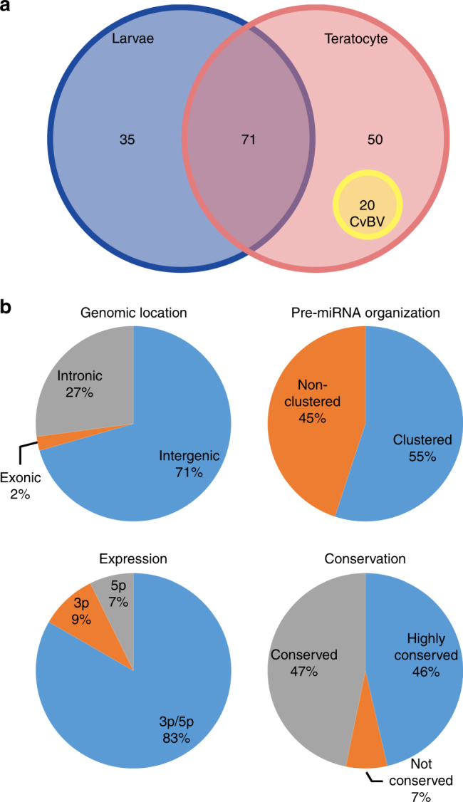
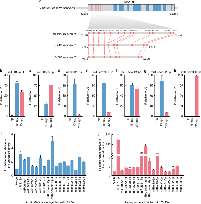
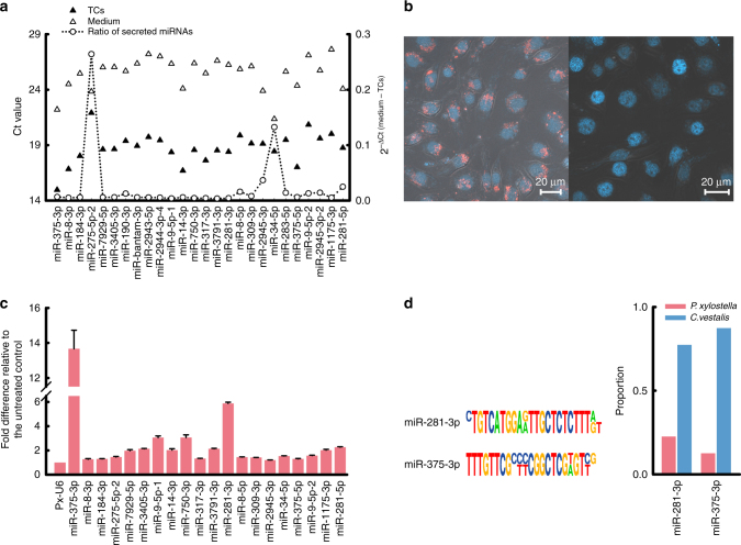
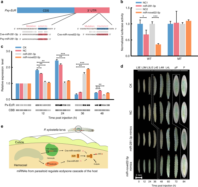
Parasitic wasps produce several factors including venom, polydnaviruses (PDVs) and specialized wasp cells named teratocytes that benefit the survival of offspring by altering the physiology of hosts. However, the underlying molecular mechanisms for the alterations remain unclear. Here we find that the teratocytes of Cotesia vestalis, an endoparasitoid of the diamondback moth Plutella xylostella, and its associated bracovirus (CvBV) can produce miRNAs and deliver the products into the host via different ways. Certain miRNAs in the parasitized host are mainly produced by teratocytes, while the expression level of miRNAs encoded by CvBV can be 100-fold greater in parasitized hosts than non-parasitized ones. We further show that one teratocyte-produced miRNA (Cve-miR-281-3p) and one CvBV-produced miRNA (Cve-miR-novel22-5p-1) arrest host growth by modulating expression of the host ecdysone receptor (EcR). Altogether, our results show the first evidence of cross-species regulation by miRNAs in animal parasitism and their possible function in the alteration of host physiology during parasitism.


Conflict of interest statement

The authors declare no competing interests.

Figures

Fig. 1
Fig. 1
Characteristic of miRNAs in C. vestalis. a miRNAs in C. vestalis larvae and teratocytes. The yellow circle indicates 20 miRNA precursors expressed in teratocytes and encoded within the CvBV proviral genome. b Characteristics of pre-miRNAs in the genome of C. vestalis, mature miRNAs, and conservation with miRNAs identified in other organisms as identified in miRBase. miRNAs in which >18 nucleotides matched miRNAs in miRBase were classified as highly conserved, while miRNAs with 10–18 matching nucleotides were classified as conserved
Fig. 2
Fig. 2
CvBV-derived miRNAs and their expression in P. xylostella. a miRNAs precursors located in CvBV segment C11. The upper bar schematically shows segment C11 in C. vestalis genome scaffold 83. Red vertical lines represent miRNA precursors while blue vertical bars identify predicted protein coding sequences. Below the upper bar is shown the domain of segment C11 that encodes miRNA precursors together with schematics showing miRNA precursors identified in CsBV segment 7 and CcBV segment 7 (Results). bh RT-qPCR analysis of eight CvBV-derived miRNAs in P. xylostella normalized to P. xylostella U6 snRNA. np: non-parasitized P. xylostella, hpp: hours post parasitized. Ct values for undetected samples are considered as 40. i RT-qPCR analysis of 17 CvBV-encoded miRNAs in P. xylostella larvae injected 24 h earlier with 0.1 FEs of CvBV particles. P. xylostella were injected during third instars. Relative expression of CvBV-encoded miRNAs was normalized to the P. xylostella U6 snRNA. j RT-qPCR analysis of 17 CvBV-encoded miRNAs in Pxem-ZJU cells (5 × 106 cells) 24 h post infection with 20 FEs of CvBV particles. The expression of CvBV-encoded miRNAs was normalized to P. xylostella U6 snRNA. Results shown are mean relative abundance ± s.e.m. of each miRNA from three independent biological replicates
Fig. 3
Fig. 3
Teratocytes release miRNAs into the host. a Ct values (left axis) for 30 miRNAs detected in primary cultures of teratocytes and medium conditioned by teratocytes. Each primary culture contained 1 × 105 teratocytes in 1 ml of medium, which were each extracted for isolation of miRNAs after 24 h in primary culture. The abundance ratio (right axis) for each miRNA detected in the medium and in teratocytes is indicated by open circles connected with a dotted line. b Uptake by Pxem_cells of exosomes labeled with Alexa flour 568 (left). Pxem_cells incubation with cultured medium without fluorescent stain was used as control. Red: Alexa flour 568 NHS; Blue: DAPI. c Detection of the 30 most highly expressed miRNAs in teratocytes in Pxem_ZJU cells after co-culturing for 6 h. The y axis shows fold differences for each miRNA in Pxem_ZJU cells co-cultured with teratocytes versus Pxem_ZJU cells cultured without teratocytes using the P. xylostella U6 snRNA as the endogenous control. d Sequence comparison of two miRNAs between C. vestalis and P. xylostella. Sequence differences of two miRNAs between C. vestalis (lower line) and P. xylostella (upper line) are shown on the left. Proportion of two different isoforms of miR-281-3p and miR-375-3p in parasitized P. xylostella larval hemocytes (n = 100) (right). The pink columns represent the proportion of miRNAs from P. xylostella and the blue ones represent that of C. vestalis miRNAs. Primers were designed based on the miR-281-3p and miR-375-3p from C. vestalis and P. xylostella. PCR-amplified fragments were cloned and sequenced, and the difference sequence numbers were calculated. Results shown are mean relative abundance ± s.e.m. of each miRNA from three independent biological replicates
Fig. 4
Fig. 4
Cve-miR-281-3p and Cve-novel22-5p delay growth and pupation of P. xylostella. a Nucleotide sequences showing the complementarity of Cve-miR-281-3p/Cve-miR-novel22-5p and Pxy-miR-281-3p with the wild-type (WT) or mutant (MT) site (red letter) in Pxy-EcR on the Cve-miR-281-3p seed-binding region, highlighted in red. b EcR 3′-UTRs are direct targets of Cve-miR-281-3p or Cve-novel22-5p. pMIR-REPOR-EcR luciferase constructs, containing a WT or MT EcR 3′-UTR were transfected into HEK293 cells. Relative fluorescence ratios in HEK293 cells co-transfected with the indicated pMIR-REPORT-EcR luciferase vector plus either a negative control miRNA (NC1 or NC2), Cve-miR-281-3p, or Cve-novel22-5p mimic. Results shown are mean of four independent experiments ± s.e.m. Differences between groups were analyzed by Student’s t test (*p < 0.05, **p < 0.01, ***p < 0.001). c Transcript (upper graph) and protein levels (lower immunoblots) for Pxy-EcR following miRNA agomir treatment. The time points of miRNA agomir injection were treated as 0 h. Transcript abundance is reported as the mean ± s.e.m. from analysis of three individuals per treatment and time point. Differences between the control and treatment groups were analyzed by Student’s t test (*p < 0.05, **p < 0.01, ***p < 0.001). Coomassie brilliant blue G-250 (CBB) staining was used as a loading control. CK: untreated larvae, NC: larvae injected with a control miRNA. d Development of P. xylostella larvae following miRNA agomir treatment. Sixty larvae were tested in each treatment and their development were monitored until pupation. In the CK and NC groups, P. xylostella larvae initiated apolysis at 24 h post injection (the first white box), while larvae treated with Cve-miR-281-3p or Cve-miR-novel22-5p mimics initiated apolysis at 36 h. At 72 h, larvae in the CK and NC groups pupated, while Cve-miR-281-3p or Cve-miR-novel22-5p treated larvae pupated 12 h later. L3E, -3M, -3L: represent early stage, middle stage, and later stage of third larvae, respectively. E: ecdysis. L4E, -4M, -4L: represent early stage, middle stage, and later stage of 4th larvae, respectively. pP: prepupa, P: pupa. e Cartoon illustrates that Cve-miR-281-3p from teratocytes and Cve-miR-novel22-5p from CvBV regulate ecdysone cascade of the host

Comment in

  • Parasitic interference.
    Zlotorynski E. Zlotorynski E. Nat Rev Mol Cell Biol. 2018 Aug;19(8):487. doi: 10.1038/s41580-018-0035-9. Nat Rev Mol Cell Biol. 2018. PMID: 29942031 No abstract available.

References

    1. Pennacchio F, Strand MR. Evolution of developmental strategies in parasitic hymenoptera. Annu. Rev. Entomol. 2006;51:233–258. doi: 10.1146/annurev.ento.51.110104.151029. - DOI - PubMed
    1. You MS, et al. A heterozygous moth genome provides insights into herbivory and detoxification. Nat. Genet. 2013;45:220–225. doi: 10.1038/ng.2524. - DOI - PubMed
    1. Strand MR, Burke GR. Polydnaviruses: nature's genetic engineers. Annu. Rev. Virol. 2014;1:333–354. doi: 10.1146/annurev-virology-031413-085451. - DOI - PubMed
    1. Asgari S, Rivers DB. Venom proteins from endoparasitoid wasps and their role in host-parasite interactions. Annu. Rev. Entomol. 2011;56:313–335. doi: 10.1146/annurev-ento-120709-144849. - DOI - PubMed
    1. Beckage NE, Gelman DB. Wasp parasitoid disruption of host development: Implications for new biologically based strategies for insect control. Annu. Rev. Entomol. 2004;49:299–330. doi: 10.1146/annurev.ento.49.061802.123324. - DOI - PubMed

Publication types

LinkOut - more resources