

Brain Structure Alterations in Respect to Tobacco Consumption and Nicotine Dependence: A Comparative Voxel-Based Morphometry Study
- PMID: 29881337
- PMCID: PMC5978277
- DOI: 10.3389/fnana.2018.00043
Brain Structure Alterations in Respect to Tobacco Consumption and Nicotine Dependence: A Comparative Voxel-Based Morphometry Study
Abstract
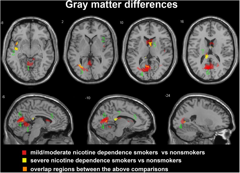
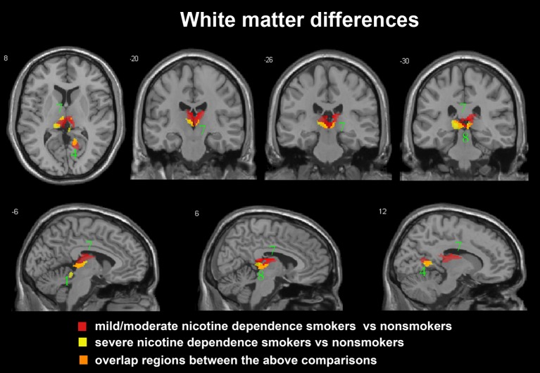
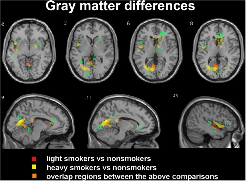
The main purpose of this study is to examine the lifetime tobacco consumption and the degree of nicotine dependence related gray matter (GM) and white matter (WM) volume alterations in young adult-male smokers. Fifty-three long-term male smokers and 53 well-matched male healthy non-smokers participated in the study, and the smokers were respectively categorized into light and heavy tobacco consumption subgroups by pack-years and into moderate and severe nicotine dependence subgroups using the Fagerström Test for Nicotine Dependence (FTND). Voxel-based morphometry analysis was then performed, and ANCOVA analysis combined with subsequent post hoc test were used to explore the between-group brain volume abnormalities related to the smoking amount and nicotine dependence. Light and heavy smokers displayed smaller GM and WM volumes than non-smokers, while heavy smokers were found with more significant brain atrophy than light smokers in GM areas of precuneus, inferior and middle frontal gyrus, superior temporal gyrus, cerebellum anterior lobe and insula, and in WM areas of cerebellum anterior lobe. However, the contrary trend was observed regarding alterations associated with severity of nicotine dependence. Severe nicotine dependence smokers rather demonstrated less atrophy levels compared to moderate nicotine dependence smokers, especially in GM areas of precuneus, superior and middle temporal gyrus, middle occipital gyrus, posterior cingulate and insula, and in WM areas of precuneus, posterior cingulate, cerebellum anterior lobe and midbrain. The results reveal that the nicotine dependence displays a dissimilar effect on the brain volume in comparison to the cigarette consumption. Our study could provide new evidences to understand the adverse effects of smoking on the brain structure, which is helpful for further treatment of smokers.
Keywords: FTND score; nicotine dependence; pack-years; smoking; voxel-based morphometry.
Figures

Similar articles
-
Brain-volume changes in young and middle-aged smokers: a DARTEL-based voxel-based morphometry study.Clin Respir J. 2017 Sep;11(5):621-631. doi: 10.1111/crj.12393. Epub 2015 Oct 13. Clin Respir J. 2017. PMID: 26404024
-
Spontaneous brain activity in chronic smokers revealed by fractional amplitude of low frequency fluctuation analysis: a resting state functional magnetic resonance imaging study.Chin Med J (Engl). 2014;127(8):1504-9. Chin Med J (Engl). 2014. PMID: 24762597
-
[Study on the mechanism of brain damage based on structural covariant network to evaluate the brain structure of nicotine addicts].Zhonghua Yi Xue Za Zhi. 2019 Mar 5;99(9):669-674. doi: 10.3760/cma.j.issn.0376-2491.2019.09.007. Zhonghua Yi Xue Za Zhi. 2019. PMID: 30831615 Chinese.
-
Severity of alcohol dependence is negatively related to hypothalamic and prefrontal cortical gray matter density in heavy drinking smokers.Am J Drug Alcohol Abuse. 2017 May;43(3):281-290. doi: 10.1080/00952990.2016.1257632. Epub 2016 Dec 20. Am J Drug Alcohol Abuse. 2017. PMID: 27996310 Free PMC article.
-
Chronic smoking and brain gray matter changes: evidence from meta-analysis of voxel-based morphometry studies.Neurol Sci. 2013 Jun;34(6):813-7. doi: 10.1007/s10072-012-1256-x. Epub 2012 Dec 4. Neurol Sci. 2013. PMID: 23207549 Review.
Cited by
-
Managing psychotic depression and diagnostic uncertainty in liaison psychiatry.BMJ Case Rep. 2019 Jan 20;12(1):e227606. doi: 10.1136/bcr-2018-227606. BMJ Case Rep. 2019. PMID: 30665931 Free PMC article.
-
Shared gray matter alterations in subtypes of addiction: a voxel-wise meta-analysis.Psychopharmacology (Berl). 2021 Sep;238(9):2365-2379. doi: 10.1007/s00213-021-05920-w. Epub 2021 Jul 27. Psychopharmacology (Berl). 2021. PMID: 34313804 Review.
-
Brain Magnetic Resonance Imaging Features of Nicotine-Dependent Individuals and Its Correlation with Polymorphisms of Dopamine D Receptor Gene.Contrast Media Mol Imaging. 2022 Aug 24;2022:2296776. doi: 10.1155/2022/2296776. eCollection 2022. Contrast Media Mol Imaging. 2022. PMID: 36082055 Free PMC article.
-
Cigarette smoking is associated with cortical thinning in anterior frontal regions, insula and regions showing atrophy in early Alzheimer's Disease.Drug Alcohol Depend. 2018 Nov 1;192:277-284. doi: 10.1016/j.drugalcdep.2018.08.009. Epub 2018 Sep 29. Drug Alcohol Depend. 2018. PMID: 30300802 Free PMC article.
-
Sex-Dependent Alterations of Regional Homogeneity in Cigarette Smokers.Front Psychiatry. 2022 Apr 25;13:874893. doi: 10.3389/fpsyt.2022.874893. eCollection 2022. Front Psychiatry. 2022. PMID: 35546937 Free PMC article.
References
-
- Chu S., Xiao D., Wang S., Peng P., Xie T., He Y., et al. (2014). Spontaneous brain activity in chronic smokers revealed by fractional amplitude of low frequency fluctuation analysis: a resting state functional magnetic resonance imaging study. 127 1504–1509. - PubMed
LinkOut - more resources
Full Text Sources
Other Literature Sources