. 2018 May 24:9:1033.
doi: 10.3389/fimmu.2018.01033. eCollection 2018.

Estrogen Receptor-Alpha (ESR1) Governs the Lower Female Reproductive Tract Vulnerability to Candida albicans

Affiliations

Estrogen Receptor-Alpha (ESR1) Governs the Lower Female Reproductive Tract Vulnerability to Candida albicans

Laura Salinas-Muñoz et al. Front Immunol. .

Abstract

Estradiol-based therapies predispose women to vaginal infections. Moreover, it has long been known that neutrophils are absent from the vaginal lumen during the ovulatory phase (high estradiol). However, the mechanisms that regulate neutrophil influx to the vagina remain unknown. We investigated the neutrophil transepithelial migration (TEM) into the vaginal lumen. We revealed that estradiol reduces the CD44 and CD47 epithelial expression in the vaginal ectocervix and fornix, which retain neutrophils at the apical epithelium through the estradiol receptor-alpha. In contrast, luteal progesterone increases epithelial expression of CD44 and CD47 to promote neutrophil migration into the vaginal lumen and Candida albicans destruction. Distinctive to vaginal mucosa, neutrophil infiltration is contingent to sex hormones to prevent sperm from neutrophil attack; although it may compromise immunity during ovulation. Thus, sex hormones orchestrate tolerance and immunity in the vaginal lumen by regulating neutrophil TEM.

Keywords: ESR1; cervix; estradiol; neutrophils; progesterone; transepithelial migration.


Figures

Figure 1
Figure 1
Neutrophils in the vaginal lavage. Hormone-treated ovariectomized mice were Candida albicans inoculated in the vagina for 6 h. (A) Representative flow cytometry plots of the neutrophils in the vaginal lavage and lower female reproductive tract tissue (n = 5). (B) Representative flow cytometry plots and quantification of the neutrophils in the vaginal lavage of estradiol treated ESR1/ and Wt mice challenged with C. albicans. Data are expressed as box and whiskers 10–90 percentile (n = 5). *p < 0.05 Mann–Whitney. Abbreviations: E2, estradiol; P4, progesterone.
Figure 2
Figure 2
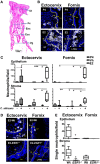
Neutrophils in the female reproductive tract (FRT). Hormone-treated ovariectomized mice were mock or Candida albicans inoculated in the vagina for 6 h. (A) Photomicrograph of the FRT. (B) FRT indicating anatomical regions with LY6G-stained neutrophils (white). (C) LY6G+ cells/field in the lower FRT epithelium and stroma. Data were calculated in at least three different sections of each sample (n = 5). (D) Neutrophils in the vaginal tissue of estradiol treated ESR1/ and Wt mice challenged with C. albicans. Representative photographs of ectocervix and fornix stained with LY6G (neutrophils white) and (E) quantification of the neutrophils. Data are expressed as box and whiskers 10–90 percentile (n = 5). *p < 0.05, **p < 0.01, and ***p < 0.001 Mann–Whitney. Yellow line indicates the lamina propia. Scale bar, 200 µm. Abbreviations: E2, estradiol; Vh, Vehicle; P4, progesterone; Ut, uterus; Fx, fornix; Enx, endocervix; Ecx, ectocervix; Vg, vagina; Lu, lumen; Ep, epithelium; St, stroma.
Figure 3
Figure 3
Hormone-treated ovariectomized mice were mock or Candida albicans inoculated in the vagina. (A) Representative pictures of sections assayed for metallomatrix proteinase 9 (MMP9) (green) and LY6G+ (red). Representative flow cytometry plots and quantification of the MMP9 expression in LY6G+ neutrophils from the lower female reproductive tract (FRT) of hormone-treated mice. MMP9 expression in LY6G+ neutrophils from in vivo hormone treated mice, (B) bone marrow, and (C) ex vivo treated neutrophils. (D) Representative pictures of sections assayed for CD11b (green) and LY6G+ (red). Representative flow cytometry plots and quantifications of the lower FRT neutrophils from hormone-treated mice. CD11b expression in LY6G+ neutrophils from in vivo hormone treated mice, (E) bone marrow, and (F) ex vivo treated neutrophils. Isotype control mAb (line) and indicated mAb (gray filled lines). Scale bar, 50 µm. Each circle represents a mouse in (A,D), n = 4 in (B,E) and n = 5 in (C,F). Abbreviations: E2, estradiol; Vh, vehicle; P4, progesterone; Lu, lumen; Ep, epithelium; St, stroma.
Figure 4
Figure 4
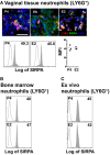
Hormone-treated ovariectomized mice were mock or Candida albicans inoculated in the vagina. (A) Representative pictures of sections assayed for SIRPA (green) and LY6G+ (red). Representative flow cytometry plots and quantification of the SIRPA expression in LY6G+ neutrophils from the lower female reproductive tract of hormone-treated mice. SIRPA expression in LY6G+ neutrophils from in vivo hormone-treated mice, (B) bone marrow, and (C) ex vivo treated neutrophils. Scale bar, 50 µm. Isotype control mAb (line) and indicated mAb (gray filled lines). Each circle represents a mouse (A), n = 4 in (B), and n = 5 in (C). Abbreviations: E2, estradiol; Vh, vehicle; P4, progesterone.
Figure 5
Figure 5
Hormone-treated ovariectomized mice were Candida albicans inoculated in the vagina. (A) Representative pictures of sections assayed for LY6G+ (green) and estradiol nuclear receptor ESR1 (red) or progesterone nuclear receptor PGR (red). (B) Representative flow cytometry plots neutrophils (LY6G+) assayed for ESR1 and PGR expression from vaginal lavage, lower female reproductive tract, blood, and bone marrow cells from hormone-treated mice. Each circle represents a mouse. (C) Bone marrow neutrophils were isolated and ex vivo hormone-treated for 12 h and assayed for ESR1 and PGR expression. A representative experiment of three independent tests is shown. Isotype control mAb (line) and indicated mAb (gray filled lines). Abbreviations: E2, estradiol; P4, progesterone; Ep, epithelium; St, stroma.
Figure 6
Figure 6
CD47 expression in the female reproductive tract (FRT). Hormone-treated ovariectomized mice were mock or Candida albicans inoculated in the vagina. (A) Representative flow cytometry plots of the lower FRT epithelial cells from hormone-treated mice (n = 3). (B) FRT with indicated anatomical regions stained for CD47 (red) or neutrophils (white). (C) CD47 expression in the basolateral epithelium. Data were calculated in at least three different points in three different sections of each sample (n = 5). (D) Estradiol treated ESR1/ and Wt mice challenged with C. albicans. Representative photographs of ectocervix and fornix stained with CD47 (red) and (E) quantification (n = 5). Data are expressed as box and whiskers 10–90 percentile. **p < 0.01 and ***p < 0.001 Mann–Whitney. Isotype control mAb (gray filled lines) and indicated mAb (lines). Yellow line indicates the lamina propia. Scale bar, 200 µm. Abbreviations: E2, estradiol; Vh, vehicle; P4, progesterone. Lu, lumen; Ep, epithelium; St, stroma; A.U., arbitrary units.
Figure 7
Figure 7
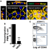
CD44 expression in the vaginal cells. (A) Confocal microscopy images of non-permeabilized VK2 monolayers co-stained with GM35 mAb (green) and CD44 (red). A representative picture of three independent tests is shown. (B) Percentage of control PMNs migrated in response to fMLF. Confluent VK2 monolayers were pre-treated apically with isotype control or GM35 mAb before PMNs were added to the basolateral surface. A representative experiment of 3 independent tests is shown. Data are expressed as mean ± SD. **p < 0.01 Unpaired t test with Welch’s correction. (C) Representative flow cytometry plots of the CD44 expression in the lower female reproductive tract epithelial cells from hormone-treated mice. Data are expressed as percentage of total cells in the gate (n = 3).
Figure 8
Figure 8
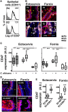
CD44 expression in the female reproductive tract (FRT). Hormone-treated ovariectomized mice were mock or Candida albicans inoculated in the vagina. (A) FRT indicated anatomical regions CD44-stained (red) and LY6G-stained neutrophils (white). (B) CD44 expression in the apical epithelium. Data were calculated in at least three different points in three different sections of each sample (n = 5). (C) Estradiol treated ESR1/ and Wt mice challenged with C. albicans. Representative photographs of ectocervix and fornix stained with CD47 (red) and (D) quantification (n = 5). (E) CD44 was assayed by ELISA in the vaginal lavage of hormone treated C. albicans inoculated mice and (F) estradiol-treated ESR1/ and Wt mice challenged with C. albicans. Data are expressed as box and whiskers 10–90 percentile (n = 5). *p < 0.05, **p < 0.01, and ***p < 0.001 Mann–Whitney. Yellow line indicates the lamina propia. Scale bar, 200 µm. E2, estradiol; Vh, vehicle; P4, progesterone. Abbreviations: Lu, lumen; Ep, epithelium; St, stroma; A.U., arbitrary units.
Figure 9
Figure 9
Bilaterally ovariectomized mice received hormone treatment subcutaneously (n = 5). (A) Bladder and gut CD47-stained. CD47 expression in the epithelium (CDH1+) cross section. (B) Bladder and gut CD44-stained. CD44 expression in the epithelium (CDH1+) cross section. Green arrow shows the cross section direction. Scale bar, 200 µm. E2, estradiol; P4, progesterone. Abbreviation: A.U., arbitrary units; Lu: lumen; Ep: Epithelium; St: Stroma.
Figure 10
Figure 10
Hormone regulation of neutrophil killing activity. Bilaterally ovariectomized mice received hormone-treatment subcutaneously and rat anti-mouse LY6G (1A8) or isotype control antibody intravenously 24 h prior to Candida albicans inoculation to deplete the neutrophils. (A) Progesterone and (B) estradiol treated mice 6 h after the infection. Number of neutrophils and fungal burden (CFU) in the vaginal lavage after the infection (n = 6–8). (C) Fungal burden (CFU) in the vaginal lavage of progesterone or estradiol-treated ESR1−/− and Wt mice challenged with C. albicans. Data are expressed as box and whiskers 10–90 percentile (n = 5). *p < 0.05, **p < 0.01, and ***p < 0.001 Mann–Whitney. Abbreviations: CFU, colony forming unit. E2, estradiol; P4, progesterone.
Figure 11
Figure 11
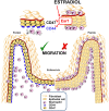
Summarizing schematic figure. Progesterone treatment promotes neutrophil migration to the vaginal lumen. In contrast, estradiol treatment downregulates CD44 and CD47 expression in the cervix and fornix epithelial cells, by an ESR1-dependent mechanism, which accumulates neutrophils in the sub- and supra-epithelial spaces of the ectocervix and vaginal fornix rather than at the vaginal lumen.

References

    1. Nicolas-Avila JA, Adrover JM, Hidalgo A. Neutrophils in homeostasis, immunity, and cancer. Immunity (2017) 46:15–28.10.1016/j.immuni.2016.12.012 - DOI - PubMed
    1. Kruger P, Saffarzadeh M, Weber AN, Rieber N, Radsak M, Von Bernuth H, et al. Neutrophils: between host defence, immune modulation, and tissue injury. PLoS Pathog (2015) 11:e1004651.10.1371/journal.ppat.1004651 - DOI - PMC - PubMed
    1. Lahoz-Beneytez J, Elemans M, Zhang Y, Ahmed R, Salam A, Block M, et al. Human neutrophil kinetics: modeling of stable isotope labeling data supports short blood neutrophil half-lives. Blood (2016) 127:3431–8.10.1182/blood-2016-03-700336 - DOI - PMC - PubMed
    1. Nemeth T, Mocsai A. Feedback amplification of neutrophil function. Trends Immunol (2016) 37:412–24.10.1016/j.it.2016.04.002 - DOI - PubMed
    1. Kim ND, Luster AD. The role of tissue resident cells in neutrophil recruitment. Trends Immunol (2015) 36:547–55.10.1016/j.it.2015.07.007 - DOI - PMC - PubMed

Publication types

MeSH terms

LinkOut - more resources