. 2018 May 24:9:1147.
doi: 10.3389/fimmu.2018.01147. eCollection 2018.

Eicosapentaenoic Acid Enhances the Effects of Mesenchymal Stromal Cell Therapy in Experimental Allergic Asthma

Affiliations

Eicosapentaenoic Acid Enhances the Effects of Mesenchymal Stromal Cell Therapy in Experimental Allergic Asthma

Soraia Carvalho Abreu et al. Front Immunol. .

Abstract

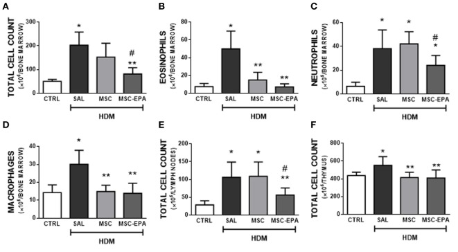
Asthma is characterized by chronic lung inflammation and airway hyperresponsiveness. Despite recent advances in the understanding of its pathophysiology, asthma remains a major public health problem and, at present, there are no effective interventions capable of reversing airway remodeling. Mesenchymal stromal cell (MSC)-based therapy mitigates lung inflammation in experimental allergic asthma; however, its ability to reduce airway remodeling is limited. We aimed to investigate whether pre-treatment with eicosapentaenoic acid (EPA) potentiates the therapeutic properties of MSCs in experimental allergic asthma. Seventy-two C57BL/6 mice were used. House dust mite (HDM) extract was intranasally administered to induce severe allergic asthma in mice. Unstimulated or EPA-stimulated MSCs were administered intratracheally 24 h after final HDM challenge. Lung mechanics, histology, protein levels of biomarkers, and cellularity in bronchoalveolar lavage fluid (BALF), thymus, lymph nodes, and bone marrow were analyzed. Furthermore, the effects of EPA on lipid body formation and secretion of resolvin-D1 (RvD1), prostaglandin E2 (PGE2), interleukin (IL)-10, and transforming growth factor (TGF)-β1 by MSCs were evaluated in vitro. EPA-stimulated MSCs, compared to unstimulated MSCs, yielded greater therapeutic effects by further reducing bronchoconstriction, alveolar collapse, total cell counts (in BALF, bone marrow, and lymph nodes), and collagen fiber content in airways, while increasing IL-10 levels in BALF and M2 macrophage counts in lungs. In conclusion, EPA potentiated MSC-based therapy in experimental allergic asthma, leading to increased secretion of pro-resolution and anti-inflammatory mediators (RvD1, PGE2, IL-10, and TGF-β), modulation of macrophages toward an anti-inflammatory phenotype, and reduction in the remodeling process. Taken together, these modifications may explain the greater improvement in lung mechanics obtained. This may be a promising novel strategy to potentiate MSCs effects.

Keywords: histology; inflammation; lung mechanics; remodeling; resolvin.


Figures

Figure 1
Figure 1
EPA-stimulated lipid body formation in MSCs and modulated secretion of biomarkers by MSCs. (A) LB (red arrow) in MSCs and EPA-MSCs stained with OsO4. (B) Quantification of LB per MSC. Levels of (C) RvD1 and (D) PGE2, assessed by EIA, and (E) IL-10 and (F) TGF-β, assessed by ELISA, in cells stimulated or not with EPA for 6 h. MSC, unstimulated MSCs; MSC-EPA, EPA-stimulated MSCs. Student’s t-test (B), Kruskal–Wallis test followed by Dunn’s test (C,D), and Mann–Whitney U (E,F) were used for statistical comparison. (B) Data presented as mean + SD of five independent experiments. (C–F) Boxes show the interquartile (25–75%) range, whiskers denote the range (minimum–maximum), and horizontal lines represent the median of five independent experiments. *Significantly different from MSC (p < 0.05). **Significantly different from MSC-EPA (p < 0.05). Abbreviations: EPA, eicosapentaenoic acid; MSCs, mesenchymal stromal cells; LB, lipid bodies; N, nucleus; C, cytoplasm; RvD1, resolvin D1; PGE2, prostaglandin E2; IL, interleukin; TGF-β, transforming growth factor-β; i15-LO, inhibitor of 15-lipoxygenase.
Figure 2
Figure 2
EPA-stimulated MSCs led to greater modulation of biomarker secretion and reduction in bronchoalveolar lavage fluid (BALF) cellularity than unstimulated MSCs. Protein levels of (A) IL-4, (B) IL-13, (C) VEGF, (D) IL-10, (E) total leukocytes, (F) eosinophils, (G) macrophages, (H) lymphocytes, and (I) neutrophil counts in BALF. CTRL, saline-challenged mice; HDM, HDM-challenged mice; SAL, HDM mice treated with saline; MSC, HDM mice treated with unstimulated MSCs; MSC-EPA, HDM mice treated with EPA-stimulated MSCs. Kruskal–Wallis test followed by Dunn’s test (A–D) and one-way ANOVA followed by Tukey’s test (E–I) were used for statistical comparison. (A–D) Boxes show the interquartile (25–75%) range, whiskers denote the range (minimum–maximum), and horizontal lines represent the median of eight animals/group. (E–G) Data presented as mean + SD of eight animals/group. *Significantly different from CTRL (p < 0.05). **Significantly different from HDM-SAL (p < 0.05). #Significantly different from HDM-MSC (p < 0.05). Abbreviations: EPA, eicosapentaenoic acid; MSCs, mesenchymal stromal cells; HDM, house dust mite; IL, interleukin; VEGF, vascular endothelial growth factor.
Figure 3
Figure 3
EPA-stimulated MSCs induced macrophage polarization toward an M2 rather than M1 profile. (A) M1-macrophage (iNOS+) and (B) M2-macrophage (CD163+) counts in lung tissue. CTRL, saline-challenge mice; HDM, HDM-challenged mice; SAL, HDM mice treated with saline; MSC, HDM mice treated with unstimulated MSCs; MSC-EPA, HDM mice treated with EPA-stimulated MSCs. The Kruskal–Wallis test followed by Dunn’s test was used for statistical comparison. Boxes show the interquartile (25–75%) range, whiskers denote the range (minimum–maximum), and horizontal lines represent the median of eight animals/group. *Significantly different from CTRL (p < 0.05). **Significantly different from HDM-SAL (p < 0.05). #Significantly different from HDM-MSC (p < 0.05). Abbreviations: EPA, eicosapentaenoic acid; HDM, house dust mite; MSCs, mesenchymal stromal cells.
Figure 4
Figure 4
EPA-stimulated MSCs led to greater reductions in bone marrow, lymph nodes, and thymus cellularity than unstimulated MSCs. (A) Total leukocytes, (B) eosinophils, (C) neutrophils, (D) macrophages in bone marrow, (E) total leukocytes in mediastinal lymph nodes, and (F) total leukocytes in thymus. CTRL, saline-challenged mice; HDM, HDM-challenged mice; SAL, HDM mice treated with saline; MSC, HDM mice treated with unstimulated MSCs; MSC-EPA, HDM mice treated with EPA-stimulated MSCs. One-way ANOVA followed by Tukey’s test was used for statistical comparison. Data are presented as mean + SD. n = 8 animals/group. *Significantly different from CTRL (p < 0.05). **Significantly different from HDM-SAL (p < 0.05). #Significantly different from HDM-MSC (p < 0.05). Abbreviations: EPA, eicosapentaenoic acid; MSCs, mesenchymal stromal cells; HDM, house dust mite.
Figure 5
Figure 5
EPA-stimulated MSCs led to greater reductions in lung remodeling and mucus hypersecretion than unstimulated MSCs. Elastic fiber content in (A) lung parenchyma and (B) airway, collagen fiber content in (C) lung parenchyma and (D) airway, (E) α-SMA expression, and (F) mucus-filled cell count in lung tissue. CTRL, saline-challenged mice; HDM, HDM-challenged mice; SAL, HDM mice treated with saline; MSC, HDM mice treated with unstimulated MSCs; MSC-EPA, HDM mice treated with EPA-stimulated MSCs. The Kruskal–Wallis test followed by Dunn’s test was used for statistical comparison. Boxes show the interquartile (25–75%) range, whiskers denote the range (minimum–maximum), and horizontal lines represent the median of eight animals/group. *Significantly different from CTRL (p < 0.05). **Significantly different from HDM-SAL (p < 0.05). #Significantly different from HDM-MSC (p < 0.05). Abbreviations: EPA, eicosapentaenoic acid; MSCs, mesenchymal stromal cells; HDM, house dust mite; SMA, smooth muscle actin.
Figure 6
Figure 6
EPA-stimulated MSCs led to greater improvement in lung mechanics than unstimulated MSCs. (A) Static lung elastance (Est,L), (B) resistive (ΔP1,L) pressure, and (C) viscoelastic (ΔP2,L) pressure. CTRL, saline-challenged mice; HMD, HMD-challenged mice; SAL, HDM mice treated with saline; MSC, HDM mice treated with unstimulated MSCs; MSC-EPA, HDM mice treated with EPA-stimulated MSCs. One-way ANOVA followed by Tukey’s test was used for statistical comparison. Data are presented as mean + SD. n = 8 animals/group. *Significantly different from CTRL (p < 0.05). **Significantly different from HDM-SAL (p < 0.05). #Significantly different from HDM-MSC (p < 0.05). Abbreviations: EPA, eicosapentaenoic acid; MSCs, mesenchymal stromal cells; HDM, house dust mite.

References

    1. Fahy JV. Type 2 inflammation in asthma – present in most, absent in many. Nat Rev Immunol (2015) 15:57–65.10.1038/nri3807 - DOI - PMC - PubMed
    1. Al-Muhsen S, Johnson JR, Hamid Q. Remodeling in asthma. J Allergy Clin Immunol (2011) 128:451–62.10.1016/j.jaci.2011.04.047 - DOI - PubMed
    1. Royce SG, Tang ML. The effects of current therapies on airway remodeling in asthma and new possibilities for treatment and prevention. Curr Mol Pharmacol (2009) 2:169–81.10.2174/1874467210902020169 - DOI - PubMed
    1. Barnes PJ. New therapies for asthma: is there any progress? Trends Pharmacol Sci (2010) 31:335–43.10.1016/j.tips.2010.04.009 - DOI - PubMed
    1. Papierniak ES, Lowenthal DT, Harman E. Novel therapies in asthma: leukotriene antagonists, biologic agents, and beyond. Am J Ther (2013) 20:79–103.10.1097/MJT.0b013e31826915c2 - DOI - PubMed

Publication types

MeSH terms