. 2018 Jun 6;23(6):1365.
doi: 10.3390/molecules23061365.

Stomatal Complex Development and F-Actin Organization in Maize Leaf Epidermis Depend on Cellulose Synthesis

Affiliations

Stomatal Complex Development and F-Actin Organization in Maize Leaf Epidermis Depend on Cellulose Synthesis

Emmanuel Panteris et al. Molecules. .

Abstract

Cellulose microfibrils reinforce the cell wall for morphogenesis in plants. Herein, we provide evidence on a series of defects regarding stomatal complex development and F-actin organization in Zea mays leaf epidermis, due to inhibition of cellulose synthesis. Formative cell divisions of stomatal complex ontogenesis were delayed or inhibited, resulting in lack of subsidiary cells and frequently in unicellular stomata, with an atypical stomatal pore. Guard cells failed to acquire a dumbbell shape, becoming rounded, while subsidiary cells, whenever present, exhibited aberrant morphogenesis. F-actin organization was also affected, since the stomatal complex-specific arrays were scarcely observed. At late developmental stages, the overall F-actin network was diminished in all epidermal cells, although thick actin bundles persisted. Taken together, stomatal complex development strongly depends on cell wall mechanical properties. Moreover, F-actin organization exhibits a tight relationship with the cell wall.

Keywords: actin; cell wall; cellulose synthesis; cytoskeleton; leaf epidermis; maize; stomata.


Conflict of interest statement

The authors declare no conflict of interest.

Figures

Figure 1
Figure 1
Median CLSM sections of young stomatal rows of control developing Z. mays leaves (labeled as “Control”), and maximum projections of serial CLSM sections through protodermal areas of 2 μΜ DCB-germinated Z. mays leaves (labeled as “DCB”). F-actin is depicted in red and nuclear DNA in blue. (a Control) Progression of stomatal complex ontogenesis, from the stage of initial SMC induction by the GMC (marked as 1), to the stage of subsidiary cell formation (2) and finally to GMC division (3) and fully formed stomatal complex (4). The intervention of a younger stage (arrow) is normal. Note the intense F-actin patches in polarized SMCs and young subsidiary cells, as well as the F-actin aggregations at the pole areas of dividing GMC (arrowheads in 3). (a DCB) Abnormal development of a stomatal row. Between two fully formed stomatal complexes (arrows), undivided GMCs (asterisks) and SMCs (arrowheads) can be observed. (b Control, c Control) Prominent F-actin patches (arrowheads) can be observed at the SMC cortical cytoplasm, at the site of SMC bulging towards the adjacent inducing GMCs (asterisks). Most SMCs appear polarized, as manifested by the juxtaposition of their nuclei to the F-actin patch (arrows). (b DCB, c DCB) SMCs adjacent to inducing GMCs (asterisks) appear unpolarized, as their nuclei reside away from the inducing GMCs (cf. b Control, c Control). F-actin patches can be observed only in mitotic/cytokinetic SMCs (arrowheads), while they are absent from SMCs with interphase nuclei. Scale bars: 10 μm.
Figure 2
Figure 2
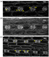
F-actin organization in kidney-shaped (a,b) and dumbbell-shaped (c) stomatal complexes of untreated Z. mays leaves. (a) Median CLSM section, depicting stomata with kidney-shaped guard cells, exhibiting strong fluorescence along either side of the ventral wall, at the middle of which a circular slot (arrowheads) can be observed. Subsidiary cells are triangular, with intense F-actin aggregations at the lateral junctions with the guard cells (arrows, see also (b)). (b) Maximum projection of serial CLSM sections of the cells depicted in (a), exhibiting a dense network of abundant actin filaments in stomatal complexes and pavement cells. (c) Median CLSM section of dumbbell-shaped stomatal complexes, with obtuse-triangular subsidiary cells (asterisks). Intense F-actin signal can be observed at the bulbous ends of ventral guard cell wall (arrows), as well as at the lateral junctions of subsidiary cells with guard cells (arrowheads). Scale bars: 10 μm.
Figure 3
Figure 3
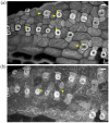
F-actin organization in epidermal areas of 2 μΜ DCB-germinated Z. mays leaves. (a) Abnormally-rounded stomatal complexes at median CLSM section. Guard cells appear swollen as semi-circles, while a longitudinal slot can be observed vertically to the ventral wall (arrowhead). Subsidiary cells appear crescent-shaped (arrows), compressed between guard cells and pavement cells. None of the F-actin aggregations, typical of this developmental stage, can be observed in these stomatal complexes (cf. Figure 2a,b). (b) Severely affected epidermal area, exhibiting highly abnormal stomatal complexes. Several unicellular stomata can be observed (arrows), most of which include an atypical stomatal pore intrusion (arrowheads). Subsidiary cells are abnormally shaped, not exhibiting the typical triangular contour (cf. Figure 2), while in several stomatal complexes there are no subsidiary cells at all (asterisks mark undivided SMCs). None of the F-actin aggregations observed in the untreated kidney- and dumbbell-shaped stomatal complexes (cf. Figure 2) can be observed. Scale bars: 10 μm.
Figure 4
Figure 4
Aberrant cell patterning, abnormal morphogenesis and bundled F-actin network can be observed in this severely affected leaf epidermal area of 2 μΜ DCB-germinated seedlings. (a) Single cortical CLSM section; (b) maximum projection of serial sections of this area. Unicellular stomata (arrows in (a)) with abnormally shaped subsidiary cells and atypical stomatal pore-like intrusions (arrowheads in (a)) are abundant. Actin filaments appear diminished, yet bundled and distorted ((b); cf. Figure 2b), also exhibiting ring-shaped configurations (arrowheads in (b)). Scale bars: 10 μm.
Figure 5
Figure 5
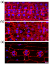
Median CLSM sections of young (a,b) and mature (c) leaf epidermal areas of Z. mays seedlings, treated with 2 μΜ DCB after initial germination. F-actin is depicted in red and nuclear DNA in blue. (a) SMCs adjacent to inducing GMCs (asterisks) appear unpolarized, as their nuclei (arrows) reside away from the inducing GMCs (cf. Figure 1 Control-labeled images). F-actin patches, like those of untreated SMCs (cf. Figure 1 Control-labeled images) are not observed. In some cases, in which SMC division has produced a young subsidiary cell, the latter appears devoid of nucleus, while the rest SMC includes two nuclei (arrowheads). (b) Abnormal development of a stomatal row. Between fully formed stomatal complexes, an undivided GMC (asterisk) flanked by unpolarized SMCs can be observed. One of the young stomatal complexes includes three subsidiary cells (arrowheads). Note that all the young subsidiary cells do not protrude towards the guard cells as in untreated leaves (cf. Figure 1c Control). (c) Abnormally-rounded stomatal complexes. Guard cells appear swollen as semi-circles, while a longitudinal slot can be observed vertically to the ventral wall (arrowhead). Subsidiary cells appear crescent-shaped (arrows), compressed between guard cells and pavement cells. None of the F-actin aggregations, typical of this developmental stage, can be observed in these stomatal complexes (cf. Figure 2a,b). Scale bars: 10 μm.
Figure 6
Figure 6
Maximum projections of serial CLSM sections through protodermal leaf areas (a,b) and single section of mature leaf epidermis (c) of Z. mays seedlings treated with 10 μΜ isoxaben after germination. F-actin is depicted in red and nuclear DNA in blue. (a) SMCs adjacent to inducing GMCs (asterisks) appear unpolarized, as their nuclei (arrows) reside away from the inducing GMCs (cf. Figure 1 Control-labeled images). A prominent F-actin patch, like those of untreated SMCs (cf. Figure 1 Control-labeled images), are observed only in two SMCs (arrowheads), one of which is undergoing cytokinesis. (b) An abnormally developed stomatal row, with an almost mature stomatal complex (arrow) between young ones. Among the latter, one with abnormally divided GMC and one with partially divided GMC (arrowheads) can be observed. (c) Stomatal complexes with kidney-shaped guard cells. Note the abnormally-shaped subsidiary cells (arrows). Scale bars: 10 μm.
Figure 7
Figure 7
Comparison of developing (a) and mature (c) untreated stomatal complexes (at single median CLSM section) with 10 μΜ isoxaben-treated mature (b,d) stomatal complexes (maximum projections of serial CLSM sections). F-actin is depicted in red and nuclear DNA in blue. Note the abnormal swelling of guard cells (b,d), as compared to the normally elongating ones of untreated leaves (a,c), and the presence of abnormally-shaped subsidiary cells (arrows in b,d). In isoxaben-treated epidermis the F-actin aggregations, typical of this developmental stage (cf. Figure 2a,b), are not organized in the stomatal complexes, while extensive F-actin bundling can be also observed in all epidermal cells. Scale bars: 10 μm.

References

    1. Cosgrove D.J. Growth of the plant cell wall. Nat. Rev. Mol. Cell Biol. 2005;6:850–861. doi: 10.1038/nrm1746. - DOI - PubMed
    1. Landrein B., Hamant O. How mechanical stress controls microtubule behavior and morphogenesis in plants: History, experiments and revisited theories. Plant J. 2013;75:324–338. doi: 10.1111/tpj.12188. - DOI - PubMed
    1. Paredez A.R., Somerville C.R., Ehrhardt D.W. Visualization of cellulose synthase demonstrates functional association with microtubules. Science. 2006;312:1491–1495. doi: 10.1126/science.1126551. - DOI - PubMed
    1. Panteris E., Galatis B. The morphogenesis of lobed plant cells in the mesophyll and epidermis: Organization and distinct roles of cortical microtubules and actin filaments. New Phytol. 2005;167:721–732. doi: 10.1111/j.1469-8137.2005.01464.x. - DOI - PubMed
    1. Martinière A., Gayral P., Hawes C., Runions J. Building bridges: Formin1 of Arabidopsis forms a connection between the cell wall and the actin cytoskeleton. Plant J. 2011;66:354–365. doi: 10.1111/j.1365-313X.2011.04497.x. - DOI - PubMed

LinkOut - more resources