. 2018 May 21;23(5):1232.
doi: 10.3390/molecules23051232.

Evaluation of Transition Metal Complexes of Benzimidazole-Derived Scaffold as Promising Anticancer Chemotherapeutics

Affiliations

Evaluation of Transition Metal Complexes of Benzimidazole-Derived Scaffold as Promising Anticancer Chemotherapeutics

Afzal Hussain et al. Molecules. .

Abstract

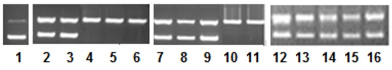
Three new transition metal complexes, Cu(II) 1, Co(II) 2, and Zn(II) 3 with ligand “bimnap” derived from 1-methyl-2-aminobenzimidazole and 2-hydroxynapthaldehyde were synthesized and characterized. The structure of the ligand was determined by single X-ray crystallography. All the three complexes, 13, were examined for the mode of interaction with biomolecule viz., calf thymus-DNA (CT-DNA) using various spectroscopic methods. The nuclease activity was performed against pBR322 DNA that exhibited concentration-dependent degradation of the nucleic acid. The mechanism of DNA cleavage was studied by the electrophoretic pattern in the presence of the radical scavengers. Also, the complexes 13 were analyzed for groove binding affinity. Moreover, in vitro cytotoxicities of the complexes 13 were tested against the five human cancer cell lines, i.e., HeLa, SK-MEL-1, HepG2, HT108, and MDA-MB 231. Also, the cell adhesion and migration properties upon treatment of cell lines with complexes 13, and consequently, their cell death pathway via apoptosis and necrosis were analyzed. Further, complexes 13 were studied in vivo for their toxicities and tolerabilities in mice. In sum, the complexes 13 showed merits of an effective anticancer agent in cell lines⁻based study while minor side effects were observed in vivo.A green solvent extraction technology involving a microwave processing method was used to increase the content of minor ginsenosides from Panax notoginseng. This article aims to investigate the optimization of preparation of the minor ginsenosides by this microwave processing method using single-factor experiments and response surface methodology (RSM), and discuss the blood-enriching activity and hemostatic activity of the extract of microwave processed P. notoginseng (EMPN) The RSM for production of the minor ginsenosides was based on a three-factor and three-level Box-Behnken design. When the optimum conditions of microwave power, temperature and time were 495.03 W, 150.68 °C and 20.32 min, respectively, results predicted that the yield of total minor ginsenosides (Y₉) would be 93.13%. The actual value of Y₉ was very similar to the predicted value. In addition, the pharmacological results of EMPN in vivo showed that EMPN had the effect of enriching blood in N-acetylphenylhydrazine (APH) and cyclophosphamide (CTX)-induced blood deficient mice because of the increasing content of white blood cells (WBCs) and hemoglobin (HGB) in blood. Hemostatic activity in vitro of EMPN showed that it had significantly shortened the clotting time in PT testing (p < 0.05). The hemostatic effect of EMPN was mainly caused by its components of Rh₄, 20(S)-Rg₃ and 20(R)-Rg₃. This microwave processing method is simple and suitable to mass-produce the minor ginsenosides from P. notoginseng.

Keywords: DNA binding/cleavage; apoptosis; cell migration/adhesion; cytotoxicity; metal complexes; toxicity profile.


Conflict of interest statement

The authors declare no conflict of interest.

Figures

Figure 1
Figure 1
Schematic representation of the ligand synthesis and proposed structures of the Cu(II) (1), Co(II) (2), and Zn(II) (3) complexes.
Figure 2
Figure 2
Ortep view of the single X-ray structure of the ligand at the 50% probability level.
Figure 3
Figure 3
Absorption spectra of complexes (A) 1, (B) 2, and (C) 3, with calf thymus-DNA (CT-DNA) in 5 mM Tris-HCl/50 mM NaCl buffer of pH 7.5 at room temperature.
Figure 4
Figure 4
Quenching spectra of EtBr-DNA adduct by the complexes (A) 1, (B) 2, and (C) 3, with CT-DNA in 5 mM Tris-HCl/50 mM NaCl buffer of pH 7.5 at room temperature.
Figure 5
Figure 5
Concentration dependent pattern of pBR322 DNA (100 ng) by complex 1 (lane 2–6), complex 2 (lane 7–11), and complex 3 (lane 12–16) (0.5–2.5 μM), in 50 mM Tris-HCl/NaCl buffer pH 7.4 after 45 min of incubation. lane 1: DNA alone (control); lane 2: DNA + 0.5 μM 1; lane 3: DNA + 1.0 μM 1; lane 4: DNA + 1.5 μM 1; lane 5: DNA + 2.0 μM 1; lane 6: DNA + 2.5 μM 1; lane 7: DNA + 0.5 μM 2; lane 8: DNA + 1.0 μM 2; lane 9: DNA + 1.5 μM 2; lane 10: DNA + 2.0 μM 2; lane 11: DNA + 2.5 μM; lane 12: DNA + 0.5 μM 3; lane 13: DNA + 1.0 μM 3; lane 14: DNA + 1.5 μM 3; lane 15: DNA + 2.0 μM 3; lane 16: DNA + 2.5 μM.
Figure 6
Figure 6
Electrophoretic pattern of pBR322DNA in the presence of scavengers/groove binders in 50 mM Tris-HCl/NaCl buffer pH 7.4 after 45 min. lane 1: DNA alone (control); lane 2: DNA + DAPI + 1; lane 3: DNA + MG + 1; lane 4: DNA + DMSO + 1; lane 5: DNA + EtOH + 1; lane 6: DNA + NaN3 + 1; lane 7: DNA + SOD + 1; lane 8: DNA + DAPI + 2; lane 9: DNA + MG + 2; lane 10: DNA + DMSO + 2; lane 11: DNA + EtOH + 2; lane 12: DNA + NaN3 + 2; lane 13: DNA + SOD + 2. lane 14: DNA + DAPI + 3; lane 15: DNA + MG + 3; lane 16: DNA + DMSO + 3; lane 17: DNA + EtOH + 3; lane 18: DNA + NaN3 + 3; lane 19: DNA + SOD + 3.
Figure 7
Figure 7
(A) Effect of complex 3 on cell adhesion against five cancer cell lines. Assays for adhesion and migration were performed with a cytoselect 24-well plate, and the absorbance of extracted samples was read at 560 nm. (B) Effect of complex 3 on cell migration against five cancer cell lines. Assays for adhesion and migration were performed with a cytoselect 24-well plate, and the absorbance of extracted samples was read at 560 nm.
Figure 8
Figure 8
The apoptotic effect of complex 3 using Annexin-V staining of tested cancer cell lines. Dots represent cells as follows: lower left quadrant, normal cells (FITC/PI); lower right quadrant, early apoptotic cells (FITC+/PI); upper left quadrant, necrotic cells (FITC/PI+); upper right quadrant, late apoptotic cells (FITC+/PI+).
Figure 9
Figure 9
Chronic toxicity profile of complexes 1, 2 and 3. Effect of complexes 1, 2 and 3 on (A) blood components, (B) biochemical parameters of liver function, (C) biochemical parameters of kidney function, and (D) biochemical parameters of lipids and heart function. M and F denote male and female mice, respectively. Statistical analysis was performed by two-way ANOVA, Dunett as post-Hoc test compared to control (C), n = 4. (* p < 0.05; ** p < 0.01; *** p < 0.005; **** p < 0.001).
Figure 9
Figure 9
Chronic toxicity profile of complexes 1, 2 and 3. Effect of complexes 1, 2 and 3 on (A) blood components, (B) biochemical parameters of liver function, (C) biochemical parameters of kidney function, and (D) biochemical parameters of lipids and heart function. M and F denote male and female mice, respectively. Statistical analysis was performed by two-way ANOVA, Dunett as post-Hoc test compared to control (C), n = 4. (* p < 0.05; ** p < 0.01; *** p < 0.005; **** p < 0.001).

References

    1. Wong E., Giandomenico C.M. Current status of platinum-based antitumor drugs. Chem. Rev. 1999;99:2451–2466. doi: 10.1021/cr980420v. - DOI - PubMed
    1. Alderden R.A., Hall M.D., Hambley T.W. The discovery and development of cisplatin. J. Chem. Educ. 2006;83:728. doi: 10.1021/ed083p728. - DOI
    1. Jamieson E.R., Lippard S.J. Structure, recognition, and processing of cisplatin-DNA adducts. Chem. Rev. 1999;99:2467–2498. doi: 10.1021/cr980421n. - DOI - PubMed
    1. Munteanu C.R., Suntharalingam K. Advances in cobalt complexes as anticancer agents. Dalton Trans. 2015;44:13796–13808. doi: 10.1039/C5DT02101D. - DOI - PubMed
    1. Bergamo A., Sava G. Ruthenium anticancer compounds: Myths and realities of the emerging metal-based drugs. Dalton Trans. 2011;40:7817–7823. doi: 10.1039/c0dt01816c. - DOI - PubMed

MeSH terms

LinkOut - more resources