. 2018 Jun 8;13(6):e0198817.
doi: 10.1371/journal.pone.0198817. eCollection 2018.

Age-related endolysosome dysfunction in the rat urothelium

Affiliations

Age-related endolysosome dysfunction in the rat urothelium

Steven T Truschel et al. PLoS One. .

Abstract

Lysosomal dysfunction is associated with a number of age-related pathologies that affect all organ systems. While much research has focused on neurodegenerative diseases and aging-induced changes in neurons, much less is known about the impact that aging has on lower urinary tract function. Our studies explored age-dependent changes in the content of endo-lysosomal organelles (i.e., multivesicular bodies, lysosomes, and the product of their fusion, endolysosomes) and age-induced effects on lysosomal degradation in the urothelium, the epithelial tissue that lines the inner surface of the bladder, ureters, and renal pelvis. When examined by transmission electron microscopy, the urothelium from young adult rats (~3 months), mature adult rats (~12 months), and aged rats (~26 months old) demonstrated a progressive age-related accumulation of aberrantly large endolysosomes (up to 7μm in diameter) that contained undigested content, likely indicating impaired degradation. Stereological analysis confirmed that aged endolysosomes occupied approximately 300% more volume than their younger counterparts while no age-related change was observed in multivesicular bodies or lysosomes. Consistent with diminished endolysosomal degradation, we observed that cathepsin B activity was significantly decreased in aged versus young urothelial cell lysates as well as in live cells. Further, the endolysosomal pH of aged urothelium was higher than that of young adult (pH 6.0 vs pH 4.6). Our results indicate that there is a progressive decline in urothelial endolysosomal function during aging. How this contributes to bladder dysfunction in the elderly is discussed.


Conflict of interest statement

The authors have declared that no competing interests exist.

Figures
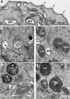
Fig 1
Fig 1. Ultrastructure of endo-lysosomal organelles in the umbrella cell of young adult rats.
(A) TEM of subapical region of umbrella cells showing the hinge regions of the apical plasma membrane (green arrows), and scattered discoidal/fusiform vesicles (DFVs). (B) Ultrastructure of multivesicular bodies (MVBs) in umbrella cells including those with angular limiting membrane (asterisk) and those with a more spherical morphology and containing numerous intraluminal vesicles (ILVs; indicated by arrows). MVBs have a relatively clear lumen. (C) The lumens of lysosomes (LYS) are electron dense and have a fine granular appearance. (D) Endolysosomes (EL) contain numerous luminally disposed vesicles that are larger than the ILVs within MVBs and are contained within a more electron dense lumen. (E) Autophagosomes (AP) are identified by the presence of a double limiting membrane (yellow arrow).
Fig 2
Fig 2. Comparison of endolysosomes in the umbrella cells of young and aged rats.
(A) Umbrella cell from young rat showing typical endolysosomes (EL) ≤ 1 μm in diameter. (B-D) Endolysosomes in aged umbrella cells are generally much larger than those observed in the young and have a tendency to cluster (D). In C, black asterisks denote large luminally disposed vesicles that may represent undigested MVBs. In C-D, white asterisks denote lysosomes that have not yet dispersed their content. In D, the red arrow marks stacked undigested membrane, whereas the dashed red circles highlight areas of possible fusion between adjacent endolysosomes (magnified in the boxed region as an inset.
Fig 3
Fig 3. Features of endolysosomes in aged animals.
(A) Luminally disposed vesicles contain particulate matter (lower white arrow), electron dense cores (upper white arrow), and stacked membrane (red arrows). (B) Some luminally disposed vesicles appeared to protrude from the limiting membrane of endolysosomes (black arrows), and based on their size and the presence of faintly stained ILVs, endolysosomes also contained MVBs. Autophagosomes were sometimes observed in direct contact with the outer limiting membrane of the endolysosome (yellow arrow). (C) A large MVB present in the lumen of the endolysosome. (D) In addition, endolysosomes contain autophagosomal content based on the presence of vesicles with a double limiting membrane (yellow arrow in inset).
Fig 4
Fig 4
Comparison of LIMP2-positive organelles in young (A), adult (B), and aged (C) rat urothelium. LIMP2-positive structures (green) shown in umbrella cells (delineated by dashed yellow line). Rhodamine phalloidin (red), which labels the cortical actin cytoskeleton, was used to show cell borders. Umbrella cells show an apparent, age-related increase in LIMP2 staining (white arrows).
Fig 5
Fig 5. The volume of endolyosomes increases in umbrella cells with age.
(A-C) TEM comparing size of MVBs (yellow structures), endolysosomes (green structures), and lysosomes (purple structures) in umbrella cells taken from young, adult, and aged rats. (D,E) Stereological analysis of the fractional volume (Vv) (D) and total organelle volume (E) of MVBs (yellow bars), endolysosomes (green bars), and lysosomes (purple bars) in umbrella cells. Values represent mean ± standard deviation. Asterisks denote significant differences (p value < 0.05).
Fig 6
Fig 6. Lysotracker Red accumulates in endolysosomes.
Immunofluorescence of young (A), adult (B), and aged (C) urothelium incubated with Lysotracker Red (red) and co-stained with antibodies to LIMP2 (green structures) and phalloidin (blue), which labels the cortical actin cytoskeleton. Large, Lysotracker Red-positive endolysosomes are marked with white arrows. Underlying intermediate (IC) and basal cells (BC) show non-specific cytoplasmic Lysotracker Red staining that did not appear in umbrella cells (UC).
Fig 7
Fig 7. Aging is associated with decreased endolysosome acidification and loss of cathepsin B activity.
(A) Lysosensor Yellow/Blue DND-160 fluorescence measurements (ratio of emission readings at 450 nm/550 nm, excitation at 365 nm) of urothelial cells from young (red inverted triangles) or aged (blue open circles) rats were converted to pH readings through use of a standard curve (black closed circles, with dotted linear regression line). Note the tendency for endolysosomal pH to trend towards neutral pH in aged rat urothelial cells as compared to cells from young animals. (B) Summary of endolysosomal pH measurements. (C,D) Activity assays demonstrate that N-acetylglucosaminidase activity (C) is unaffected by age but Cathepsin B activity (D) is significantly decreased in urothelial cell lysates. (E) Magic Red Cathepsin activity in young and aged cells shows time-dependent increase in enzyme activity with young, but not aged cells. Results are quantified in (F). Values in B,C,D, and F represent mean ± standard deviation. Asterisks denote significant differences (p value < 0.05).
Fig 8
Fig 8. Point sampled intercept method for measuring volume weighted mean umbrella cell volume.
Representative image taken from adult urinary bladder stained with Rhodamine Phalloidin (to outline cell borders). After the grayscale image was inverted (and randomly rotated), a grid (shown as vertical and horizontal intersecting blue lines) was superimposed onto each image and intersections of grid lines were counted as vertices when they fell within the profile of an umbrella cell (examples illustrated by red dots). The horizontal distance along each grid line that spanned the boundary of the umbrella cell (shown by yellow lines) was measured for each cell. The distance of the yellow line in the top umbrella cell was counted twice because two profiles (shown by red dots) fell within that cell. As described in the methods, the length of the intersection was converted to volume-weighted mean cell volume using the following formula: V¯v=π3(l3¯).

References

    1. Xu H, Ren D. Lysosomal physiology. Annu Rev Physiol. 2015;77:57–80. Epub 2015/02/11. doi: 10.1146/annurev-physiol-021014-071649 ; PubMed Central PMCID: PMCPMC4524569. - DOI - PMC - PubMed
    1. Bright NA, Davis LJ, Luzio JP. Endolysosomes Are the Principal Intracellular Sites of Acid Hydrolase Activity. Curr Biol. 2016;26(17):2233–45. doi: 10.1016/j.cub.2016.06.046 ; PubMed Central PMCID: PMCPMC5026700. - DOI - PMC - PubMed
    1. Kundu M, Thompson CB. Autophagy: basic principles and relevance to disease. Annu Rev Pathol. 2008;3:427–55. Epub 2007/11/28. doi: 10.1146/annurev.pathmechdis.2.010506.091842 . - DOI - PubMed
    1. Zoncu R, Bar-Peled L, Efeyan A, Wang S, Sancak Y, Sabatini DM. mTORC1 senses lysosomal amino acids through an inside-out mechanism that requires the vacuolar H(+)-ATPase. Science. 2011;334(6056):678–83. Epub 2011/11/05. doi: 10.1126/science.1207056 ; PubMed Central PMCID: PMCPMC3211112. - DOI - PMC - PubMed
    1. Forgac M. Vacuolar ATPases: rotary proton pumps in physiology and pathophysiology. Nat Rev Mol Cell Biol. 2007;8(11):917–29. Epub 2007/10/04. doi: 10.1038/nrm2272 . - DOI - PubMed

Publication types

MeSH terms