

Relationship of In Vivo MR Parameters to Histopathological and Molecular Characteristics of Newly Diagnosed, Nonenhancing Lower-Grade Gliomas
- PMID: 29883968
- PMCID: PMC6041571
- DOI: 10.1016/j.tranon.2018.05.005
Relationship of In Vivo MR Parameters to Histopathological and Molecular Characteristics of Newly Diagnosed, Nonenhancing Lower-Grade Gliomas
Abstract
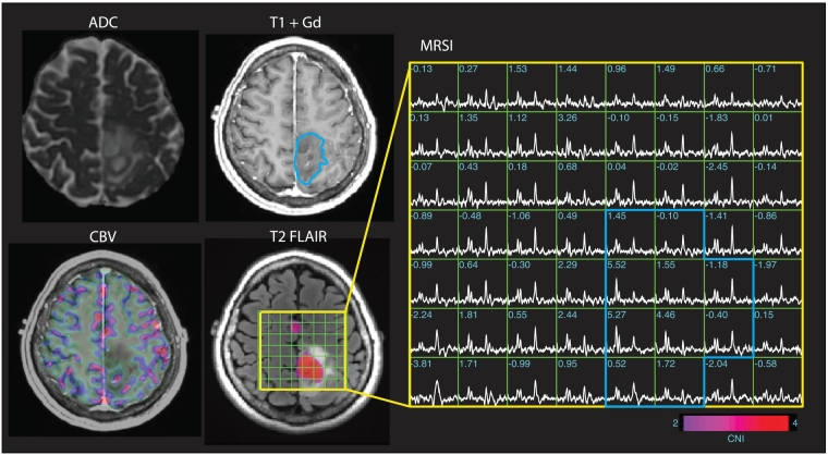
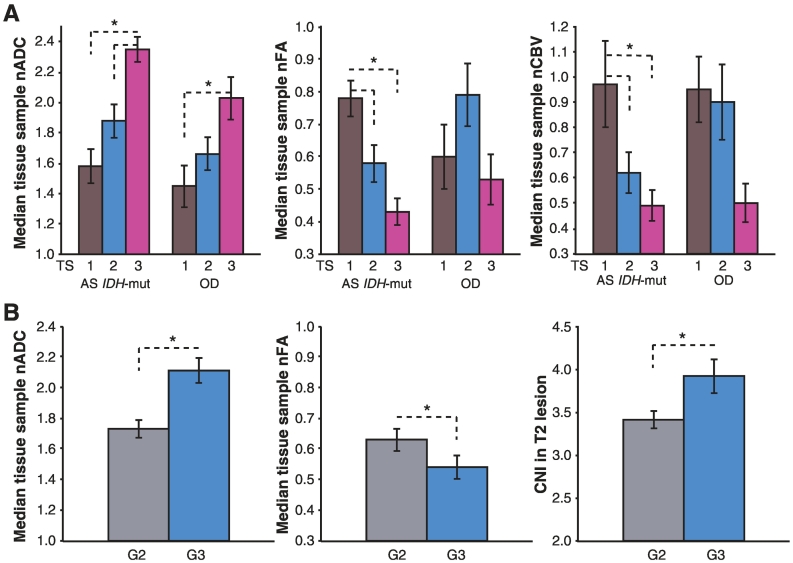
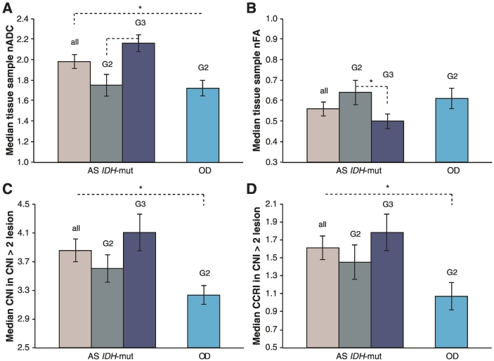
The goal of this research was to elucidate the relationship between WHO 2016 molecular classifications of newly diagnosed, nonenhancing lower grade gliomas (LrGG), tissue sample histopathology, and magnetic resonance (MR) parameters derived from diffusion, perfusion, and 1H spectroscopic imaging from the tissue sample locations and the entire tumor. A total of 135 patients were scanned prior to initial surgery, with tumor cellularity scores obtained from 88 image-guided tissue samples. MR parameters were obtained from corresponding sample locations, and histograms of normalized MR parameters within the T2 fluid-attenuated inversion recovery lesion were analyzed in order to evaluate differences between subgroups. For tissue samples, higher tumor scores were related to increased normalized apparent diffusion coefficient (nADC), lower fractional anisotropy (nFA), lower cerebral blood volume (nCBV), higher choline (nCho), and lower N-acetylaspartate (nNAA). Within the T2 lesion, higher tumor grade was associated with higher nADC, lower nFA, and higher Cho to NAA index. Pathological analysis confirmed that diffusion and metabolic parameters increased and perfusion decreased with tumor cellularity. This information can be used to select targets for tissue sampling and to aid in making decisions about treating residual disease.
Copyright © 2018 The Authors. Published by Elsevier Inc. All rights reserved.
Figures
Similar articles
-
Apparent diffusion coefficient and fractional anisotropy of newly diagnosed grade II gliomas.NMR Biomed. 2009 May;22(4):449-55. doi: 10.1002/nbm.1357. NMR Biomed. 2009. PMID: 19125391 Free PMC article.
-
Spatial characteristics of newly diagnosed grade 3 glioma assessed by magnetic resonance metabolic and diffusion tensor imaging.Transl Oncol. 2012 Feb;5(1):10-8. doi: 10.1593/tlo.11208. Epub 2012 Feb 1. Transl Oncol. 2012. PMID: 22348171 Free PMC article.
-
Measurements of diagnostic examination performance using quantitative apparent diffusion coefficient and proton MR spectroscopic imaging in the preoperative evaluation of tumor grade in cerebral gliomas.Eur J Radiol. 2011 Nov;80(2):462-70. doi: 10.1016/j.ejrad.2010.07.017. Epub 2010 Aug 13. Eur J Radiol. 2011. PMID: 20708868
-
Imaging biomarkers guided anti-angiogenic therapy for malignant gliomas.Neuroimage Clin. 2018 Jul 5;20:51-60. doi: 10.1016/j.nicl.2018.07.001. eCollection 2018. Neuroimage Clin. 2018. PMID: 30069427 Free PMC article. Review.
-
Differentiating surgical from non-surgical lesions using perfusion MR imaging and proton MR spectroscopic imaging.Technol Cancer Res Treat. 2004 Dec;3(6):557-65. doi: 10.1177/153303460400300605. Technol Cancer Res Treat. 2004. PMID: 15560713 Review.
Cited by
-
Radiological differences between subtypes of WHO 2016 grade II-III gliomas: a systematic review and meta-analysis.Neurooncol Adv. 2020 Apr 4;2(1):vdaa044. doi: 10.1093/noajnl/vdaa044. eCollection 2020 Jan-Dec. Neurooncol Adv. 2020. PMID: 32642698 Free PMC article. Review.
-
Advanced MR Techniques for Preoperative Glioma Characterization: Part 1.J Magn Reson Imaging. 2023 Jun;57(6):1655-1675. doi: 10.1002/jmri.28662. Epub 2023 Mar 3. J Magn Reson Imaging. 2023. PMID: 36866773 Free PMC article. Review.
-
T2 FLAIR Hyperintensity Volume Is Associated With Cognitive Function and Quality of Life in Clinically Stable Patients With Lower Grade Gliomas.Front Neurol. 2022 Jan 28;12:769345. doi: 10.3389/fneur.2021.769345. eCollection 2021. Front Neurol. 2022. PMID: 35153976 Free PMC article.
-
Longitudinal MR spectroscopy to detect progression in patients with lower-grade glioma in the surveillance phase.Neurooncol Adv. 2022 Nov 16;4(1):vdac175. doi: 10.1093/noajnl/vdac175. eCollection 2022 Jan-Dec. Neurooncol Adv. 2022. PMID: 36479058 Free PMC article.
-
Spectroscopic imaging of D-2-hydroxyglutarate and other metabolites in pre-surgical patients with IDH-mutant lower-grade gliomas.J Neurooncol. 2022 Aug;159(1):43-52. doi: 10.1007/s11060-022-04042-3. Epub 2022 Jun 8. J Neurooncol. 2022. PMID: 35672531 Free PMC article.
References
-
- Louis DN, Perry A, Reifenberger G, von Deimling A, Figarella-Branger D, Cavenee WK, Ohgaki H, Wiestler OD, Kleihues P, Ellison DW. The 2016 World Health Organization Classification of Tumors of the Central Nervous System: a summary. Acta Neuropathol. 2016;131:803–820. - PubMed
-
- Field KM, Rosenthal MA, Khasraw M, Sawkins K, Nowak AK. Evolving management of low grade glioma: No consensus amongst treating clinicians. J Clin Neurosci. 2016;23:81–87. - PubMed
-
- Le Rhun E, Taillibert S, Chamberlain MC. Current Management of Adult Diffuse Infiltrative Low Grade Gliomas. Curr Neurol Neurosci Rep. 2016;16:15. - PubMed
Grants and funding
LinkOut - more resources
Full Text Sources
Other Literature Sources
