. 2018 Jun 8;19(1):114.
doi: 10.1186/s12931-018-0816-x.

The therapeutic effect of mesenchymal stem cells on pulmonary myeloid cells following neonatal hyperoxic lung injury in mice

Affiliations

The therapeutic effect of mesenchymal stem cells on pulmonary myeloid cells following neonatal hyperoxic lung injury in mice

Ali Al-Rubaie et al. Respir Res. .

Abstract

Background: Exposure to high levels of oxygen (hyperoxia) after birth leads to lung injury. Our aims were to investigate the modulation of myeloid cell sub-populations and the reduction of fibrosis in the lungs following administration of human mesenchymal stem cells (hMSC) to neonatal mice exposed to hyperoxia.

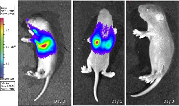
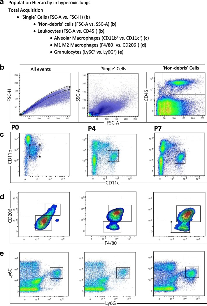
Method: Newborn mice were exposed to 90% O2 (hyperoxia) or 21% O2 (normoxia) from postnatal days 0-4. A sub-group of hyperoxia mice were injected intratracheally with 2.5X10⁵ hMSCs. Using flow cytometry we assessed pulmonary immune cells at postnatal days 0, 4, 7 and 14. The following markers were chosen to identify these cells: CD45+ (leukocytes), Ly6C+Ly6G+ (granulocytes), CD11b+CD11c+ (macrophages); macrophage polarisation was assessed by F4/80 and CD206 expression. hMSCs expressing enhanced green fluorescent protein (eGFP) and firefly luciferase (fluc) were administered via the trachea at day 4. Lung macrophages in all groups were profiled using next generation sequencing (NGS) to assess alterations in macrophage phenotype. Pulmonary collagen deposition and morphometry were assessed at days 14 and 56 respectively.

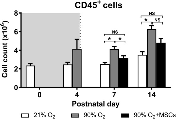
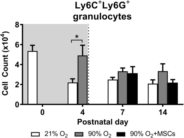
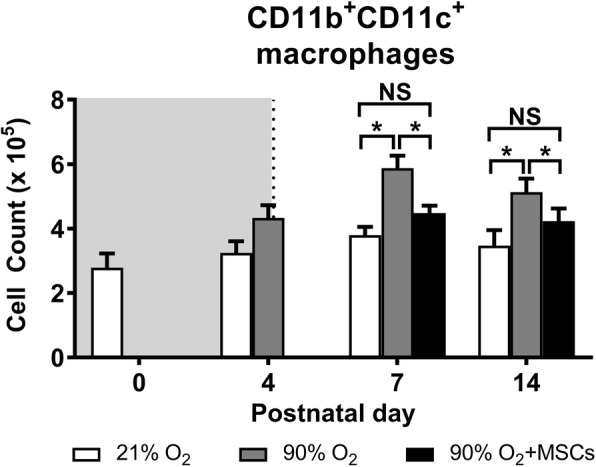
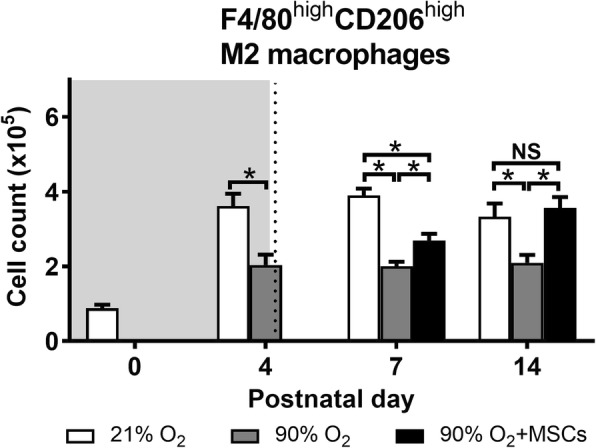
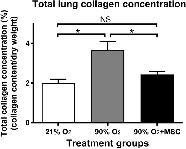
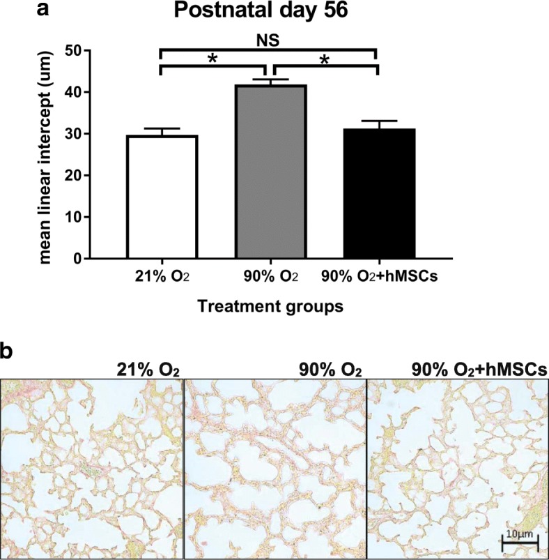
Results: At day 4, hyperoxia increased the number of pulmonary Ly6C+Ly6G+ granulocytes and F4/80lowCD206low macrophages but decreased F4/80highCD206high macrophages. At days 7 and 14, hyperoxia increased numbers of CD45+ leukocytes, CD11b+CD11c+ alveolar macrophages and F4/80lowCD206low macrophages but decreased F4/80highCD206high macrophages. hMSCs administration ameliorated these effects of hyperoxia, notably reducing numbers of CD11b+CD11c+ and F4/80lowCD206low macrophages; in contrast, F4/80highCD206high macrophages were increased. Genes characteristic of anti-inflammatory 'M2' macrophages (Arg1, Stat6, Retnla, Mrc1, Il27ra, Chil3, and Il12b) were up-regulated, and pro-inflammatory 'M1' macrophages (Cd86, Stat1, Socs3, Slamf1, Tnf, Fcgr1, Il12b, Il6, Il1b, and Il27ra) were downregulated in isolated lung macrophages from hyperoxia-exposed mice administered hMSCs, compared to mice without hMSCs. Hydroxyproline assay at day 14 showed that the 2-fold increase in lung collagen following hyperoxia was reduced to control levels in mice administered hMSCs. By day 56 (early adulthood), hMSC administration had attenuated structural changes in hyperoxia-exposed lungs.

Conclusions: Our findings suggest that hMSCs reduce neonatal lung injury caused by hyperoxia by modulation of macrophage phenotype. Not only did our cell-based therapy using hMSC induce structural repair, it limited the progression of pulmonary fibrosis.

Keywords: Alveolar macrophages; Mesenchymal stem cells; Neonatal hyperoxia.


Conflict of interest statement

Ethics approval

The study was conducted under animal ethics number MARP2014/092 issued by Monash Animal Ethics Committee.

Competing interests

The authors declare that they have no competing interests.

Publisher’s Note

Springer Nature remains neutral with regard to jurisdictional claims in published maps and institutional affiliations.

Figures

Fig. 1
Fig. 1
Luciferin/GFP + MSCs were injected into mice after hyperoxia n = 8, and the bioluminescence fluc signal was examined at time 0 and day 1 and 3 after luciferase injection. Bioluminescence imaging showing the intensity of fluc signal representing MSC localization to lungs. Red signals indicate high intensity of MSCs and blue indicates low intensity – see heatmap panel
Fig. 2
Fig. 2
The representative gating strategy for assessing myeloid cell sub-populations in the lung. The population hierarchy shows the gating strategy (a). ‘Single’ cells (excluding doublets and triplets) were selected with a polygon gate on a FSC-A vs. FSC-H dot plot (b). Single cells were gated on the resulting daughter population on a FSC-A vs. SCA-A dot plot (b). A ‘Live’ cell gate (which excludes debris) was created with the aid of the CD45+ cells on FSC-A vs. CD45+ (b). CD45+CD11b+CD11c+ alveolar macrophages were selected with a gate (b), CD45+ F4/80+ CD206+ M1 vs. M2 macrophages were selected with a gate (d) and CD45+Ly6C+Ly6G+ granulocytes (e) were selected for further analysis of myeloid cell subsets. Plots in c, d and e are from a hyperoxic lung taken at day 0, day 4, and day 7 from left to right
Fig. 3
Fig. 3
CD45+ myeloid cell count in lungs from mice following normoxia (), 90% O2 () and 90% O2 with administration of hMSC () delivered at day 4. The shaded area (grey) depicts period of hyperoxia exposure (postnatal days 0–4) prior to delivery of hMSCs (dotted line) on day 4. For all treatment groups, n = 8 per group. Data are shown as mean ± SEM. * = p < 0.05
Fig. 4
Fig. 4
Ly6C+Ly6G+ granulocyte cell count in lungs from mice following normoxia , 90% O2 () and 90% O2 with administration of hMSC () delivered at day 4. The shaded area (gray) depicts period of hyperoxia exposure (postnatal days 0–4) prior to delivery of hMSCs (dotted line) on day 4. For all treatment groups, n = 8 per group. Data are shown as mean ± SEM. * = p < 0.05
Fig. 5
Fig. 5
CD11b+CD11c+ macrophage cell count in lungs from mice following normoxia (), 90% O2 () and 90% O2 with administration of hMSC () delivered at day 4. The shaded area (gray) depicts period of hyperoxia exposure (postnatal days 0–4) prior to delivery of hMSCs (dotted line) on day 4. For all treatment groups, n = 8 per group. Data are shown as mean ± SEM. * = p < 0.05 and NS = not significant
Fig. 6
Fig. 6
F4/80lowCD206low M1 macrophage cell count in lungs from mice following normoxia (), 90% O2 () and 90% O2 with administration of hMSC () delivered at day 4. The shaded area (gray) depicts period of hyperoxia exposure (postnatal days 0–4) prior to delivery of hMSCs (dotted line) on day 4. For all treatment groups, n = 8 per group. Data are shown as mean ± SEM. * = p < 0.05 and NS = not significant
Fig. 7
Fig. 7
F4/80highCD206high M2 macrophage cell count in lungs from mice following normoxia (), 90% O2 () and 90% O2 with administration of hMSC () delivered at day 4. The shaded area (gray) depicts period of hyperoxia exposure (postnatal days 0–4) prior to delivery of hMSCs (dotted line) on day 4. For all treatment groups, n = 8 per group. Data are shown as mean ± SEM. * = p < 0.05 and NS = not significant
Fig. 8
Fig. 8
hMSCs reduce collagen accumulation in the lung following hyperoxic injury total lung collagen concentration (% collagen content/dry weight tissue) in normoxia, 90% O2 and 90% O2 with administration of hMSC on postnatal day 14. For all treatment groups n = 8. Data are shown as mean ± SEM. * = p < 0.05 and NS = not significant
Fig. 9
Fig. 9
a MLI following normoxia, 90% O2 () and 90% O2 + hMSC (). 90% O2 was given between postnatal days 0–4 and MSCs were given on postnatal day 4. For all treatment groups at day 56 (n = 8 per group). Data were analysed using a one-way ANOVA and shown as mean ± SEM. p < 0.05. b representative histological images of the three treatment groups stained with picrosirius red

References

    1. O'Reilly M, Sozo F, Harding R. Impact of preterm birth and bronchopulmonary dysplasia on the developing lung: long-term consequences for respiratory health. Clin Exp Pharmacol Physiol. 2013;40(11):765–773. doi: 10.1111/1440-1681.12068. - DOI - PubMed
    1. Stockwell S. Benefits of kangaroo Care for Premature Babies Continue into young adulthood. Am J Nurs. 2017;117(3):15. doi: 10.1097/01.NAJ.0000513272.38141.e3. - DOI - PubMed
    1. Maitre NL, et al. Respiratory consequences of prematurity: evolution of a diagnosis and development of a comprehensive approach. J Perinatol. 2015;35(5):313–321. doi: 10.1038/jp.2015.19. - DOI - PMC - PubMed
    1. Iyer NP, Mhanna MJ. The role of surfactant and non-invasive mechanical ventilation in early management of respiratory distress syndrome in premature infants. World J Pediatr. 2014;10(3):204–210. doi: 10.1007/s12519-014-0494-9. - DOI - PubMed
    1. Malkar MB, et al. Respiratory severity score on day of life 30 is predictive of mortality and the length of mechanical ventilation in premature infants with protracted ventilation. Pediatr Pulmonol. 2015;50(4):363–369. doi: 10.1002/ppul.23020. - DOI - PubMed

Publication types

MeSH terms