. 2018 Jul 2;145(13):dev157222.
doi: 10.1242/dev.157222.

Amniotic ectoderm expansion in mouse occurs via distinct modes and requires SMAD5-mediated signalling

Affiliations

Amniotic ectoderm expansion in mouse occurs via distinct modes and requires SMAD5-mediated signalling

Mariya P Dobreva et al. Development. .

Erratum in

Abstract

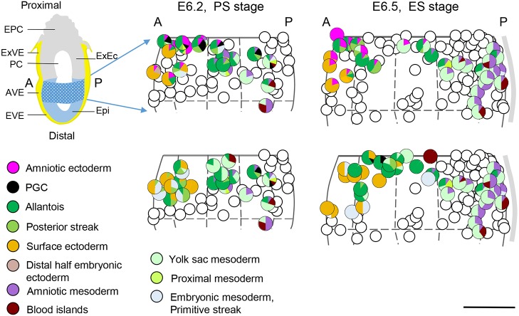
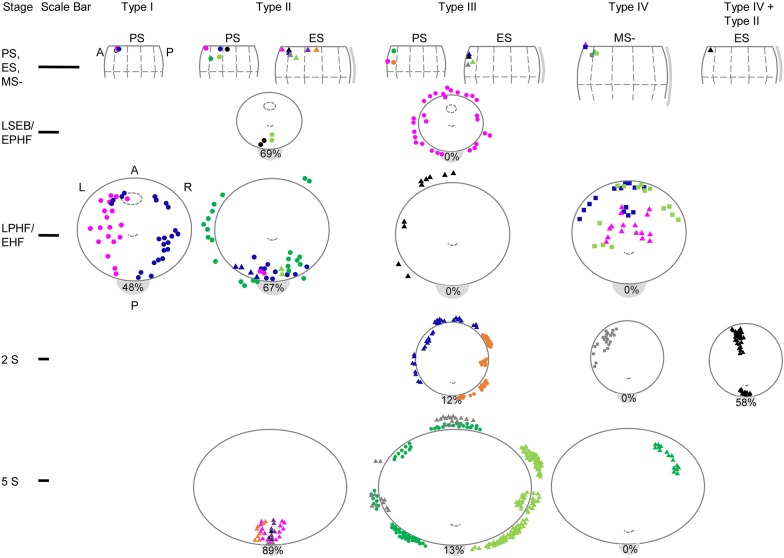
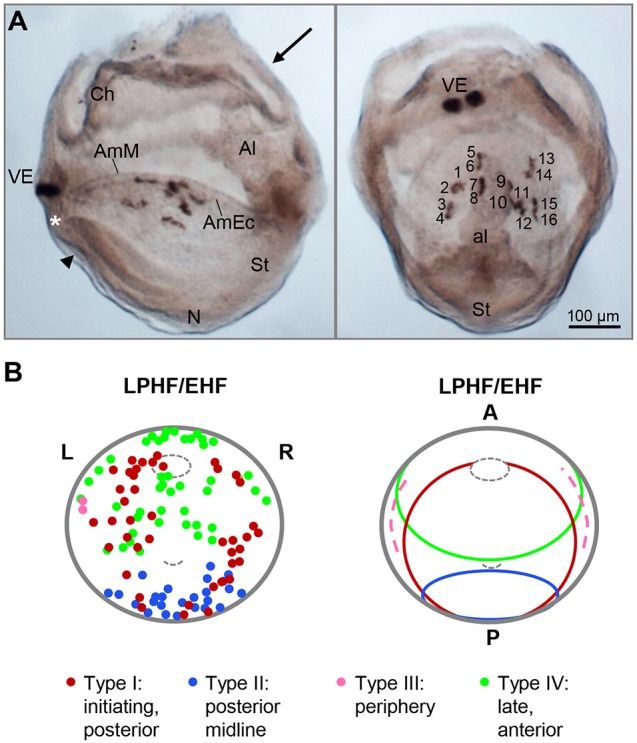
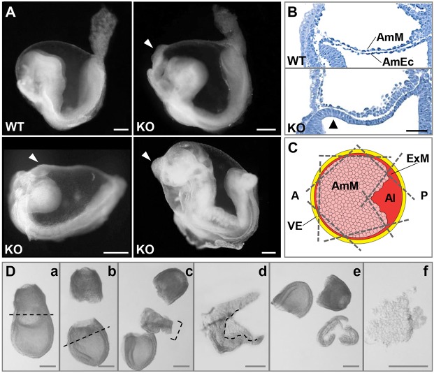
Upon gastrulation, the mammalian conceptus transforms rapidly from a simple bilayer into a multilayered embryo enveloped by its extra-embryonic membranes. Impaired development of the amnion, the innermost membrane, causes major malformations. To clarify the origin of the mouse amnion, we used single-cell labelling and clonal analysis. We identified four clone types with distinct clonal growth patterns in amniotic ectoderm. Two main types have progenitors in extreme proximal-anterior epiblast. Early descendants initiate and expand amniotic ectoderm posteriorly, while descendants of cells remaining anteriorly later expand amniotic ectoderm from its anterior side. Amniogenesis is abnormal in embryos deficient in the bone morphogenetic protein (BMP) signalling effector SMAD5, with delayed closure of the proamniotic canal, and aberrant amnion and folding morphogenesis. Transcriptomics of individual Smad5 mutant amnions isolated before visible malformations and tetraploid chimera analysis revealed two amnion defect sets. We attribute them to impairment of progenitors of the two main cell populations in amniotic ectoderm and to compromised cuboidal-to-squamous transition of anterior amniotic ectoderm. In both cases, SMAD5 is crucial for expanding amniotic ectoderm rapidly into a stretchable squamous sheet to accommodate exocoelom expansion, axial growth and folding morphogenesis.

Keywords: Amnion; Amnion fate map; BMP-SMAD; Chorion; Clonal analysis; Extra-embryonic ectoderm; Extra-embryonic–embryonic interface.


Conflict of interest statement

Competing interestsThe authors declare no competing or financial interests.

Figures

Fig. 1.
Fig. 1.
Amnion fate map. Fate maps of amniotic ectoderm and amniotic mesoderm. Top left: cartoon of a midsagittal section of an E6.2, prestreak stage (PS) embryo. The proximal half of the epiblast cup (blue pattern) is expanded in the right panels and projected on the sagittal midline of PS and early-streak (ES) stages. Left and right halves of the epiblast are superimposed. Half the circumference of the normalized epiblast is flattened and fitted to its diameter (D), i.e. πD/2 is reduced to D. The primitive streak is represented by a grey stripe. The composition (to the nearest 10%) of clones contributing to amnion is shown as pie charts at the positions of clone initiation in the two upper panels. Clones not contributing to the amnion are represented by empty circles. Lower panel: composition of the clones not contributing to amniotic ectoderm in the same region as in the upper panel. Clones contributing to amniotic ectoderm are not represented for clarity. Scale bar: 50 µm. A, anterior; AVE, anterior visceral endoderm; Epi, epiblast; EPC, ectoplacental cone; EVE, embryonic visceral endoderm; ExEc, extra-embryonic ectoderm; ExVE, extra-embryonic visceral endoderm; P, posterior; PC, proamniotic cavity.
Fig. 2.
Fig. 2.
Spread of clone descendants in amniotic ectoderm. Clonal spread in amniotic ectoderm and associated surface ectoderm. Each clone within a type is identified by a colour/shape combination (Table 1). Top row: the positions of clone progenitors are plotted, as in Fig. 1, on scaled representations of the proximal half of the epiblast at different initial stages (PS, ES and MS-). Scale bars: 100 µm. The top edge of the diagram is at the junction with the apposing extra-embryonic (prechorionic) ectoderm. The position of the progenitor of the unplotted Type I PS clone #3 is shown as an empty circle. Lower rows: the view of clone distribution in the amniotic ectoderm (solid line ellipses) is from dorsal with anterior (A) at the top. The projected positions of the closing/closed proamniotic canal and node are indicated by an ellipse (dashed line) and an arc (dashed line), respectively. Cells plotted outside the amnion are in the adjacent non-neural ectoderm (mainly surface ectoderm). The number in the grey crescent at the posterior edge of the amnion is the total number of extra-embryonic mesoderm and posterior streak cells in the clones expressed as a percentage of the total labelled cells in the embryos contributing to the plot. Clones #2 (Type I, dark blue), #17 (Type III, dark blue) and #27 (Type IV, light green) are shown in LR mirror image to reduce overlapping in display.
Fig. 3.
Fig. 3.
Illustration and summary of amniotic ectoderm clones. (A) Lateral view, anterior facing left, and posteriodorsal view (indicated by arrow in lateral view) of a mouse embryo with HRP-labelled Type IV clone (#23) in the amniotic ectoderm (Table 1, Fig. 2). The clone was initiated close to the midline at the extra-embryonic–embryonic junction (Fig. 2), as indicated by the VE marker clone. As the epiblast clone is a pure clone, there is no supporting evidence about the time of entry of descendants into the amniotic ectoderm. The pattern of four files of four cells [(1-4)(5-8)(9-12)(13-16)] oriented anterior-posteriorly indicates that the progenitor divided twice before all four descendants left the periphery of the amniotic ectoderm during the same cell cycle and went through two additional cycles of oriented cell division. Two other Type IV clones (#24 and #27) indicate that cells may divide once or twice at the anterior periphery while releasing descendants to start populating the anterior amniotic ectoderm. The asterisk and arrowhead mark the amniotic ectoderm/embryonic ectoderm junction and the incipient foregut pocket, respectively. Scale bar: 100 µm. (B) Combined distribution (left) of the four types of amniotic ectoderm clones (Fig. 2) in an LPHF/EHF amnion, and simplified territories (right). Anterior (A) to the top. The projected positions of the closed PAC and node are indicated by a small ellipse and arc (dashed lines), respectively. Type I descendants initiate the primordium and establish the amniotic ectoderm posterior to the anterior separation point, whereas Type II descendants populate the most posterior midline subregion. The lineage-restricted Type IV descendants produce amniotic ectoderm anterior to the ASP, and also contribute to the already established Type I territory. Type III descendants expand the amnion only at the periphery. Al, allantois; AmEc, amniotic ectoderm; AmM, amniotic mesoderm; Ch, chorion; N, node; St, streak; PAC, proamniotic canal; VE, visceral endoderm.
Fig. 4.
Fig. 4.
Phenotype of Smad5 mutants and amnion microdissection procedure. (A) Appearance of early somite stage wild-type (WT) and Smad5 knockout (KO) embryos. The mutant amnion contains an anteriorly localized tissue aggregate (arrowhead). Scale bars: 200 μm. (B) Longitudinal sections of E7.5 LPHF-stage WT and Smad5 KO embryos stained with Haematoxylin. Amniotic ectoderm (AmEc) is thickened in mutants (arrowhead). Scale bar: 50 μm. AmM, amniotic mesoderm. (C) Scheme of a dorsal view of an amnion positioned to trim it free (see D) from neighbouring tissues; broken lines represent cuts. Al, allantoic bud; AmM, amniotic mesoderm; ExM, extra-embryonic mesoderm; VE, visceral endoderm. Anterior to the left. (D) Amnion microdissection procedure for an E7.5 LPHF embryo. Following removal of the proximal (a) and distal (b) parts of the conceptus (cuts at the broken lines), the extra-embryonic–embryonic junction region (bracket in c) is flipped 90° and the borders and allantoic bud are trimmed from the amniotic tissue (d). The remainder of the conceptus (e) and the isolated amniotic tissue (f) are used for genotyping and transcriptome analysis, respectively. Scale bars: 200 μm.
Fig. 5.
Fig. 5.
Differential expression analysis shows two sets of mutants with distinct signatures. (A) Clustering of control (Ctrl) and Smad5 mutant (KO) amnion samples (details on identity tags of samples are provided in Table S1) by principal component analysis (PCA). The KO samples robustly segregated into two distinct groups. (B) Volcano plots showing the DESeq2-estimated log2-ratios versus the significance as the negative log10 adjusted P-value (FDR). Green lines correspond to the used thresholds on the FDR (<0.05) and on the log fold change (<−1 and >1). (C) Expression heat maps for selected transcripts. The scale indicates variance-stabilized values from minimum to maximum limits of expression values. The mid-values represent the median. Streak/mesoderm markers are enriched in KO-SetA, and extra-embryonic ectoderm markers (ExEc) are enriched in KO-SetB. (D) Independent validation of RNA-seq findings. Distribution of mutant expression signatures among 25 littermate knockout/control pair amnion samples based on RT-qPCR for the KO-SetA markers Nodal, Lefty2, T and Fgf5 (orange) and KO-SetB markers Cldn4, Elf5, Esrrb and Sox2 (green). A knockout was considered to belong to a particular set if it overexpressed (>3-fold) at least two of the set markers and not more than one marker of the other set. Knockouts with mixed signature (KO-SetA+KO-SetB) overexpressed two or more markers of each set. Noncategorized samples are in grey.
Fig. 6.
Fig. 6.
Ectopic presence of the epiblast marker Fgf5 and the extra-embryonic marker Elf5 in the mutant amnion. (A) In situ hybridization of control (EPHF) and two Smad5 knockout (EHF) embryos for Fgf5, a KO-SetA-enriched transcript. The Fgf5+ segment had a variable extension in the mutant amniotic ectoderm (KO-SetB embryos, arrowheads). The anterior separation point (ASP, asterisks), where the proamniotic canal closes and amnion and chorion separate, shifted posteriorly in mutants. Scale bars: 100 μm. (B) Longitudinal sections of control and Smad5 knockout embryos stained with anti-Oct4/Pou5f1 and anti-Elf5 antibodies. Oct4 is present in amnion and Elf5 in chorion in control embryos and in 80% of Smad5 knockout embryos (KO-SetA embryos), while 20% of the mutants have an Oct4, Elf5+ segment in the amnion (KO-SetB embryos, arrowheads). Magnifications of these areas are shown. The magnified region in the lower right embryo is bracketed by arrows. Scale bars: 75 μm; 25 μm in magnifications.
Fig. 7.
Fig. 7.
Tetraploid chimera production confirms two separate amnion defects in Smad5 mutants. (A) Morula aggregation of control GFP+ ESC↔4n WT and Smad5−/−;GFP+ ESC↔4n WT assays with (potential) outcome. The mutant amnion can be completely epiblast (Epi) derived or can contain an extra-embryonic ectoderm (ExEc) inclusion (arrowhead). The latter would result in a GFP section in the mutant amnion that might or might not further transdifferentiate into the aggregate. (B-E) Chimeric embryos derived from GFP+ WT (B, E7.5) or Smad5−/− (C-E, left panels) ESCs; and longitudinal sections of the same embryos stained with anti-GFP antibody (green). See also Fig. S5. Epiblast ectoderm inclusion (C,D) as well as ExEc inclusion (E, arrowhead) were observed. Amniotic aggregates (D, arrow) were always GFP+. Scale bars: 100 μm in whole mounts; 50 μm in sections.
Fig. 8.
Fig. 8.
Model of normal and abnormal amniogenesis. (A) Schematic representation of normal amnion expansion. (B) Expression of BMP ligands and spatiotemporal region of the progenitors of the different clone types contributing to amniotic ectoderm (regions in respective colour) at PS, ES and MS stages. Bmp2 (Madabhushi and Lacy, 2011) and Bmp4 (Lawson et al., 1999) are expressed in the three stages; the expression of Bmp8b (Ying and Zhao, 2001) in ExEc is only reported at ES (not shown). Representation of amniotic contribution of descendants of the four different clone types (Figs 2 and 3B). (C,D) Two sets of amnion defects occur in Smad5 mutants. Posteriorized closure of the PAC and ASP (circle with asterisk) are common to both, indicating a deficiency in Type I descendants (burgundy arrows). In a few mutants (KO-SetB), a major deficiency in Type I, descendants, and presumably also a reduced Type II amniotic ectoderm contribution, causes an early posterior amniotic ectoderm deficiency leading to cuboidal chorionic extra-embryonic ectoderm trapping into the amniotic environment at PAC closure (arrowhead) (C). In most mutants (KO-SetA), the Type IV (and III) population is reduced (green arrow) with consequent inclusion of nonamniotic epiblast in amnion, or, alternatively, anterior amniotic ectoderm differentiation stalls, preventing the cuboidal-to-squamous transition of this epithelium (both represented by light-blue stripes) (D). In these mutants, the atypical ectoderm will transdifferentiate into an amniotic aggregate with posterior streak mesoderm features. Epiblast (D) and extra-embryonic inclusion (C) defects might occur together, but our data are not conclusive on this point. Scale bars: 100 µm. AC, amniotic cavity; ACF, amniochorionic fold; Am, amnion; ASP, anterior separation point; Ch, chorion; EHF, early headfold; EPC, ectoplacental cavity; EPHF, early preheadfold; ExC, exocoelomic cavity; LHF, late headfold; LSEB, late streak early allantoic bud; PAC, proamniotic canal.

References

    1. Anders S. and Huber W. (2010). Differential expression analysis for sequence count data. Genome Biol. 11, R106 10.1186/gb-2010-11-10-r106 - DOI - PMC - PubMed
    1. Armit C., Richardson L., Venkataraman S., Graham L., Burton N., Hill B., Yang Y. and Baldock R. A. (2017). eMouseAtlas: an atlas-based resource for understanding mammalian embryogenesis. Dev. Biol. 423, 1-11. 10.1016/j.ydbio.2017.01.023 - DOI - PMC - PubMed
    1. Arnold S. J., Hofmann U. K., Bikoff E. K. and Robertson E. J. (2008). Pivotal roles for eomesodermin during axis formation, epithelium-to-mesenchyme transition and endoderm specification in the mouse. Development 135, 501-511. 10.1242/dev.014357 - DOI - PMC - PubMed
    1. Beddington R. S. P. and Lawson K. A. (1990). Clonal analysis of cell lineage. In Postimplantation Mammalian Embryos: A Practical Approach (ed. Copp A. J. and Cockroft D. L.), pp. 267-316. Oxford: IRL Press.
    1. Bosman E. A., Lawson K. A., Debruyn J., Beek L., Francis A., Schoonjans L., Huylebroeck D. and Zwijsen A. (2006). Smad5 determines murine amnion fate through the control of bone morphogenetic protein expression and signalling levels. Development 133, 3399-3409. 10.1242/dev.02497 - DOI - PubMed

Publication types