An official website of the United States government
The .gov means it’s official.
Federal government websites often end in .gov or .mil. Before
sharing sensitive information, make sure you’re on a federal
government site.
The site is secure.
The https:// ensures that you are connecting to the
official website and that any information you provide is encrypted
and transmitted securely.
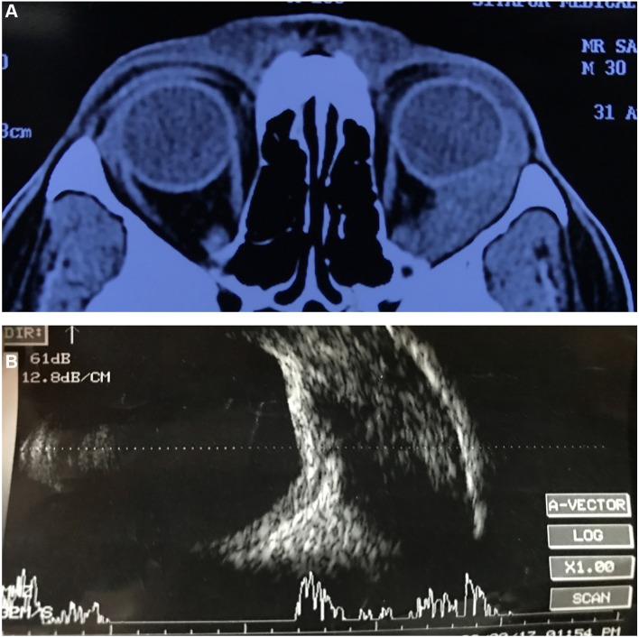
A young male patient presented to our ocular emergency department with chief complaints of progressive pain, redness, diplopia and a right-sided face turn. Ocular examination revealed severely restricted extraocular movements along with retinal folds in the left eye. Initial orbital ultrasound and CT findings were equivocal; however, serology favoured an infective cause. Considering the endemicity of the disease and equivocal investigation findings, a diagnosis of orbital cysticercosis with an atypical presentation was made. The patient was managed medically with a combination of oral albendazole and steroids over a period of 6 weeks to achieve optimal results.
Keywords:
medical education; medical management; ophthalmology.
Nine gaze clinical pictures revealing an exodeviation in the primary gaze with restriction…
Figure 1
Nine gaze clinical pictures revealing an exodeviation in the primary gaze with restriction of adduction, elevation and depression in the presence of moderate ptosis.
Figure 2
(A) Axial sections of CT…
Figure 2
(A) Axial sections of CT showing extensive fairly well-defined mass lesion extending from…
Figure 2
(A) Axial sections of CT showing extensive fairly well-defined mass lesion extending from the orbital apex to just anterior to the orbital rim. Lateral rectus muscle could not be made out. (B) Scan ultrasound showing extensively thickened lateral rectus muscle with a large hypoechoic area within.
Figure 3
At the end of 8…
Figure 3
At the end of 8 weeks, there was an improvement in ptosis along…
Figure 3
At the end of 8 weeks, there was an improvement in ptosis along with significant improvement in extraocular motility.
Figure 4
B-scan ultrasound at the end…
Figure 4
B-scan ultrasound at the end of 8 weeks showed significantly reduced lateral rectus…
Figure 4
B-scan ultrasound at the end of 8 weeks showed significantly reduced lateral rectus muscle thickness.
Rath S, Honavar SG, Naik M, et al. . Orbital cysticercosis: clinical manifestations, diagnosis, management, and outcome. Ophthalmology 2010;117:600–5. 10.1016/j.ophtha.2009.07.030
-
DOI
-
PubMed
Pushker N, Bajaj MS, Betharia SM. Orbital and adnexal cysticercosis. Clin Exp Ophthalmol 2002;30:322–33. 10.1046/j.1442-9071.2002.00550.x
-
DOI
-
PubMed
Sekhar GC, Lemke BN. Orbital cysticercosis. Ophthalmology 1997;104:1599–604. 10.1016/S0161-6420(97)30090-6
-
DOI
-
PubMed
Damani M, Mehta VC, Baile RB, et al. . Orbital cysticercosis: A case report. Saudi J Ophthalmol 2012;26:457–8. 10.1016/j.sjopt.2012.07.002
-
DOI
-
PMC
-
PubMed
Dhiman R, Devi S, Duraipandi K, et al. . Cysticercosis of the eye. Int J Ophthalmol 2017;10:1319–24. 10.18240/ijo.2017.08.21
-
DOI
-
PMC
-
PubMed