. 2018 Jul 27;293(30):11901-11912.
doi: 10.1074/jbc.RA117.001690. Epub 2018 Jun 8.

Novel hyperoxidation resistance motifs in 2-Cys peroxiredoxins

Affiliations

Novel hyperoxidation resistance motifs in 2-Cys peroxiredoxins

Jesalyn A Bolduc et al. J Biol Chem. .

Abstract

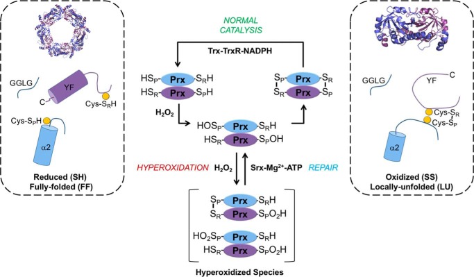
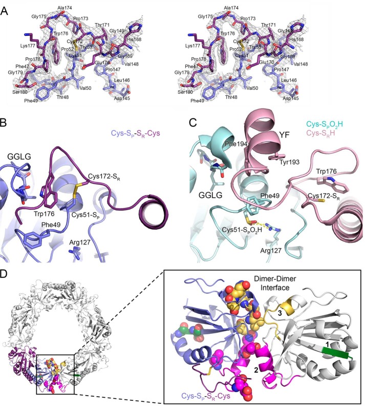
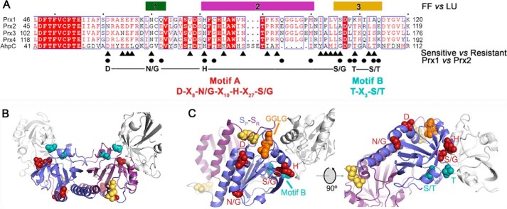
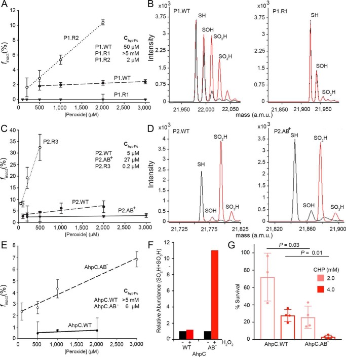
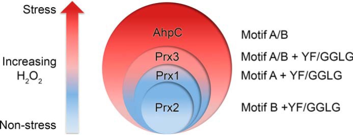
2-Cys peroxiredoxins (Prxs) modulate hydrogen peroxide (H2O2)-mediated cell signaling. At high H2O2 levels, eukaryotic Prxs can be inactivated by hyperoxidation and are classified as sensitive Prxs. In contrast, prokaryotic Prxs are categorized as being resistant to hyperoxidation and lack the GGLG and C-terminal YF motifs present in the sensitive Prxs. Additional molecular determinants that account for the subtle differences in the susceptibility to hyperoxidation remain to be identified. A comparison of a new, 2.15-Å-resolution crystal structure of Prx2 in the oxidized, disulfide-bonded state with the hyperoxidized structure of Prx2 and Prx1 in complex with sulfiredoxin revealed three structural regions that rearrange during catalysis. With these regions in hand, focused sequence analyses were performed comparing sensitive and resistant Prx groups. From this combinatorial approach, we discovered two novel hyperoxidation resistance motifs, motifs A and B, which were validated using mutagenesis of sensitive human Prxs and resistant Salmonella enterica serovar Typhimurium AhpC. Introduction and removal of these motifs, respectively, resulted in drastic changes in the sensitivity to hyperoxidation with Prx1 becoming 100-fold more resistant to hyperoxidation and AhpC becoming 800-fold more sensitive to hyperoxidation. The increased sensitivity of the latter AhpC variant was also confirmed in vivo These results support the function of motifs A and B as primary drivers for tuning the sensitivity of Prxs to different levels of H2O2, thus enabling the initiation of variable signaling or antioxidant responses in cells.

Keywords: Cys sulfinic acid; X-ray crystallography; cell signaling; enzyme kinetics; oxidation-reduction (redox); peroxiredoxin; protein oxidation; redox biology; structural biology.


Conflict of interest statement

The authors declare that they have no conflicts of interest with the contents of this article

Figures

Figure 1.
Figure 1.
Catalytic cycle and hyperoxidation of typical, 2-Cys Prxs. These Prxs exist as obligate homodimers with each subunit (blue or purple) containing Cys-SP (α2 helix) and Cys-SR (near the C terminus) residues (highlighted as yellow circles). For most members of this family, including human Prx1, Prx2, and bacterial AhpC, the dimers can assemble into a decameric toroid with 52-point group symmetry. Reaction of the Cys-SP residue with H2O2 creates the Cys-SPOH and releases a water molecule (not shown for clarity). During normal catalysis and low H2O2 levels, the Cys-SR residue from the adjacent subunit condenses with Cys-SPOH to form an intermolecular disulfide (Cys-SP–SR-Cys), releasing a second water molecule (not shown). For this disulfide bond to occur, the α2 helix, near the GGLG and YF motifs, and the C terminus of the adjacent subunit, must transition from the FF to the LU state. The disulfide can then be reduced by the NADPH-dependent Trx-TrxR system. If the H2O2 levels are high, the Cys-SPOH moiety can alternatively react with a second H2O2 molecule to form Cys-SPO2H. This modification or hyperoxidation can occur on one or both Cys-SP residues of the dimer. The inactivated Prx can be repaired in an ATP-dependent manner by the enzyme sulfiredoxin (Srx).
Figure 2.
Figure 2.
Crystal structures of Prx2 help to identify regions implicated in hyperoxidation. A, stereoview of the 2FoFc map contoured at the 1 σ level (chains E and F), including the Cys-SP–SR-Cys disulfide bond between Prx2 subunits. B, active-site structure of oxidized Prx2, Prx2-SPSR. The new structure reveals the necessary reorganization of the active site for disulfide bond formation. In this locally unfolded conformation, the C terminus is present as a coil that positions Trp176 (purple subunit) so that it packs into a crevice generated by the disulfide bond and the GGLG motif (blue subunit). Importantly, Trp176 is >20 Å away from this position in the Prx2-SPO2H structure as shown in the next panel. C, active-site structure of hyperoxidized Prx2 (chains A and B). In this fully folded conformation, the Cys51-SPO2H moiety (cyan) has an electrostatic interaction with Arg127 (Protein Data Bank code 1QMV) (35). The C terminus of the adjacent subunit (pink) places the Cys172-SR residue ∼14 Å away from the Cys51-SP residue. The YF motif (residues 193 and 194) is present in a helix that packs against the GGLG motif. D, summary of structural changes between Prx oxidation states. Left, one Prx dimer is highlighted in its decameric context (other Prx dimers are colored white). Right, location of regions that have the potential to influence the resistance to hyperoxidation. One subunit of the Prx dimer is shown in reference to the adjacent subunit (white) across the dimer-dimer interface of the toroid. Regions 1–3 are shown in green, magenta, and yellow, respectively. Region 1 is located within the β-strand and turn that precede the helix on which the Cys51-SP residue is located. Regions 2 and 3 interact with the same regions on the adjacent subunit. The residues denoted by spheres are those that differ in sequence between Prx1 and Prx2 (see Fig. 3).
Figure 3.
Figure 3.
Structure and sequence comparisons identify resistance motifs. A, summary of the structural and sequence information that led to the identification of resistance motifs A and B. The bars for regions 1–3 indicate the largest structural changes between the reduced (FF) and oxidized (LU) conformations (see Fig. 2 and Fig. S2). The triangles below the alignment highlight the residues, identified by WebLogo analysis, that differ between sensitive and resistant Prxs (Fig. S3 and Table S2). The circles indicate the residue differences in regions 1–3 for Prx1 and Prx2. The putative resistance motifs A and B are highlighted in red and cyan, respectively. The same coloring scheme for the residues within these motifs is used in the subsequent panels. B, location of motifs A and B in the Prx toroid context (see Fig. 2D for entire toroid). Both the Cys-SP and Cys-SR residues involved in disulfide bonds are colored yellow for the central Prx dimer. One subunit for each of the two adjacent dimers is shown in white. Motif A is predominantly located on the exterior of the toroid. Motif B lines the interior of the toroid. C, proximity of resistance motifs A and B to the Cys-SP residue, the GGLG motif, and the dimer-dimer interface.
Figure 4.
Figure 4.
Changes in motifs A and B alter resistance to hyperoxidation in vitro and in vivo. A, assessment of sensitivity to hyperoxidation for Prx1 variants. The Chyp1% value was obtained by generating the plot of the fraction of inactivated Prx molecules per catalytic cycle (finact) versus the H2O2 concentration. Changes in sequence for motif A and B residues (see Table 1) can either sensitize (P1.R2 variant, DNQS-KA; bold indicates change from WT) or prevent hyperoxidation (P1.R1 variant, DGHS-KA) relative to the WT protein (DNHS-KA). The presented data are the mean and S.D. (error bars) for the finact values determined on 3 or more separate days with fresh aliquots of reduced enzyme and each peroxide concentration tested in triplicate (see Fig. S1 and associated method description for details). B, cross-validation of Prx1 variants using ESI-TOF MS. Spectra for samples without and with peroxide treatment (1 mm H2O2 and 10 mm DTT for 3 h) are shown in black and red, respectively. The theoretical and observed mass values for the different oxidation states are presented in Table S4. C, Chyp1% value determination for Prx2 variants, WT (NGQA-TS), P2.R3 (NGQS-KA), and P2.AB+ (DNHS-TS). D, ESI-TOF MS analysis of the Prx2 variants with H2O2 treatment as in B. E, Chyp1% value determination for S. enterica Typhimurium AhpC variants, WT (DGHG-TT) and AhpC.AB (NNQA-KA). F, relative abundance of the Cys-SP-SO2H/SO3H species determined by LC-MS/MS before and after treatment of AhpC.WT or AhpC.AB with 3 mm H2O2. G, assessment of AhpC variant inactivation in vivo. The E. coli AhpC knockout strain was transformed with plasmids containing either AhpC.WT or AhpC.AB. Cells were challenged with two concentrations of cumene hydroperoxide (CHP), rinsed with fresh medium, allowed to recover for 1 h, and plated to determine percent survival. The data are presented as mean ± S.D. (error bars). p values were derived from an unpaired Student's two-tailed t test (each measurement was performed in triplicate with four independent cultures). a.m.u., atomic mass units.
Figure 5.
Figure 5.
Model for the hierarchy of Prx resistance. Stratification of Prx susceptibility to hyperoxidation is shown. Low levels of H2O2 are associated with nonstress cell signaling, and high levels can lead to oxidative stress–induced signaling. The Prxs most sensitive to hyperoxidation in this study each contain only one resistance motif (Prx1, motif A; Prx2, motif B). Prx3 contains both motifs A and B and is more resistant than Prx1 and Prx2 but is not as resistant as AhpC. AhpC also contains both motifs A and B but lacks the YF and GGLG motifs. Based on the proposed grading of resistance, each eukaryotic Prx will be inactivated at a different threshold of H2O2 concentration, enabling a diversity of cellular responses.

References

    1. Winterbourn C. C. (2013) The biological chemistry of hydrogen peroxide. Methods Enzymol. 528, 3–25 10.1016/B978-0-12-405881-1.00001-X - DOI - PubMed
    1. Choi M. H., Lee I. K., Kim G. W., Kim B. U., Han Y. H., Yu D. Y., Park H. S., Kim K. Y., Lee J. S., Choi C., Bae Y. S., Lee B. I., Rhee S. G., and Kang S. W. (2005) Regulation of PDGF signalling and vascular remodelling by peroxiredoxin II. Nature 435, 347–353 10.1038/nature03587 - DOI - PubMed
    1. Rhee S. G., and Kil I. S. (2016) Mitochondrial H2O2 signaling is controlled by the concerted action of peroxiredoxin III and sulfiredoxin: linking mitochondrial function to circadian rhythm. Free Radic. Biol. Med. 99, 120–127 10.1016/j.freeradbiomed.2016.07.029 - DOI - PubMed
    1. Perkins A., Nelson K. J., Parsonage D., Poole L. B., and Karplus P. A. (2015) Peroxiredoxins: guardians against oxidative stress and modulators of peroxide signaling. Trends Biochem. Sci. 40, 435–445 10.1016/j.tibs.2015.05.001 - DOI - PMC - PubMed
    1. Rabilloud T., Heller M., Gasnier F., Luche S., Rey C., Aebersold R., Benahmed M., Louisot P., and Lunardi J. (2002) Proteomics analysis of cellular response to oxidative stress: evidence for in vivo overoxidation of peroxiredoxins at their active site. J. Biol. Chem. 277, 19396–19401 10.1074/jbc.M106585200 - DOI - PubMed

Publication types