. 2018 Jun 6;50(6):1-14.
doi: 10.1038/s12276-018-0101-6.

The EGF/hnRNP Q1 axis is involved in tumorigenesis via the regulation of cell cycle-related genes

Affiliations

The EGF/hnRNP Q1 axis is involved in tumorigenesis via the regulation of cell cycle-related genes

Yu-Chu Wang et al. Exp Mol Med. .

Abstract

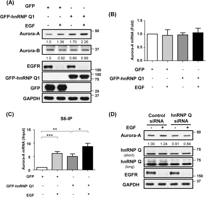
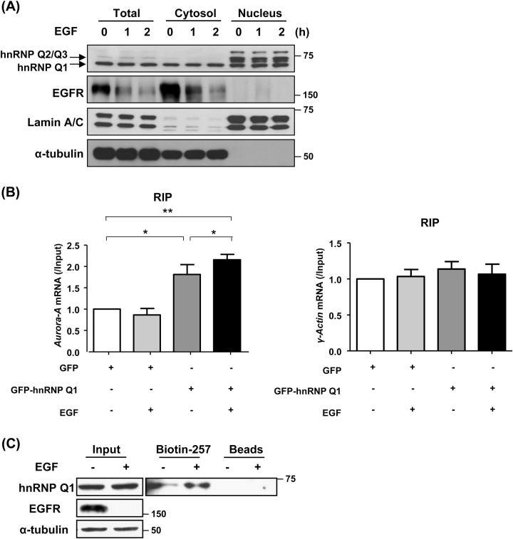
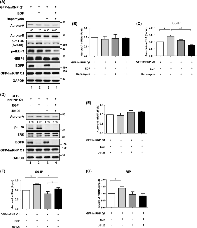
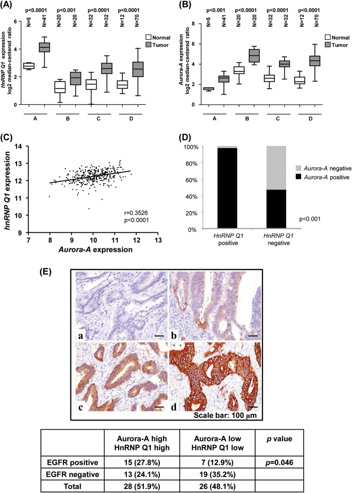
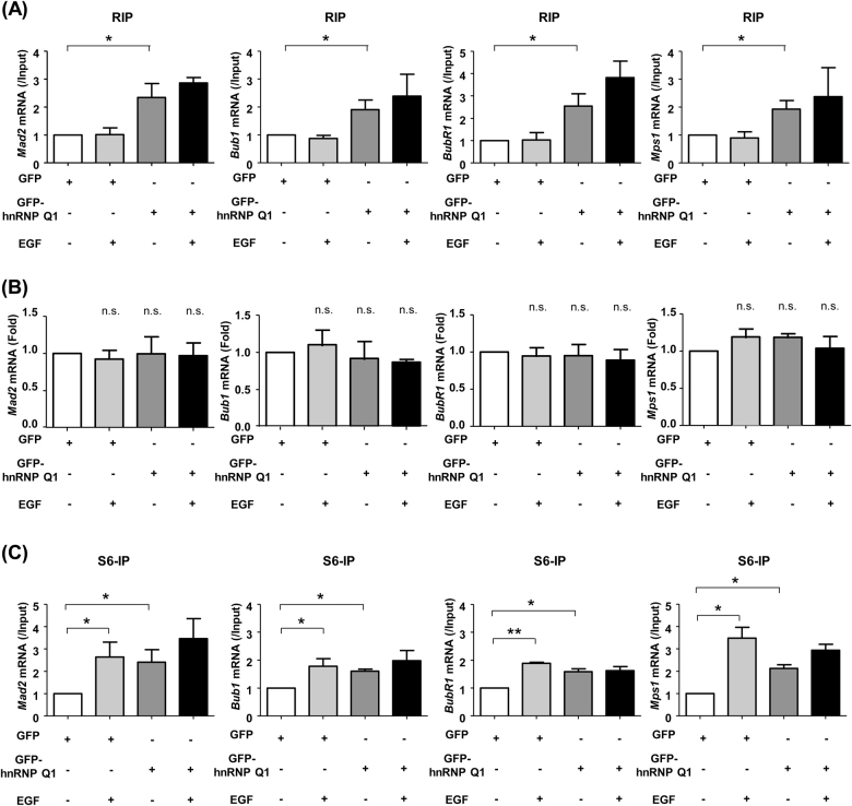
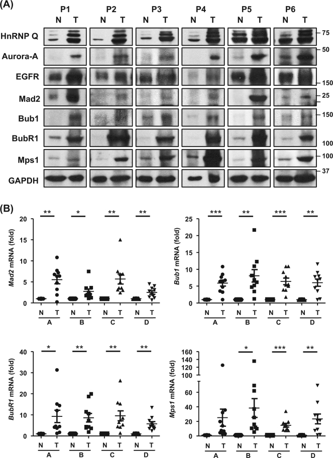
Heterogeneous nuclear ribonucleoprotein (hnRNP) Q1, an RNA-binding protein, has been implicated in many post-transcriptional processes, including RNA metabolism and mRNA splicing and translation. However, the role of hnRNP Q1 in tumorigenesis remains unclear. We previously performed RNA immunoprecipitation (RIP)-seq analysis to identify hnRNP Q1-interacting mRNAs and found that hnRNP Q1 targets a group of genes that are involved in mitotic regulation, including Aurora-A. Here, we demonstrate that altering the hnRNP Q1 level influences the expression of the Aurora-A protein, but not its mRNA. Stimulation with epidermal growth factor (EGF) enhances both binding between hnRNP Q1 and Aurora-A mRNA as well as the efficacy of the hnRNP Q1-induced translation of Aurora-A mRNA. The EGF/hnRNP Q1-induced translation of Aurora-A mRNA is mediated by the mTOR and ERK pathways. In addition, we show that hnRNP Q1 up-regulates the translation of a group of spindle assembly checkpoint (SAC) genes. hnRNP Q1 overexpression is positively correlated with the levels of Aurora-A and the SAC genes in human colorectal cancer tissues. In summary, our data suggest that hnRNP Q1 plays an important role in regulating the expression of a group of cell cycle-related genes. Therefore, it may contribute to tumorigenesis by up-regulating the translation of these genes in colorectal cancer.


Conflict of interest statement

The authors declare that they have no conflict of interest.

Figures

Fig. 1
Fig. 1. EGF enhances the hnRNP Q1- mediated increase in Aurora-A mRNA translation.
a SW480 cells were transfected with GFP or GFP-hnRNP Q1 and were incubated for 24 h. After serum starvation, cells were treated with EGF (10 nM) for 2 h, and total cell lysates were then harvested for western blot analysis. After EGF treatment, EGFR underwent endocytosis and ubiquitination. b The levels of Aurora-A mRNA in GFP- and GFP-hnRNP-Q1-expressing SW480 cells were quantified following treatment with EGF. c Cell lysates were harvested for the ribosomal S6-IP assay. The amount of Aurora-A mRNA in the S6-IP complex was analyzed using RT-qPCR. The mean ± S.D. was obtained from three independent experiments (*p < 0.05; **p < 0.01; ***p < 0.001). d The expression level of the Aurora-A protein in hnRNP Q siRNA-transfected SW480 cells treated with EGF was determined by western blot analysis. GAPDH was used as the loading control. The expression level of Aurora-A is shown as a ratio
Fig. 2
Fig. 2. EGF enhances the ability of hnRNP Q1 to bind to the Aurora-A mRNA 5′-UTR.
a Total cell lysate, the cytosolic and nuclear fractions were obtained from SW480 cells after treatment with 10 nM EGF and were used to perform western blot analysis with the indicated antibodies. b Cytoplasmic extracts obtained from GFP- or GFP-hnRNP Q1-expressing cells treated with EGF were used to perform RIP using anti-GFP antibodies. The bound Aurora-A mRNA was measured using RT-qPCR. γ-Actin was used as the negative control for RIP. The mean ± S.D. was obtained from three independent experiments (*p < 0.05; **p < 0.01). c SW480 cells were treated with (+) or without (−) EGF for 2 h, and then total cell lysates were harvested to perform biotin-pull down assays using the biotin-labeled Aurora-A mRNA 5′-UTR 257 nt probe (Biotin-257). Beads were used as the negative control
Fig. 3
Fig. 3. The mTOR and ERK pathways regulate the hnRNP Q1-induced translation of Aurora-A mRNA following stimulation with EGF.
a SW480 cells were transfected with GFP-hnRNP Q1 for 24 h. Serum-starved cells were pre-treated with rapamycin (100 nM) for 2 h and then were treated with EGF (10 nM) for an additional 2 h. The level of Aurora-A protein was evaluated using western blot analysis and was quantified as a ratio. b Quantification of the expression level of Aurora-A mRNA in GFP-hnRNP Q1-expressing cells treated with rapamycin and EGF, as described in (a). c The translational efficiency of the Aurora-A mRNA was determined using S6-IP and was quantitated using RT-qPCR. d, e GFP-hnRNP Q1-expressing SW480 cells were serum-starved and then pre-treated with U0126 (10 μM) for 30 min followed by treatment with EGF for another 2 h. Cell lysates were collected to determine the protein expression of Aurora-A by western blot analysis (d) and the mRNA expression of Aurora-A using RT-qPCR (e). f, g GFP-hnRNP Q1-expressing cells were treated with U0126 and then EGF, as described above. The cell lysates were then collected for S6-IP (f) or RIP assays using anti-GFP antibodies (g). The bound Aurora-A mRNA was measured using RT-qPCR. The mean ± S.D. was obtained from three independent experiments. (*p < 0.05; **p < 0.01)
Fig. 4
Fig. 4. hnRNP Q1 expression is positively correlated with Aurora-A expression in human colorectal cancer.
a, b Four cohorts of colorectal cancer from the “Oncomine” public database (https://www.oncomine.org) were used to analyze the expression patterns of hnRNP Q1 (SYNCRIP, a) and Aurora-A (b). c Spearman’s correlation was used to analyze the relationship between Aurora-A and hnRNP Q1 expression in clinical colorectal cancer samples obtained from the TCGA dataset. d The expression levels of the hnRNP Q1 and Aurora-A mRNAs were quantitated using RT-qPCR in 56 paired colorectal cancer and normal tissue samples, and the correlation between their expression levels was analyzed using Pearson’s Chi-square analysis (p < 0.001). e Colorectal cancer tissues were collected to analyze the expression of EGFR by IHC in 28 tissues that overexpressed Aurora-A and hnRNP Q1 and 26 tissues that expressed low levels of Aurora-A and hnRNP Q1. Representative images demonstrate the intensity of EGFR as follows: a score = 0, no staining visible; b score = 1, weak membranous staining in <10% of all tumor cells; c score = 2, moderate continuous membranous staining in >10% of all tumor cells; and d score = 3, strong and continuous membranous staining in >10% of all tumor cells. Positive staining ≥30% indicated high levels of Aurora-A and hnRNP Q1, and scores <30% indicated low levels of Aurora-A and hnRNP Q1. The results of the IHC were used to determine the correlation among EGFR, Aurora-A and hnRNP Q1 expression by Pearson’s Chi-square analysis, p = 0.046
Fig. 5
Fig. 5. hnRNP Q1 plays an important role in translationally regulating a group of spindle assembly checkpoint genes.
Serum-starved GFP- or GFP-hnRNP Q1-expressing SW480 cells were treated with or without EGF for 2 h and then were harvested to perform RIP using anti-GFP antibodies (a), RT-qPCR to quantify the expression levels of the Mad2, Bub1, BubR1 and Mps1 mRNAs (b), or S6-IP assay (c). The mean ± S.D. was obtained from three independent experiments. (*p < 0.05; **p < 0.01; n.s. not significant)
Fig. 6
Fig. 6. Parallel changes in the overexpression of EGFR, hnRNP Q1, Aurora-A and spindle assembly checkpoint genes in colorectal cancer tissues.
a Western blot analysis was used to determine the expression levels of the EGFR, hnRNP Q1, Aurora-A, Mad2, Bub1, BubR1, and Mps1 proteins in human colorectal tumors (T) and adjacent normal colon mucosal tissues (N). Six representative specimens (P1–P6) are shown. b The expression levels of the Mad2, Bub1, BubR1, and Mps1 mRNAs in human colorectal cancer tissues (T) and their normal colon mucosal tissues (N) were analyzed using RT-qPCR. (N = 40; *p < 0.05; **p < 0.01; ***p < 0.001)

References

    1. Pino MS, Chung DC. The chromosomal instability pathway in colon cancer. Gastroenterology. 2010;138:2059–2072. doi: 10.1053/j.gastro.2009.12.065. - DOI - PMC - PubMed
    1. Marumoto T, Zhang D, Saya H. Aurora-A - a guardian of poles. Nat. Rev. Cancer. 2005;5:42–50. doi: 10.1038/nrc1526. - DOI - PubMed
    1. Fu J, Bian M, Jiang Q, Zhang C. Roles of Aurora kinases in mitosis and tumorigenesis. Mol. Cancer Res. 2007;5:1–10. doi: 10.1158/1541-7786.MCR-06-0208. - DOI - PubMed
    1. Landen CN, et al. Overexpression of the centrosomal protein Aurora-A kinase is associated with poor prognosis in epithelial ovarian cancer patients. Clin. Cancer Res. 2007;13:4098–4104. doi: 10.1158/1078-0432.CCR-07-0431. - DOI - PubMed
    1. Yan M, et al. Aurora-A kinase: a potent oncogene and target for cancer therapy. Med. Res. Rev. 2016;36:1036–1079. doi: 10.1002/med.21399. - DOI - PubMed

Publication types

MeSH terms

Substances