. 2018 Aug;48(9-10):773-783.
doi: 10.1016/j.ijpara.2018.04.002. Epub 2018 Jun 6.

Prospects of developing a prophylactic vaccine against human lymphatic filariasis - evaluation of protection in non-human primates

Affiliations

Prospects of developing a prophylactic vaccine against human lymphatic filariasis - evaluation of protection in non-human primates

Vishal Khatri et al. Int J Parasitol. 2018 Aug.

Erratum in

Abstract

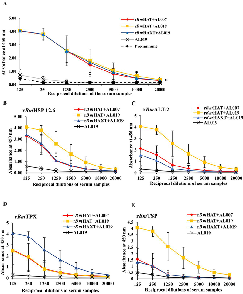
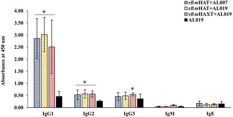
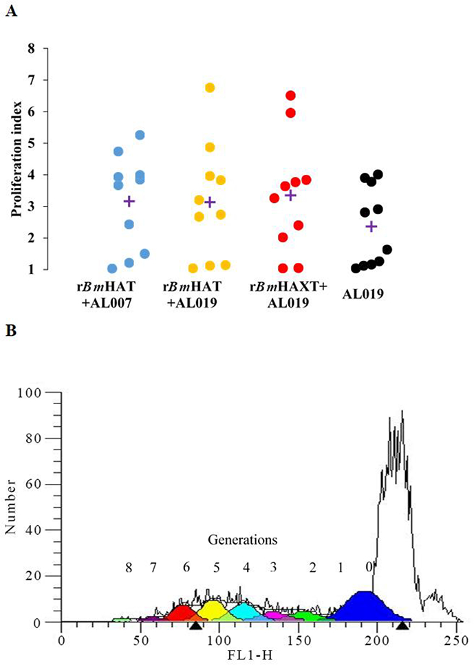
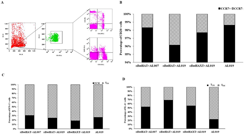
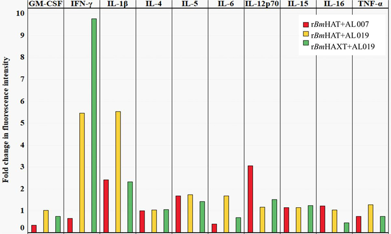
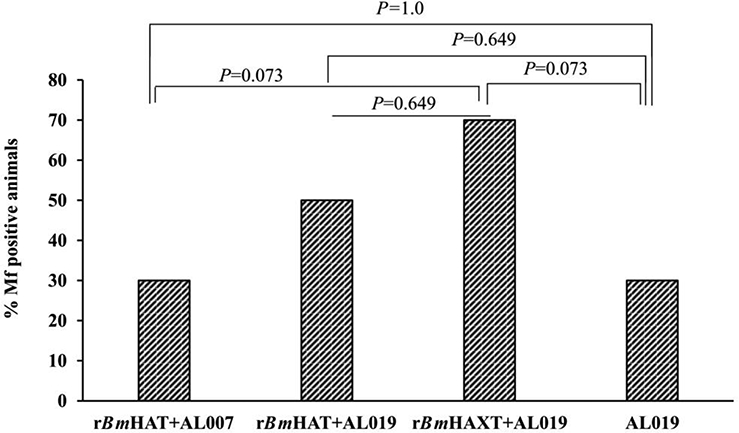
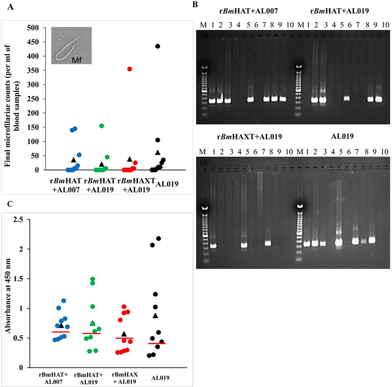
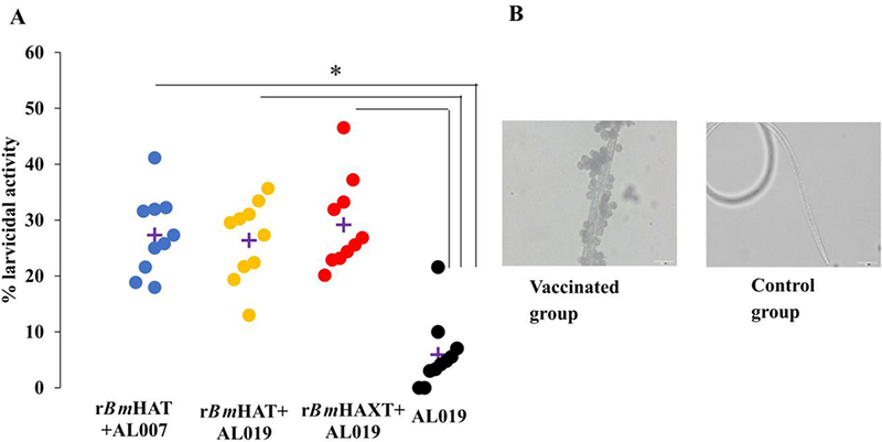
Lymphatic filariasis (LF) affects 120 million people around the world and another 856 million people are at risk of acquiring the infection. Mass Drug Administration (MDA) spearheaded by the World Health Organization is the only current strategy to control this infection. Recent reports suggest that despite several rounds of MDA, elimination has not been achieved and there is a need for more stringent control strategies for control of LF. An effective prophylactic vaccine combined with MDA has significant potential. Initial trials using a prophylactic trivalent recombinant Brugia malayi heat shock protein 12.6, abundant larval transcript -2 and tetraspanin large extra-cellular loop (rBmHAT) vaccine developed in our laboratory conferred only 35% protection in macaques. Therefore, the focus of the present study was to improve the current vaccine formulation to obtain better protection in non-human primates. We made two modifications to the current formulation: (i) the addition of another antigen, thioredoxin peroxidase-2 (TPX-2) to make it a tetravalent vaccine (rBmHAXT) and (ii) the inclusion of an adjuvant; AL019 (alum plus glucopyranosyl lipid adjuvant-stable emulsion) that is known to promote a balanced Th1/Th2 response. A double-blinded vaccination trial was performed with 40 macaques that were divided into three treatment groups and one control group (n = 10/group). Vaccinated animals received 4 immunisations at 1 month intervals with 150 µg/ml of rBmHAT plus alum, rBmHAT plus AL019 or rBmHAXT plus AL019. Control animals received AL019 only. All vaccinated macaques developed significant (P ≤ 0.003) titers of antigen-specific IgG antibodies (1:20,000) compared with the controls. One month after the last dose, all macaques were challenged s.c. with 130-180 B. malayi L3s. Our results showed that seven out of 10 (70%) of macaques given the improved rBmHAXT vaccine did not develop the infection compared with AL019 controls, of which seven out of 10 macaques developed the infection. Titers of antigen-specific IgG1 and IgG2 antibodies were significantly (P ≤ 0.01) higher in vaccinated animals and there was an increase in the percentage of IL-4 and IFN-γ secreting antigen-responding memory T cells. These studies demonstrated that the improved formulation (rBmHAXT plus AL019) is a promising vaccine candidate against human lymphatic filariasis.

Keywords: Adjuvant; Lymphatic filariasis; Multivalent vaccine; Non-human primates; TLR-4 agonist; Vaccine.


Figures

Fig. 1.
Fig. 1.
Titer of antigen-specific total IgG antibodies against recombinant Brugia malayi (rBm)HAXT, rBmHAT, rBmHAP 12.6, rBmALT-2, rBmTPX and rBmTSP antigens in the sera of macaques. Macaques were immunized (s.c.) with four doses (150 μg each dose) of respective protein in AL007 (alum) or A1019 (alum plus glucopyranosyl lipid adjuvant-stable emulsion) as adjuvant on days 0, 28, 56 and 84. Blood samples were collected prior to each immunization and the levels of antigen-specific total IgG antibodies were analyzed in all the groups of immunized animals using sera after the fourth immunization. (A) Titer of anti-rBmHAXT (rBm HSP12.6+ALT-2+TPX+TSPLEL) IgG antibodies. (B-E) Titer of IgG antibodies against each component antigen of the vaccine formulation. The variables are mean ± S.D. n=10 macaques per group. *P≤0.003 in comparison with the AL019 group as analyzed by a Kruskal-Wallis test followed by Bonferroni correction for multiple tests.
Fig. 2.
Fig. 2.
Levels of antigen-specific IgG1, IgG2, IgG3, IgM and IgE antibody isotypes in the sera of macaques. The levels of antigen-specific IgG1, IgG2, IgG3, IgM and IgE antibody isotypes were measured using an ELISA. The variables are mean ± S.D. n=10 macaques per group *P≤0.01 in comparison with the AL019 (alum plus glucopyranosyl lipid adjuvant-stable emulsion) group as analyzed by a Kruskal-Wallis test followed by Bonferroni correction for multiple tests.
Fig. 3.
Fig. 3.
Proliferative response of antigen-responding peripheral blood mononuclear cells (PBMCs)) in the blood of vaccinated macaques. (A) PBMCs (1×10⁶) were labeled with carboxyfluorescein diacetate succinimidyl ester (CFSE) and stimulated with 1 ug/ml of rBmHAT or rBmHAXT (recombinant Brugia malayi HSP12.6+ALT-2+TPX+TSPLEL) or ConA for 5 days. Dividing cells were counted in a flow cytometer. Each data point represents a proliferation index. ‘+’indicates the mean proliferation index for each group. (B) Representative flow cytometer data from the experimental group. Up to eight generations of dividing cells were present in the PBMC cultures that were stimulated with rBmHAXT. n=10 macaques per group. AL007, alum; AL019, alum plus glucopyranosyl lipid adjuvant-stable emulsion; FL1-H, fluorescence relative intensity.
Fig. 4.
Fig. 4.
T cell population and their cytokine profile in the peripheral blood mononuclear cells (PBMCs) from rBmHAT and rBmHAXT (recombinant Brugia malayi HSP/ALT-2/TPX/TSPLEL) vaccinated macaques. PBMCs (1×10⁷) were incubated with rBmHAT or rBmHAXTfor 3 days. Following incubation, cells were stained with fluorescent antibodies and processed for flow cytometry. Cells were initially gated for CD3 and the population of CD4, CD8 and CD28 positive cells was identified as indicated in (A). The CD28 positive cells were then further gated to determine the population of CCR7 positive cells with intracellular IFN-γ or IL-4. (B) Percentage of CCR7+ and CCR7− cells among CD28+ cell populations. (C) Percentage of IFN-γ+ and (D) IL-4+ cells among CCR7-CD28+ cell populations. The values represent percentage of cell population in each group (n = 10 macaques per group). (SC-H, side scatter-height; FSC-H, forward scatter-height; PE, phycoerythrin; TCM, T central memory cells; TEM, T effector memory cells; Interferon IFN)-γ; Interleukin (IL)-4; CD, cluster of differentiation; CCR, C-C chemokine receptor type.
Fig. 5.
Fig. 5.
Secreted levels of cytokines in the culture supernatants of peripheral blood mononuclear cells. (PBMCs) (1×10⁶) were cultured with rBmHAT and rBmHAXT (recombinant Brugia malayi HSP/ALT-2/TPX/TSPLEL) for 5 days. Secreted levels of cytokines in the culture supernatants were determined using an antibody slide array. Median fluorescence intensities were calculated after adjusting the background fluorescence values and normalizing the data with intensity of fluorescence in the positive control. The values represent fold changes in the mean fluorescence intensity relative to the AL019 (alum plus glucopyranosyl lipid adjuvant-stable emulsion) control group (n=10 per group). GM-CSF, Granulocyte-macrophage colony-stimulating factor.
Fig. 6.
Fig. 6.
Vaccine-induced protection against Brugia malayi infection in macaques. All animals (vaccinated and control) were challenged with 130–180 L3s of Brugia malayi 1 month after the last immunization. In weeks 5, 10, 15 and 18 post challenge, 10 ml of blood were collected from each macaque between 18:00 and 22:00 h and screened for the presence of microfilariae using a modified Knott technique (Dakshinamoorthjy et al., 2014) and analysed by PCR for the Hha-1 gene (Hoti et al., 2001). Absence of infection in microfilaria (Mf)-negative animals was further confirmed by SXP-1 (B. malayi diagnostic antigen) ELISA (Abdul Rehman et al., 2007). Results show that rBmHAXT+AL019 (alum plus glucopyranosyl lipid adjuvant-stable emulsion) is a better vaccine formulation than the other formulations tested. (n=10 per group). Chi-square test and Fisher’s exact test were used to compare the proportions across the groups.
Fig. 7.
Fig. 7.
Screening macaques for the presence of filarial infection. One month after the last dose of the vaccine, all macaques were challenged s.c. with 130–180 Brugia malayi L3s. Blood was collected from all the animals between 18:00 and 22:00 h in weeks 5, 10, 15 and 18 post challenge and were screened for the presence of microfilariae using the Knott’s technique. (A) Microfilaria (Mf) counts in per ml of final blood collection samples. (B) DNA was isolated from the blood samples and analyzed by PCR to detect the Mf-specific Hha-1 gene in a 1% agarose gel. Lanes: M, 100 bp DNA ladder; lanes 1 to 10, Hha-1 PCR amplified sample from each macaque. (C) An ELISA was performed to detect the levels of anti-rBmSXP-1 (recombinant B. malayi diagnostic antigen) IgG antibodies in the sera of animals. Each data point represents an animal within the group and the bar indicates the average value of Mf-negative animals. Values above the bar were considered positive and the values below the bar were considered negative. (n=10 per group). AL007, alum; AL019, alum plus glucopyranosyl lipid adjuvant-stable emulsion; rBmHAXT, recombinant B. malyi HSP/ALT-2/TPX/TSPLEL.
Fig. 8.
Fig. 8.
Antibody-dependent cell mediated cytotoxicity (ADCC) assay. Approximately 10 Brugia malayi larvae were incubated for 72 h at 37°C with 2×10⁵ peripheral blood mononuclear cells (PBMCs) and 50 μl of sera samples from each macaque. Larval death in each well was monitored under a light microscope. (A) Each data point indicates the percent larval death using a serum sample from one animal. ‘+’ indicates the average percent larvicidal activity for that group. (B) Note the large number of cells attached to the dead larva when a serum sample from a vaccinated macaque was used. No cells were found attached to the live larvae when serum sample from control macaque were used. n=10 macaques per group. *P≤0.005 compared with the AL019 (alum plus glucopyranosyl lipid adjuvant-stable emulsion) group. Statistical analysis was performed by a Kruskal-Wallis test followed by Bonferroni correction for multiple tests. rBmHAXT, recombinant B. malyi HSP/ALT-2/TPX/TSPLEL.
Fig. 9.
Fig. 9.
Myeloperoxidase (MPO) activity in the culture supernatants of the cells from the antibody dependent cell mediated cytotoxicity (ADCC) assay. Culture supernatants from the ADCC assay were analyzed for the MPO activity using a colorimetric assay. Data (mean± S.D.) are presented in box plots showing the upper (75%) and lower (25%) quartiles, with the horizontal line as the median and the whiskers as the maximum and minimum values observed, ‘+’ indicates mean myeloperoxidase activity per ml in the culture supernatant (n=10 per group). rBmHAXT, recombinant B. malyi HSP/ALT-2/TPX/TSPLEL.
Fig. 10.
Fig. 10.
Representative data from the lymphoscintigraphy analyses of macaques 16 weeks post challenge with Brugia malayi L3s. Lymphoscintigraphy analysis was performed in all the groups of macaques on weeks 12 and 16 post challenge to assess the lymph flow and associated lymphatic pathology. All macaques were challenged on the right thigh. Data was compared between the left and right thighs of the same animal. Representative data are shown for one animal from each group at 16 weeks post challenge. In the AL019 (alum plus glucopyranosyl lipid adjuvant-stable emulsion) control animal, lymph flow in the right leg was significantly blocked. However, in all vaccinated animals, the lymph flow appeared to be comparable between the left and right legs. Lymph flow was least affected in the rBmHAXT+AL019 vaccinated animals. rBmHAXT, recombinant B. malyi HSP/ALT-2/TPX/TSPLEL; CPM, counts per minute.

References

    1. Abdul Rahman R, Hwen-Yee C, Noordin R, 2007. Pan LF-ELISA using BmR1 and BmSXP recombinant antigens for detection of lymphatic filariasis. Filaria J 6, 10. - PMC - PubMed
    1. Ahmad G, Zhang W, Torben W, Ahrorov A, Damian RT, Wolf RF, White GL, Carey DW, Mwinzi PN, Ganley-Leal L, Kennedy RC, Siddiqui AA, 2011. Preclinical prophylactic efficacy testing of Sm-p80-based vaccine in a nonhuman primate model of Schistosoma mansoni infection and immunoglobulin G and E responses to Sm-p80 in human serum samples from an area where schistosomiasis is endemic. J Infect Dis 204, 1437–1449. - PMC - PubMed
    1. Anand SB, Kodumudi KN, Reddy MV, Kaliraj P, 2011. A combination of two Brugia malayi filarial vaccine candidate antigens (BmALT-2 and BmVAH) enhances immune responses and protection in jirds. J Helminthol 85, 442–452. - PubMed
    1. Anand SB, Murugan V, Prabhu PR, Anandharaman V, Reddy MV, Kaliraj P, 2008. Comparison of immunogenicity, protective efficacy of single and cocktail DNA vaccine of Brugia malayi abundant larval transcript (ALT-2) and thioredoxin peroxidase (TPX) in mice. Acta Trop 107, 106–112. - PubMed
    1. Anil NS, 2012. Assessing Coverage of Mass Drug Administration against Lymphatic Filariasis in Gulbarga District, Karnataka. Int J Med Public Health 2, 25–28.

Publication types

MeSH terms