. 2018 Jun 27;6(6):679-691.e4.
doi: 10.1016/j.cels.2018.05.008. Epub 2018 Jun 6.

Dissection of Influenza Infection In Vivo by Single-Cell RNA Sequencing

Affiliations

Dissection of Influenza Infection In Vivo by Single-Cell RNA Sequencing

Yael Steuerman et al. Cell Syst. .

Abstract

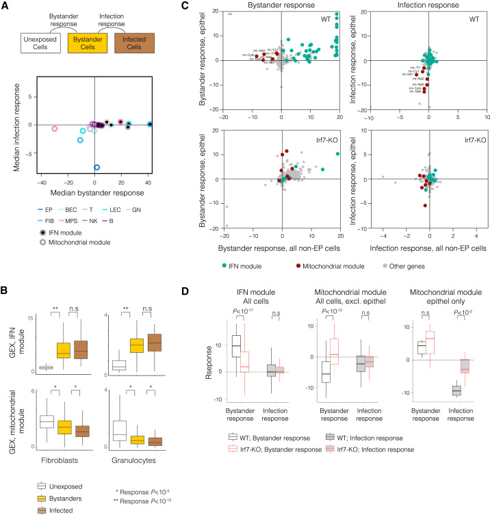
The influenza virus is a major cause of morbidity and mortality worldwide. Yet, both the impact of intracellular viral replication and the variation in host response across different cell types remain uncharacterized. Here we used single-cell RNA sequencing to investigate the heterogeneity in the response of lung tissue cells to in vivo influenza infection. Analysis of viral and host transcriptomes in the same single cell enabled us to resolve the cellular heterogeneity of bystander (exposed but uninfected) as compared with infected cells. We reveal that all major immune and non-immune cell types manifest substantial fractions of infected cells, albeit at low viral transcriptome loads relative to epithelial cells. We show that all cell types respond primarily with a robust generic transcriptional response, and we demonstrate novel markers specific for influenza-infected as opposed to bystander cells. These findings open new avenues for targeted therapy aimed exclusively at infected cells.

Keywords: bystander versus infected cells; immune and non-immune cell types; influenza infection in vivo; single-cell RNA sequencing.


Conflict of interest statement

The authors declare no competing interests.

Figures

None
Graphical abstract
Figure 1
Figure 1
Comprehensive Influenza Infection Map of Bystander and Infected Cells Identified by Single-Cell RNA Sequencing (A) Schematic illustration of the experimental workflow. Immune and non-immune single cells were isolated from the whole lung of control and influenza-treated mice, 48 hr post infection, for massively parallel single-cell RNA-seq (MARS-seq). In each single cell, the host and the vmRNA were simultaneously measured, allowing identification of infected as opposed to bystander cells as well as retrospective annotation of cell types based on transcriptional identities. (B) Transcriptional signatures of single cells associated with nine major cell types. Gene expression of annotated cell-type-specific genes (rows) is shown across 4,064 single cells (columns). The expression matrix displays the clustering of cells into nine cell-type groups (first row), where each cell type consists of cells derived from different treatments (light/dark gray respectively denote control/influenza-treated animals; second row) and shows heterogeneity of cell infection states (white/yellow/brown respectively denote unexposed/bystander/infected cells, third row). (C−E) Visualization of single cells across different cell types, treatments, and intracellular infection states. t-SNE plots show the cell-type annotation of single cells (C); the mouse to which the cell belongs (D), and the intracellular infection state (E). Color-coding is as in (B). See also Figure S1 and Table S1.
Figure 2
Figure 2
Combined Single-Cell Analysis of Virus and Host Reveals High Prevalence of Infected Cells in All Major Lung-Derived Cell Types (A) Prevalence of infected cells in each of the immune and non-immune cell types. The percentage of infected cells (y axis) in each cell type (x axis) is reported for cells derived from either the control (light gray) or the influenza-treated mice (dark gray). The expected percentages of false positives are marked in white error bars. (B) Extent to which the abundance of infected cells is an intrinsic property of the cell type. Shown is the percentage of infected cells in each cell type from an influenza-treated wild-type mouse (x axis) compared to its biological replicate (y axis). (C) Analysis of potential implications of the type I IFN pathway in cell-type-specific infection. Shown are the percentages of infected cells in an Irf7 wild-type (x axis) versus an Irf7-KO mouse (y axis). (D) Single-cell heterogeneity of intracellular viral load within the influenza-treated host. Shown are the percentages of low (yellow), medium (light brown), and high (dark brown) viral-load states (y axis) within the population of infected cells, as identified for each of the nine cell types (x axis; total numbers of infected cells are indicated). See also Figures S1–S4, Tables S2 and S3.
Figure 3
Figure 3
Characterization of the Generic Host Response to Influenza (A) Generic regulatory programs across all cell types. Differential expression in influenza-treated and control mice (color bar) of nuclear-encoded (top) and mitochondrial DNA-encoded (bottom) genes (rows) across the nine major cell types (columns). Only 450 nuclear genes that are differentially expressed (DE) in at least one cell type (p < 10−6) and 21 mitochondrial genes are shown. Right column indicates membership in four type I IFN-related categories. Generically induced genes (the “IFN module,” 101 genes), as well as generically repressed genes (the “mitochondrial module,” 7 genes), are marked on the left. (B) Shown are the distributions of single-cell expression levels (y axis) of selected generic-response genes (subpanels) in each cell type (x axis) for cells derived from control (light gray) and influenza-treated mice (dark gray). Asterisks denote the significance of differential expression. (C) Antiviral cell trajectory. Shown is the expression of the IFN module of genes (rows, Z-normalized for each gene) across single cells ranked by their position along the antiviral trajectory (columns). Cells of different types appear in separate panels. (D) Opposite dynamics of the generic IFN and mitochondrial modules. Expression of the modules (y axis) in each cell type (color-coded) across the antiviral trajectory (x axis) is shown. See also Figures S5 and S6, and Tables S4 and S5.
Figure 4
Figure 4
Distinct and Shared Regulatory Programs Among Bystander and Infected Cells (A) Examination of the type I interferon (“IFN”) and mitochondrial modules in bystander and infected cells. Shown are the median bystander response (x axis) and median infection response (y axis) across all genes in the IFN modules (filled circles) and mitochondrial modules (empty circles) of each cell type (color-coded). Bystander and infection types of responses are illustrated (top), and are shown as signed log10 p value (positive/negative, induced/repressed): the “bystander response” refers to the bystander versus unexposed differential expression and “infection response” refers to the infected versus bystander differential expression. (B) Distribution of average expression of genes (denoted “GEX”) per cell (y axis) in unexposed cells (white), bystander cells (yellow), and infected cells (brown). Shown is the average expression across the IFN module (top) and the mitochondrial module (bottom) for representative cell types. Asterisks denote significant bystander response or infection response (p < 10−3, ∗∗p < 10−13). (C) Comparison of gene response between epithelial cells (y axis) and other cell types (x axis) in wild-type (top) and Irf7-KO (bottom) mice. Shown are bystander response (left) and infection response (right) of individual genes (dots), color-coded by their module (green/maroon/gray, respectively, for IFN module/mitochondrial module/other genes). (D) Summary of transcriptional response in bystander as opposed to infected cells. Shown are distributions of bystander response (white fill) and infection response (gray fill) in wild-type (black border) and Irf7-KO (red border). Plots show data for all cells across the IFN module (left), except for epithelial cells across the mitochondrial module (middle), and epithelial cells across the mitochondrial module (right). See also Figures S7 and S8.
Figure 5
Figure 5
Cell-Type-Specific Transcriptional Changes after Intracellular Infection (A) List of the top 34 genes manifesting significant infection response (false discovery rate, p < 0.01) in a particular cell type. The bystander response (bystanders versus unexposed) and infection response (infected versus bystanders) of each gene in its relevant cell type are shown on a red/blue color scale (signed log10 p value ranging from −50 to +50; positive/negative, induced/repressed). The three bottom rows indicate membership in functional categories. Directionality of effects; enhanced induction, enhanced repression, and other dynamics are marked. (B) Representative genes displaying significant infection response in a particular cell type. Shown are the moving averages of expression levels in bystander (yellow) and infected (brown) cells (y axis) along the antiviral trajectory (x axis) for representative gene-cell type pairs from A. See also Figure S8 and Table S6.

References

    1. Altboum Z., Steuerman Y., David E., Barnett-Itzhaki Z., Valadarsky L., Keren-Shaul H., Meningher T., Mendelson E., Mandelboim M., Gat-Viks I. Digital cell quantification identifies global immune cell dynamics during influenza infection. Mol. Syst. Biol. 2014;10:720. - PMC - PubMed
    1. Avraham R., Haseley N., Brown D., Penaranda C., Jijon H.B., Trombetta J.J., Satija R., Shalek A.K., Xavier R.J., Regev A. Pathogen cell-to-cell variability drives heterogeneity in host immune responses. Cell. 2015;162:1309–1321. - PMC - PubMed
    1. Brandes M., Klauschen F., Kuchen S., Germain R.N. A systems analysis identifies a feedforward inflammatory circuit leading to lethal influenza infection. Cell. 2013;154:197–212. - PMC - PubMed
    1. Breen M., Nogales A., Baker S., Martínez-Sobrido L. Replication-competent influenza A viruses expressing reporter genes. Viruses. 2016;8:179. - PMC - PubMed
    1. Brooke C.B., Ince W.L., Wrammert J., Ahmed R., Wilson P.C., Bennink J.R., Yewdell J.W. Most influenza A virions fail to express at least one essential viral protein. J. Virol. 2013;87:3155–3162. - PMC - PubMed

Publication types

MeSH terms