

Prevalence of knee osteoarthritis features on magnetic resonance imaging in asymptomatic uninjured adults: a systematic review and meta-analysis
- PMID: 29886437
- PMCID: PMC6837253
- DOI: 10.1136/bjsports-2018-099257
Prevalence of knee osteoarthritis features on magnetic resonance imaging in asymptomatic uninjured adults: a systematic review and meta-analysis
Abstract
Background: Knee MRI is increasingly used to inform clinical management. Features associated with osteoarthritis are often present in asymptomatic uninjured knees; however, the estimated prevalence varies substantially between studies. We performed a systematic review with meta-analysis to provide summary estimates of the prevalence of MRI features of osteoarthritis in asymptomatic uninjured knees.
Methods: We searched six electronic databases for studies reporting MRI osteoarthritis feature prevalence (ie, cartilage defects, meniscal tears, bone marrow lesions and osteophytes) in asymptomatic uninjured knees. Summary estimates were calculated using random-effects meta-analysis (and stratified by mean age: <40 vs ≥40 years). Meta-regression explored heterogeneity.
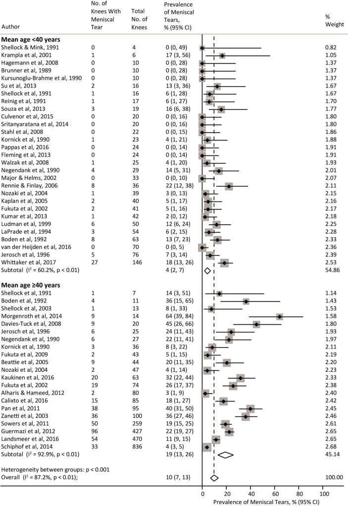
Results: We included 63 studies (5397 knees of 4751 adults). The overall pooled prevalence of cartilage defects was 24% (95% CI 15% to 34%) and meniscal tears was 10% (7% to 13%), with significantly higher prevalence with age: cartilage defect <40 years 11% (6%to 17%) and ≥40 years 43% (29% to 57%); meniscal tear <40 years 4% (2% to 7%) and ≥40 years 19% (13% to 26%). The overall pooled estimate of bone marrow lesions and osteophytes was 18% (12% to 24%) and 25% (14% to 38%), respectively, with prevalence of osteophytes (but not bone marrow lesions) increasing with age. Significant associations were found between prevalence estimates and MRI sequences used, physical activity, radiographic osteoarthritis and risk of bias.
Conclusions: Summary estimates of MRI osteoarthritis feature prevalence among asymptomatic uninjured knees were 4%-14% in adults aged <40 years to 19%-43% in adults ≥40 years. These imaging findings should be interpreted in the context of clinical presentations and considered in clinical decision-making.
Keywords: cartilage; knee; mri; osteoarthritis.
© Author(s) (or their employer(s)) 2019. Re-use permitted under CC BY-NC. No commercial re-use. See rights and permissions. Published by BMJ.
Conflict of interest statement
Competing interests: AG is president of Boston Imaging Core Lab, LLC, and a consultant to Merck Serono, Genzyme, OrthoTrophix and TissueGene.
Figures


References
Publication types
MeSH terms
Grants and funding
LinkOut - more resources
Full Text Sources
Other Literature Sources
Medical