. 2018 Jul 5;23(1):46-59.e5.
doi: 10.1016/j.stem.2018.05.002. Epub 2018 Jun 7.

Paneth Cell Multipotency Induced by Notch Activation following Injury

Affiliations

Paneth Cell Multipotency Induced by Notch Activation following Injury

Shiyan Yu et al. Cell Stem Cell. .

Abstract

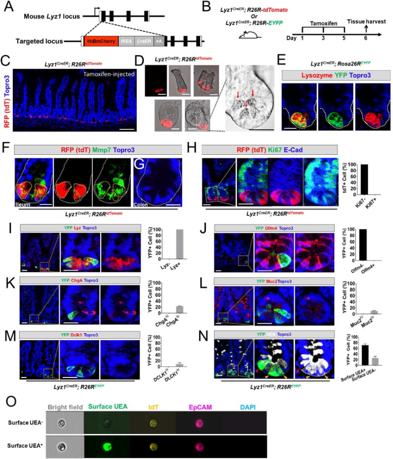
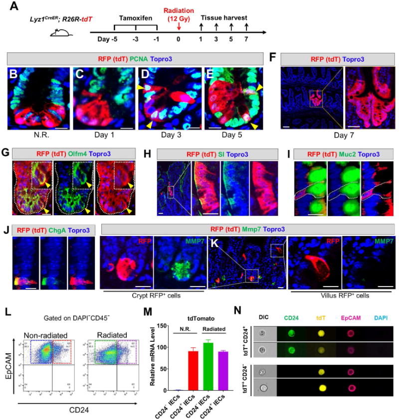
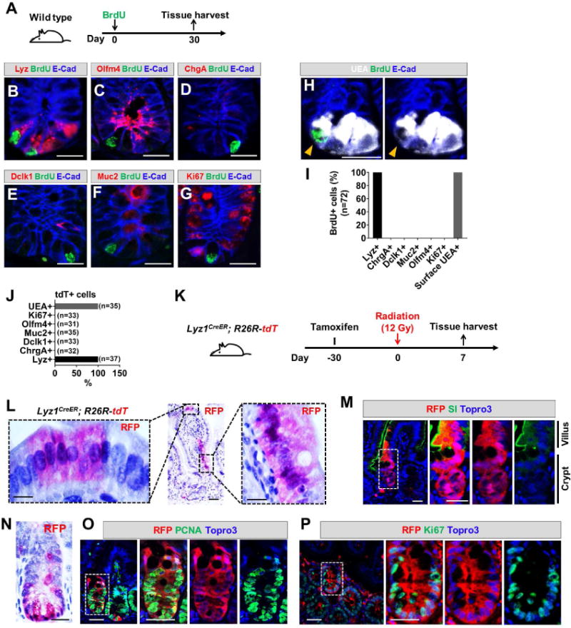
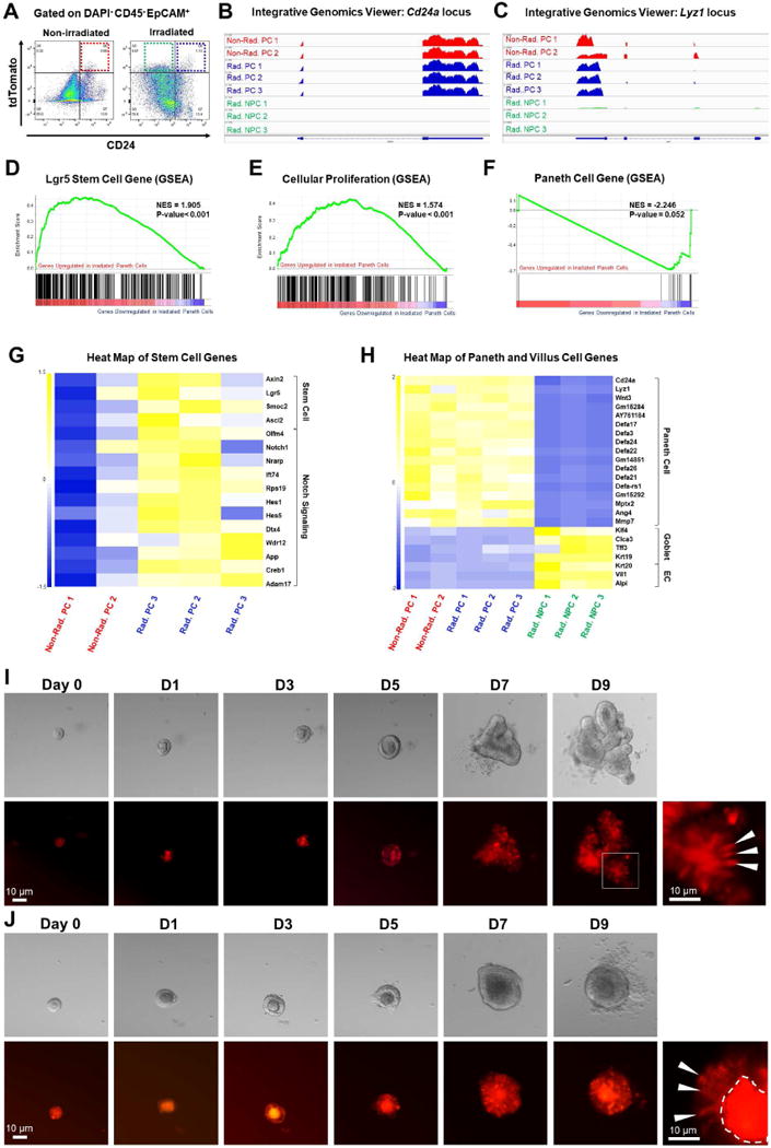
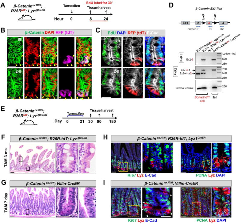
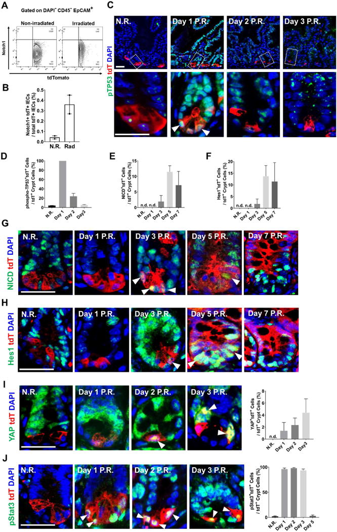
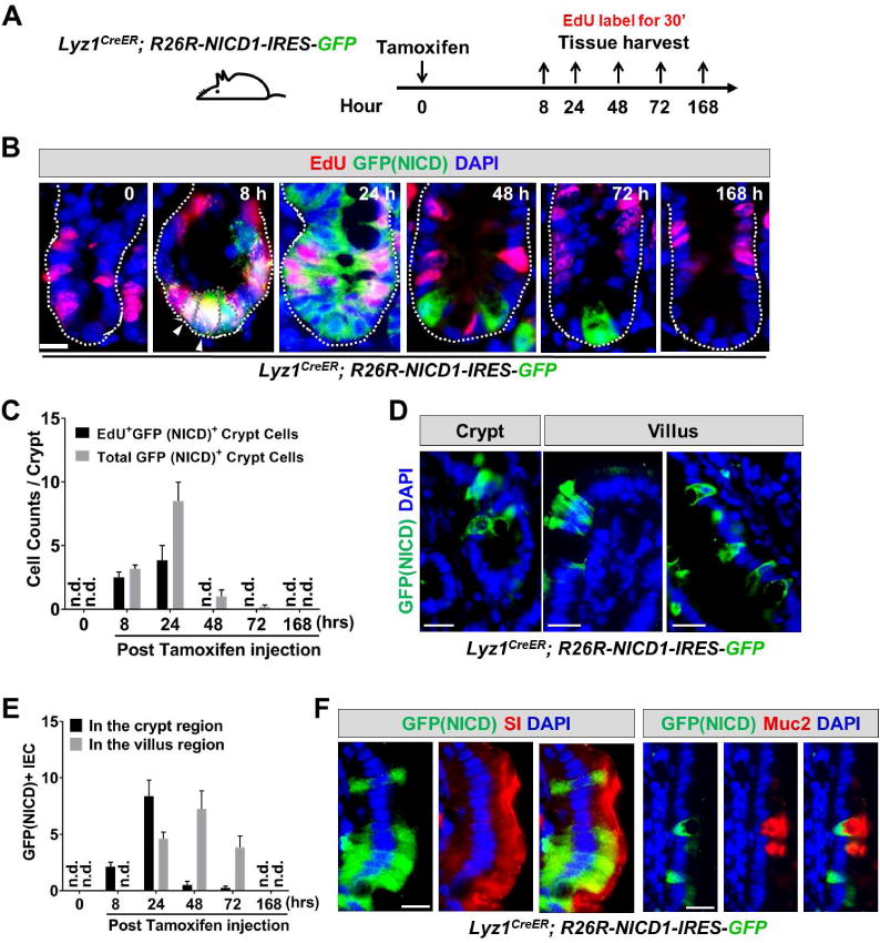
Paneth cells are post-mitotic intestinal epithelial cells supporting the stem cell niche and mucosal immunity. Paneth cell pathologies are observed in various gastrointestinal diseases, but their plasticity and response to genomic and environmental challenges remain unclear. Using a knockin allele engineered at the mouse Lyz1 locus, we performed detailed Paneth cell-lineage tracing. Irradiation induced a subset of Paneth cells to proliferate and differentiate into villus epithelial cells. RNA sequencing (RNA-seq) revealed that Paneth cells sorted from irradiated mice acquired a stem cell-like transcriptome; when cultured in vitro, these individual Paneth cells formed organoids. Irradiation activated Notch signaling, and forced expression of Notch intracellular domain (NICD) in Paneth cells, but not Wnt/β-catenin pathway activation, induced their dedifferentiation. This study documents Paneth cell plasticity, particularly their ability to participate in epithelial replenishment following stem cell loss, adding to a growing body of knowledge detailing the molecular pathways controlling injury-induced regeneration.

Keywords: Lyz1; Notch; Paneth cell; RNA-seq; Wnt; intestinal stem cell; lysozyme; plasticity.


Conflict of interest statement

DECLARATION OF INTERESTS

The authors declare no competing interests.

Figures

Figure 1
Figure 1. Lyz1CreER labels Paneth cells
(A) Schematic diagram of the H2Bmcherry-IRES-CreER knock-in allele referred to here as Lyz1CreER. (B) Lyz1CreER mice were crossed with R26R-tdTomato or R26R-EYFP mice to enable cell lineage labeling. (C) Tamoxifen-injected Lyz1CreER/+; R26R-tdTomato mice showed recombined Paneth cells that were strongly positive for tdTomato (tdT) at crypt base of small intestines. Scale bar, 50 μm. (D) Isolated crypts (top panels) and organoids (bottom panels) showed tdT+ Paneth cells (red) with large granules of typical Paneth cells (bright field panel). (E) All recombined YFP+ cells expressed endogenous lysozyme (red) in small intestine. (F) All recombined tdT+ cells expressed Paneth cell marker Mmp7 (green) in small intestine. Scale bar, 10 μm. (G) No tdT+ cell was found in colon. (H) Recombined tdT+ cells were negative for Ki67. (I-M) Tamoxifen-injected Lyz1CreER/+; R26R-EYFP mice were analyzed for the co-expression of various cell type specific markers in the recombined EYFP+ cells. All EYFP+ cells expressed lysozyme (100%, H), none expressed Olfm4 (0%, J), and a subset of them co-expressed low levels of ChgA (22%, K), Muc2 (10%, L), Dclk1 (9%, M). (N) UEA binding to α (1,2)-linked fucosylated proteins at cell surface indicates mature Paneth cells. 70% of EYFP+ cells exhibited surface UEA positivity (yellow arrowheads), while 30% of EYFP+ cells were negative for surface UEA (red arrowheads). Of note, intracellular vesicles were stained positive for UEA due to cell membrane permeabilization. Scale bars, 10 μm. (O) Image String analysis of FACS-sorted single live tdT+ cells showed 2 populations of tdT+ cells: surface UEA (top) and UEA+ (bottom). Live cells were stained without membrane permeabilization, therefore intracellular UEA was not detected. See also Figures S1 and S2.
Figure 2
Figure 2. Radiation induces proliferation and villus differentiation in a subset of Paneth cells
(A) Experimental scheme showing lineage labeling of adult Lyz1CreER; R26R-tdT mice by tamoxifen injection, followed by radiation and tissue analyses (3 mice for each time point). (B-E) Non-irradiated (N.R.) tdT+ Paneth cells do not express PCNA, however after irradiation tdT+ cells became positive for PCNA on day 3 and 5 (yellow arrowheads). Scale bar, 10 μm. (F) Radiated Lyz1CreER; R26R-tdT mice showed red epithelial stripes (a representative image of day 7 was shown). (G-J) Some irradiated tdT+ Paneth cells co-expressed Olfm4 (yellow arrowheads in G) in the crypts, while some villus tdT+ cells co-expressed SI (H), or Muc2 (I). or ChgA (J). Scale bar, 10 μm. (K) Some irradiated tdT+ cells retained Paneth cell marker (Mmp7, left panel), while villus tdT+ cells lost Paneth cell marker (right panel). Scale bar, 10 μm. (L) CD24+EpCAM+CD45 and CD24EpCAM+CD45 cells were FACS-sorted from non-irradiated (N.R.) and irradiated mice. (M) qPCR showed that CD24EpCAM+CD45 cells sorted from irradiated mice (green box in L) expressed the cell lineage marker tdT (green bar). CD24EpCAM+CD45 cells from N.R. mice (blue box in L) did not express tdT (blue bar). (N) ImageStream analysis of FACS-sorted single tdT+ cells from irradiated mice showed 2 distinct cell populations: CD24+ (top) and CD24 (bottom). Scale bar, 10 μm.
Figure 3
Figure 3. Radiation induced mature Paneth cells to proliferate and re-differentiate
(A) Wild type mice were administered a single BrdU injection to label cycling cells. After a month, the tissue was analyzed. (B-I) After one month, none of the remaining BrdU+ cells expressed markers of stem cell (C, G) or secretory precursor (D-F). All BrdU+ cells were positive for lysozyme (B) and surface UEA (H). Scale bar, 10 μm. (J) One month after tamoxifen injection of Lyz1CreER/+; R26R-tdT mice, all remaining tdT+ cells were positive only for Paneth cell markers. (K) Experimental scheme: one month after tamoxifen injection of Lyz1CreER/+; R26R-tdT mice, animals were irradiated then sacrificed 7 days after irradiation to determine whether radiation induces mature Paneth cell to proliferate. (L, N) Radiated Lyz1CreER/+; R26R-tdT mice showed villus and crypt localized tdT+ stripes. (M) Villus tdT+ cells in irradiated animals expressed enterocyte marker SI. Scale bar, 10 μm. (O, P) Crypt tdT+ cells in irradiated animals showed PCNA+ (O) and Ki67+ (P) cells. Scale bar, 10 μm.
Figure 4
Figure 4. Radiated Paneth cells gain stem cell features
(A) Gated on DAPICD45EpCAM+, FACS-sorting was performed to obtain non-irradiated tdT+CD24+ Paneth cells (red box), irradiated tdT+CD24+ Paneth cells (blue box), and irradiated tdT+CD24 non-Paneth cells (green box), for RNA-Seq analyses. (B-C) Integrative Genomics Viewer of RNA-Seq data at Cd24a and Lyz1 loci. Non-Rad. PC: non-radiated Paneth cells; Rad. PC: radiated Paneth cells; Rad. NPC: radiated non-Paneth cells. (D-F) Gene Set Enrichment Analysis (GSEA) showed that compared to non-irradiated Paneth cells, irradiated Paneth cells gained stem cell gene signature (D), exhibited a proliferative status (E), and reduced expression levels of Paneth cell genes (F). (G-H) Heat map showed that irradiated Paneth cells had higher levels of stem cell gene and Notch target gene expression (G), and that irradiated tdT+CD24 non-Paneth cells showed a loss of all Paneth cell markers and an expression of villus cell genes (H). (I-J) Single tdT+ cells sorted from irradiated mice formed organoids after 9 days. White arrowheads pointed to tdT+ cells within the organoids. See also Figures S3-5.
Figure 5
Figure 5. Constitutive β-Catenin activation did not induce Paneth cell to proliferate
(A) Adult β-Catenin ex3fl/fl; R26R-tdT; Lyz1CreER mice were injected by tamoxifen and assessed for proliferation after 8 and 24 hours. Animals were labeled with EdU for 30 minutes before sacrifice. (B) 8 and 24 hours after tamoxifen injection, tdT+ Paneth cells (purple) were readily detected with nuclear β-Catenin signals (green, pointed by arrowheads). β-Catenin remained largely at junctions in tdT cells. (C) 8 and 24 hours after tamoxifen injection to induce β-Catenin activation, tdT+ Paneth cells (red) did not show EdU incorporation (green). E-Cad (in white) was used to outline cell boundaries. (D) Genomic PCR using two sets of PCR primers (F+R1; F+R2) revealed robust recombination (excision of exon 3) in FACS-sorted tdT+ cells from tamoxifen-injected ex3fl/fl; R26R-tdT; Lyz1CreER and ex3fl/+; R26R-tdT; Lyz1CreER mice. Water and tail DNA served as controls. (E) Adult β-Catenin ex3fl/fl; R26R-tdT; Lyz1CreER mice was subcutaneously embedded with 21-day slow-releasing tamoxifen pellets, and assessed after a prolonged tracing up to 1, 3, and 6 months. (F) Tamoxifen-treated β-Catenin ex3fl/fl; R26R-tdT; Lyz1CreER mice, after a long-term tracing did not show epithelial hyperplasia or adenoma formation at 3 and 6 months (total of 8 mice). (G) Tamoxifen-treated β-Catenin ex3fl/fl; Villin-CreER mice exhibited formation of tubular adenoma within 1 week. (H-I), Lysozyme-expressing Paneth cells of above tamoxifen-treated animals did not express cell proliferation marker Ki67 (G, I) and PCNA (H, J). Scale bars, 10 μm.
Figure 6
Figure 6. Radiation activated Notch1 signaling in a subset of Paneth cells
(A-B) Flow cytometry analyzing surface Notch1 receptor expression showed that irradiated tdT+ cells had higher level of surface Notch1 (approximately 8-fold higher than N.R. cells). (C-D) Radiation immediately increased phospho-p53 (pTP53) levels in all tdT+ Paneth cells 1 day after the treatment. (E-H) Non-irradiated Paneth cells did not express NICD or Hes1; after 3 days following radiation, tdT+ cells became positive for both NICD and Hes1. (I) Non-irradiated Paneth cells did not express nuclear Yap; after 2 days following radiation, tdT+ cells became positive for nuclear Yap. (J) Following radiation, tdT+ cells became positive for nuclear phospho-Stat3. See also Figure S6.
Figure 7
Figure 7. Ectopic NICD expression induced Paneth cells to proliferate and differentiate
(A) Experimental scheme: Lyz1CreER; R26R-NICD1-IRES-GFP mice were injected with tamoxifen, and sacrificed at 8, 24, 48, 72, and 168 hours for tissue analyses. Mice were labeled with EdU for 30 minutes before sacrifice. (B-C) In non-tamoxifen treated mice, there was no GFP+/NICD+ cell detected (0 hour). After 8 hours GFP+ cells became readily detectable at crypt bottom, and the majority of GFP+ cells were EdU+. The number of EdU+GFP+ cells expanded at 24 hours, then reduced and disappeared from 48 hours onwards. (D-E) Villus GFP+/NICD+ cells were detected approximately 1 day after the initial detection of GFP+ cells in the crypts. At 168 hours, GFP+/NICD+ cell was no longer observed suggesting a transient NICD-activated proliferation and differentiation. n.d. not detected. (F) Villus GFP+NICD+ cells showed expression of enterocyte marker (SI) and goblet cell marker (Muc2). Scale bar, 10 μm.

References

    1. Barker N. Adult intestinal stem cells: critical drivers of epithelial homeostasis and regeneration. Nat Rev Mol Cell Biol. 2014;15:19–33. - PubMed
    1. Barker N, Ridgway RA, van Es JH, van de Wetering M, Begthel H, van den Born M, Danenberg E, Clarke AR, Sansom OJ, Clevers H. Crypt stem cells as the cells-of-origin of intestinal cancer. Nature. 2009;457:608–611. - PubMed
    1. Barker N, van Es JH, Kuipers J, Kujala P, van den Born M, Cozijnsen M, Haegebarth A, Korving J, Begthel H, Peters PJ, et al. Identification of stem cells in small intestine and colon by marker gene Lgr5. Nature. 2007;449:1003–1007. - PubMed
    1. Basak O, van de Born M, Korving J, Beumer J, van der Elst S, van Es JH, Clevers H. Mapping early fate determination in Lgr5+ crypt stem cells using a novel Ki67-RFP allele. EMBO J 2014 - PMC - PubMed
    1. Batlle E, Henderson JT, Beghtel H, van den Born MM, Sancho E, Huls G, Meeldijk J, Robertson J, van de Wetering M, Pawson T, et al. Beta-catenin and TCF mediate cell positioning in the intestinal epithelium by controlling the expression of EphB/ephrinB. Cell. 2002;111:251–263. - PubMed