. 2018 Aug 3;293(31):12054-12067.
doi: 10.1074/jbc.RA118.003756. Epub 2018 Jun 10.

Insight into the evolution of nidovirus endoribonuclease based on the finding that nsp15 from porcine Deltacoronavirus functions as a dimer

Affiliations

Insight into the evolution of nidovirus endoribonuclease based on the finding that nsp15 from porcine Deltacoronavirus functions as a dimer

Anjun Zheng et al. J Biol Chem. .

Abstract

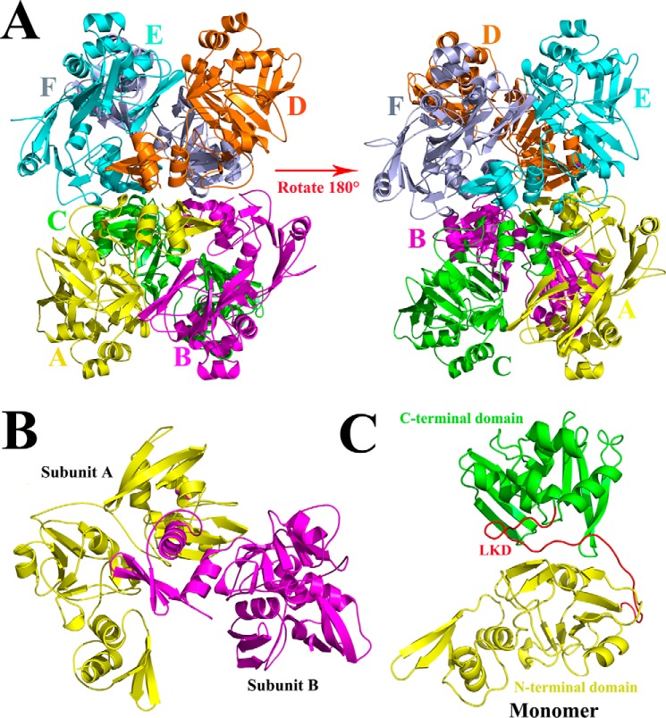
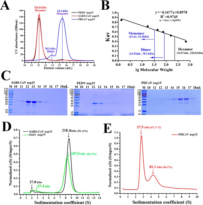
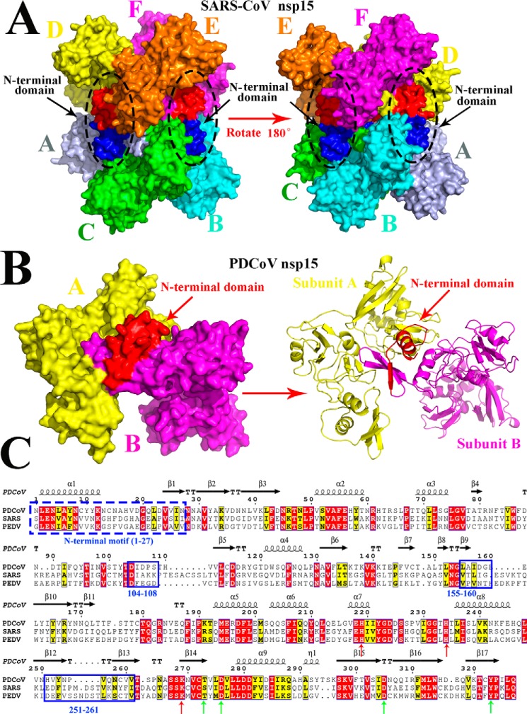
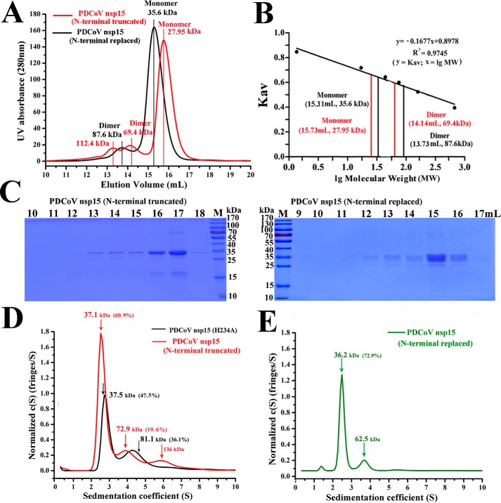
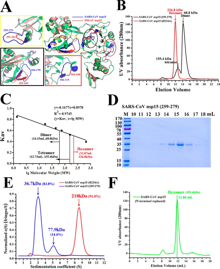
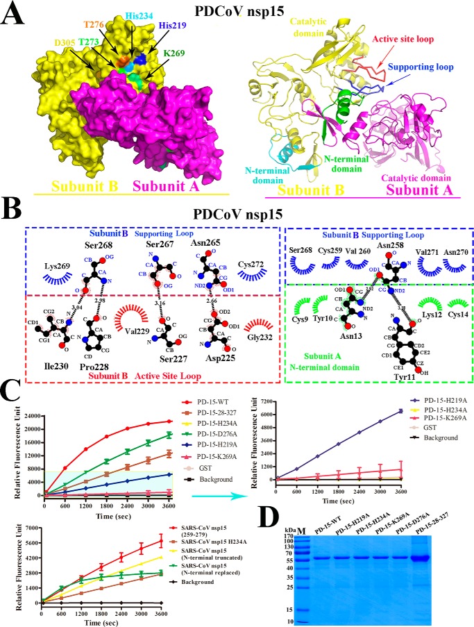
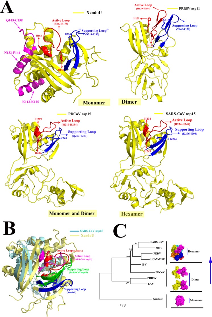
Nidovirus endoribonucleases (NendoUs) include nonstructural protein 15 (nsp15) from coronaviruses and nsp11 from arteriviruses, both of which have been reported to participate in the viral replication process and in the evasion of the host immune system. Results from a previous study of coronaviruses SARS-CoV, HCoV-229E, and MHV nsp15 indicate that it mainly forms a functional hexamer, whereas nsp11 from the arterivirus PRRSV is a dimer. Here, we found that porcine Deltacoronavirus (PDCoV) nsp15 primarily exists as dimers and monomers in vitro Biological experiments reveal that a PDCoV nsp15 mutant lacking the first 27 amino acids of the N-terminal domain (Asn-1-Asn-27) forms more monomers and displays decreased enzymatic activity, indicating that this region is important for its dimerization. Moreover, multiple sequence alignments and three-dimensional structural analysis indicated that the C-terminal region (His-251-Val-261) of PDCoV nsp15 is 10 amino acids shorter and forms a shorter loop than that formed by the equivalent sequence (Gln-259-Phe-279) of SARS-CoV nsp15. This result may explain why PDCoV nsp15 failed to form hexamers. We speculate that NendoUs may have originated from XendoU endoribonucleases (XendoUs) forming monomers in eukaryotic cells, that NendoU from arterivirus gained the ability to form dimers, and that the coronavirus variants then evolved the capacity to assemble into hexamers. We further propose that PDCoV nsp15 may be an intermediate in this evolutionary process. Our findings provide a theoretical basis for improving our understanding of NendoU evolution and offer useful clues for designing drugs and vaccines against nidoviruses.

Keywords: PDCoV nsp15; endoribonuclease; evolution; nidovirus; oligomerization; protein evolution; protein purification; viral protein.


Conflict of interest statement

The authors declare that they have no conflicts of interest with the contents of this article

Figures

Figure 1.
Figure 1.
Predicted three-dimensional structure of PDCoV nsp15. A, from the side face, the predicted three-dimensional structure of PDCoV nsp15 was built in the SWISS MODEL website. It consists of six monomers that are depicted and marked in PyMOL software with yellow, magenta, green, orange, cyan, and light blue for monomers A–F, respectively. B, two monomers interact with each other closely forming into a dimer with subunit A and subunit B. C, monomer of PDCoV nsp15 consists of three parts: the N-terminal domain (Asn-1 to Arg-167) in yellow, the C-terminal domain (Pro-193 to Gln-327) in green, and a middle linker domain (LKD; Tyr-168 to Thr-192) in red.
Figure 2.
Figure 2.
Oligomerization of PDCoV nsp15 is different from other CoV nsp15s. A, size-exclusion experiment of PDCoV nsp15 (blue), SARS-CoV nsp15 (red), and PEDV nsp15 (black). The calculated molecular masses were determined by fitting to the calibration curve as described in B. B, calculated molecular masses of these nsp15s peaks with the values obtained for known calibration standards (Bio-Rad and GE Healthcare). The calculated molecular mass of nsp15 peaks was determined by fitting to the calibration curve (Kav = volumes of elution (Ves/24)); volumes of elution of 12.07 ml (∼226.8 kDa) in SARS-CoV and PEDV nsp15s (black vertical line) and 13.92 ml (∼78.5 kDa) and 15.43 ml (∼33.3 kDa) in PDCoV nsp15 (blue vertical line) are depicted. C, SDS-PAGE analysis of SARS-CoV nsp15, PEDV nsp15, and PDCoV nsp15. The elution volume is labeled as described in A. Molecular mass markers are shown. D and E, sedimentation velocity analysis of SARS-CoV nsp15 (black), PEDV nsp15 (green), and PDCoV nsp15 (red) with their major peaks of hexamers (∼218.0 and 187.0 kDa), dimers (∼81.1 kDa), and monomers (∼37.5 kDa), respectively. The sedimentation coefficient (s20, w) and the calculated molecular masses are shown in Table 1.
Figure 3.
Figure 3.
Predicted residues involved in the dimerization of PDCoV nsp15 determined by sequence and structural alignments. A, structure of SARS-CoV nsp15 (PDB code 2rhb) is a homohexamer with six monomers A (light blue), B (cyan), C (green), D (yellow), E (orange), and F (magenta), and three NTDs are shown in red, and the other three NTDs are shown in blue and are indicated by black arrows. B, predicted three-dimensional structure of PDCoV nsp15 that was built in the SWISS MODEL website. Monomer A (yellow) and monomer B (magenta) form a dimer via the interaction with the N-terminal domain (red), and the right panel is the cartoon formation of PDCoV nsp15 depicted in PyMOL. Monomer A (yellow) and monomer B (magenta) interact with each other through the NTD (red) that are indicated by red arrows. C, amino acid sequence alignment of PDCoV, SARS-CoV, and PDCoV nsp15s. The formal 27 amino acids of these two nsp15s are depicted with a blue dotted line; Asp-104 to Ser-108, Leu-155 to Glu-160, and His-251 to Val-261 residues on PDCoV nsp15 are depicted with the blue solid lines. The conservative sites of three catalytic sites and four binding sites are indicated with red and green arrows, respectively.
Figure 4.
Figure 4.
NTD of PDCoV nsp15 is important for its dimerization. A, size-exclusion experiment with PDCoV nsp15 (N-terminal truncated) (red) and PDCoV nsp15 (N-terminal replaced) (black). The calculated molecular masses are indicated by a red vertical line for the N-terminal truncated mutant with three peak volumes corresponding to predicted masses of ∼112.4, 69.4, and 27.9 kDa; and the predicted masses of N-terminal replaced mutant were ∼87.6 and 35.6 kDa; they were determined by fitting to the calibration curve as described for B. B, calculated molecular masses of the nsp15 protein peaks with the values obtained for known calibration standards (Bio-Rad and GE Healthcare). The calculated molecular mass of nsp15 peaks was determined by fitting to the calibration curve (Kav = volumes of elution (Ves/24)). C, SDS-PAGE analysis of PDCoV nsp15 (N-terminally truncated) and PDCoV nsp15 (N-terminally replaced). The elution volume is labeled as described for A. Molecular mass markers are shown. D and E, sedimentation velocity analysis of PDCoV nsp15 (N-terminally truncated) (red), PDCoV nsp15 (H234A) (black), and PDCoV nsp15 (N-terminally replaced) (green) with their major peaks is depicted in D and E. The sedimentation coefficient (s20, w) and the calculated masses are shown in Table 1.
Figure 5.
Figure 5.
Region (His-251–Val-261) of PDCoV nsp15 rendering the PDCoV nsp15 fails to form a hexamer. A, three-dimensional structural alignment of SARS-CoV nsp15 (PDB 2H85) (green) and PDCoV nsp15 (salmon). The predicted residues participating in dimerization are depicted with three ellipses, and they are drawn in rectangles; the residues of Asp-104 to Ser-108, Leu-155 to Glu-160, and His-251 to Val-261 regions of PDCoV nsp15 are highlighted in red and located in red, black, and yellow rectangles, respectively. The corresponding residues of Asp-106 to Ala-115, Thr-166 to Tyr-178, and Gln-259 to Phe-279 regions on SARS-CoV nsp15 are depicted in blue. B, size-exclusion experiment with SARS-CoV nsp15Asp-259–Phe-279 and SARS-CoV nsp15 (H234A). The calculated molecular masses were determined by fitting to the calibration curve as described in C. C, calculated molecular masses of nsp15 protein peaks with the values obtained for known calibration standards (Bio-Rad and GE Healthcare). The calculated molecular mass of nsp15 peaks was determined by fitting to the calibration curve (Kav = volumes of elution (Ves/24)), and the two peak volumes of SARS-CoV nsp15Asp-259–Phe-279 were ∼12.72 and 14.15 ml indicated by black vertical lines with the predicted masses of ∼155.4 and 68.8 kDa, respectively, in B. D, SDS-PAGE analysis of SARS-CoV nsp15Asp-259–Phe-279. E, sedimentation velocity analysis of SARS-CoV nsp15Asp-259–Phe-279 (blue) and SARS-CoV nsp15 (H234A) (red) with their major peaks in E. The sedimentation coefficient (s20, w) and the calculated molecular masses are shown in Table 1. F, size-exclusion experiment result of SARS-CoV nsp15 (N-terminally replaced) (green) with the elution volume of ∼11.84 ml and the calculated molecular mass of ∼259.4 kDa were determined by fitting to the calibration curve as described in C.
Figure 6.
Figure 6.
NendoU activity of PDCoV nsp15 and SARS-CoV nsp15. A, predicted structure of dimeric PDCoV nsp15. The subunit A (magenta) and subunit B (yellow) are tightly interacted with each other, and the catalytic sites of His-234, His-219, and Lys-269 and binding sites of Thr-273, Asp-276, and Asp-305 are marked with arrows with the cyan, blue, green, light green, orange, and yellow, respectively. The right panel is the cartoon picture of dimeric PDCoV nsp15 with subunit A (magenta) and B (yellow) interactions via the interplay of NTD (green) on subunit A and the supporting loop (Asn-257 to Cys-273) in blue to stabilize the active-site loop (His-219 to His-234) in red on subunit B. The NTD of subunit B is in cyan. B, detailed molecular interactions of the supporting loop (blue) and the active-site loop (red) on subunit B with the NTD (green) on subunit A were determined using LIGPLOT. Carbon, oxygen, and nitrogen atoms are shown as black, red, and blue circles, respectively. Hydrogen bonds are shown with black dashed lines between the donor and acceptor atoms with the bond distance. Hydrophobic interactions are shown by arcs with spokes in blue for the supporting loop, red for the active site loop on subunit A, and green for the NTD on subunit B, which are radiating toward the atoms with which they interact. C, FRET-based enzyme activity experiment. The enzymatic activity of the WT PDCoV nsp15 and mutants (H219A, H234A, K269A, D276A, and N-terminally truncated) is depicted with different colors. The values of the triplicate experiment results are shown. The enzymatic activity of SARS-CoV nsp15 mutants (H234A, N-terminally truncated, N-terminally replaced, and Asp-259–Phe-279) is depicted. The values of the triplicate experiment results are shown. D, SDS-PAGE analysis of PDCoV nsp15 mutants.
Figure 7.
Figure 7.
Possible model of the evolutionary process for the nidorivirus NendoU. A, monomeric forms of the three-dimensional structures of XendoU (PDB code 2C1W), PRRSV nsp11 (PDB code 5DA1), PDCoV nsp15 (predicted structure), and SARS-CoV nsp15 (PDB code 2H85) are depicted in yellow, and the catalytic sites of XendoU (His-162, His-178, and Lys-224), PRRSV nsp11 (His-129, His-144, and Lys-173), PDCoV nsp15 (His-219, His-234, and Lys-269), and SARS-CoV nsp15 (His-234, His-249, and Lys-289) are indicated with red arrows. The common active loop and supporting loop on XendoU (His-162 to His-178 and Asn-214 to Phe-230), PRRSV nsp11 (His-129 to His-144 and Val-162 to Thr-179), PDCoV nsp15 (His-219 to His-234 and Gln-257 to Thr-273), and SARS-CoV nsp15 (His-234 to His-249 and Lys-276 to Ile-295) are indicated with red and blue, respectively. Moreover, residues of Lys-113 to Lys-125, Asn-133 to Phe-144, and Gln-145 to Cys-158 on XendoU are shown in magenta. B, three-dimensional structural alignment of SARS-CoV nsp15 (light blue) and XendoU (light yellow) are depicted. The active loop and supporting loop of SARS-CoV nsp15 are shown in magenta and green, respectively. The active loop and supporting loop of XendoU are shown in red and blue, respectively. C, XendoU is a monomer shown in magenta. The dimeric nsp11 is in yellow and magenta, and the hexameric Alpha-, Beta-, and Gammacoronaviruses nsp15s, including six monomers, are shown in different colors. PDCoV nsp15 is an intermediate and depicted as a dimer in yellow and magenta. The phylogenetic tree was analyzed using the distance-based neighbor-joining method in the MEGA package. The different subgenotypes are indicated.

References

    1. Nga P. T., Parquet Mdel C., Lauber C., Parida M., Nabeshima T., Yu F., Thuy N. T., Inoue S., Ito T., Okamoto K., Ichinose A., Snijder E. J., Morita K., and Gorbalenya A. E. (2011) Discovery of the first insect nidovirus, a missing evolutionary link in the emergence of the largest RNA virus genomes. PLoS Pathog. 7, e1002215 10.1371/journal.ppat.1002215 - DOI - PMC - PubMed
    1. Vasilakis N., Guzman H., Firth C., Forrester N. L., Widen S. G., Wood T. G., Rossi S. L., Ghedin E., Popov V., Blasdell K. R., Walker P. J., and Tesh R. B. (2014) Mesoniviruses are mosquito-specific viruses with extensive geographic distribution and host range. Virol. J. 11, 97 10.1186/1743-422X-11-97 - DOI - PMC - PubMed
    1. Cong Y., Verlhac P., and Reggiori F. (2017) The interaction between nidovirales and autophagy components. Viruses 9, E182 - PMC - PubMed
    1. Woo P. C., Lau S. K., Lam C. S., Lau C. C., Tsang A. K., Lau J. H., Bai R., Teng J. L., Tsang C. C., Wang M., Zheng B. J., Chan K. H., and Yuen K. Y. (2012) Discovery of seven novel mammalian and avian coronaviruses in the genus Deltacoronavirus supports bat coronaviruses as the gene source of Alphacoronavirus and Betacoronavirus and avian coronaviruses as the gene source of Gammacoronavirus and Deltacoronavirus. J. Virol. 86, 3995–4008 10.1128/JVI.06540-11 - DOI - PMC - PubMed
    1. Nedialkova D. D., Ulferts R., van den Born E., Lauber C., Gorbalenya A. E., Ziebuhr J., and Snijder E. J. (2009) Biochemical characterization of arterivirus nonstructural protein 11 reveals the nidovirus-wide conservation of a replicative endoribonuclease. J. Virol. 83, 5671–5682 10.1128/JVI.00261-09 - DOI - PMC - PubMed

Publication types

MeSH terms

Substances