. 2018 Aug 3;293(31):12177-12185.
doi: 10.1074/jbc.RA118.002018. Epub 2018 Jun 10.

Scaffold attachment factor B suppresses HIV-1 infection of CD4+ T cells by preventing binding of RNA polymerase II to HIV-1's long terminal repeat

Affiliations

Scaffold attachment factor B suppresses HIV-1 infection of CD4+ T cells by preventing binding of RNA polymerase II to HIV-1's long terminal repeat

Li Ma et al. J Biol Chem. .

Abstract

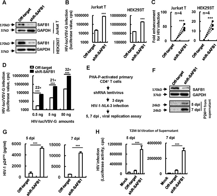
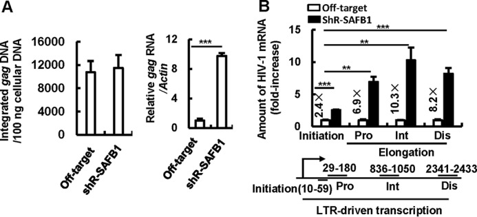
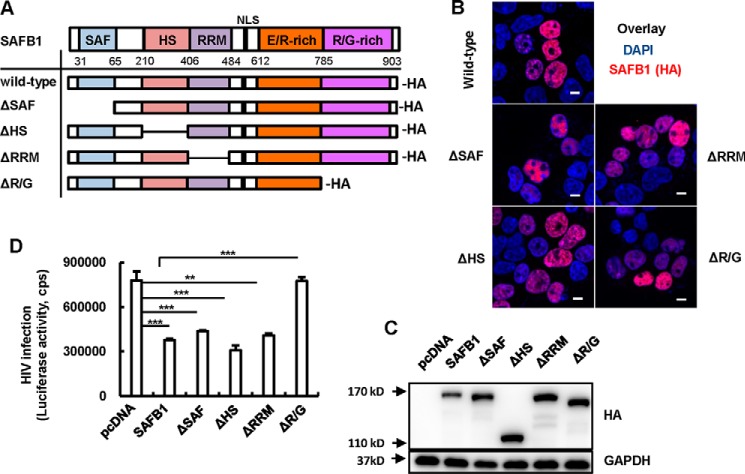
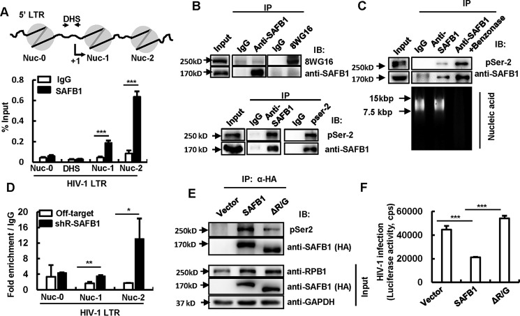
The 5' end of the HIV, type 1 (HIV-1) long terminal repeat (LTR) promoter plays an essential role in driving viral transcription and productive infection. Multiple host and viral factors regulate LTR activity and modulate HIV-1 latency. Manipulation of the HIV-1 LTR provides a potential therapeutic strategy for combating HIV-1 persistence. In this study, we identified an RNA/DNA-binding protein, scaffold attachment factor B (SAFB1), as a host cell factor that represses HIV-1 transcription. We found that SAFB1 bound to the HIV-1 5' LTR and significantly repressed 5' LTR-driven viral transcription and HIV-1 infection of CD4+ T cells. Mechanistically, SAFB1-mediated repression of HIV-1 transcription and infection was independent of its RNA- and DNA-binding capacities. Instead, by binding to phosphorylated RNA polymerase II, SAFB1 blocked its recruitment to the HIV-1 LTR. Of note, SAFB1-mediated repression of HIV-1 transcription from proviral DNA maintained HIV-1 latency in CD4+ T cells. In summary, our findings reveal that SAFB1 binds to the HIV-1 LTR and physically interacts with phosphorylated RNA polymerase II, repressing HIV-1 transcription initiation and elongation. Our findings improve our understanding of host modulation of HIV-1 transcription and latency and provide a new host cell target for improved anti-HIV-1 therapies.

Keywords: HIV; host–pathogen interaction; viral transcription; virology; virus.


Conflict of interest statement

The authors declare that they have no conflicts of interest with the contents of this article. The content is solely the responsibility of the authors and does not necessarily represent the official views of the National Institutes of Health

Figures

Figure 1.
Figure 1.
SAFB1 inhibits HIV-1 infection in HEK293T and CD4+ T cells. A–D, knockdown of SAFB1 facilitates viral infection. A, SAFB1 stable knockdown Jurkat T and HEK293T cells were generated by using lentiviruses containing SAFB1-specific shRNAs. SAFB1 expression was detected by Western blotting. B, cells were infected with HIV-luc/VSV-G (5 ng of p24gag) for an additional 48 h and monitored for viral replication by measuring luciferase activity. C, data from four independent repeats were summarized and analyzed. D, SAFB1 stable knockdown (or off-target) Jurkat T cells were infected with different amounts of HIV-luc/VSV-G for 48 h. E–H, SAFB1 knockdown enhances HIV-1 replication in primary CD4+ T cells. E, lentiviruses containing SAFB1-specific shRNAs were used to knock down SAFB1 in PHA-P–activated primary CD4+ T cells, which were then infected by replication-competent HIV-1NL4-3 (10 ng of p24gag) for 5 or 7 days. The cell culture supernatants were harvested and used for detection of the produced HIV-1 particles by p24gag Western blotting (F) and with p24gag capture ELISA (G) or by titration in TZM-bl indicator cells (H). Data are presented as mean ± S.D. One representative result from at least three independent experiments is shown. ***, p < 0.001 as determined by an unpaired Student's t test. dpi, days post infection; GAPDH, glyceraldehyde-3-phosphate dehydrogenase.
Figure 2.
Figure 2.
SAFB1 represses HIV-1 transcription. A, SAFB1 knockdown promotes HIV-1 transcription. SAFB1 stable knockdown HEK293T cells were infected with HIV-luc/VSV-G (5 ng of p24gag) for 24 h. Then the integrated HIV-1 gag DNA was quantified with Alu-PCR (left panel), and the transcribed gag mRNA levels were quantified with quantitative reverse transcription PCR. The Actin gene was used for normalization. B, SAFB1 knockdown significantly increased HIV-1 5′ LTR–driven RNA transcription initiation and elongation. The total amounts of mRNAs, as assessed by qPCR with specific primers, were used to quantify the initiation and elongation of HIV-1 transcription. Data are presented as mean ± S.D. Results are representative of at least three independent experiments. **, p < 0.01; ***, p < 0.001. Pro, proximal; Int, intermediate; Dis, distal.
Figure 3.
Figure 3.
The C-terminal Arg/Gly-rich domain is required for SAFB1-mediated HIV-1 inhibition. A, schematics of the WT SAFB1 protein motif and its mutants. An HA tag was added to the C terminus of constructs. B, cellular location of SAFB1 and its mutants. HEK293T cells were seeded on poly-l-lysine–coated microscope slides and transfected with the indicated plasmids. Anti-HA antibody was used for immunostaining to indicate SAFB1 (red), and the nucleus was indicated with 4′,6-diamidino-2-phenylindole staining (DAPI, blue). The stained cells were observed under confocal microscopy. Scale bars = 10 μm. C and D, inhibition of HIV-1 infection mediated by SAFB1 or its mutants. HEK293T cells were transfected with pCDNA3.1-HA/SAFB1 or mutant plasmids for 48 h. The expression of SAFB1 or mutants was measured by Western blotting (C), and then the cells were infected with HIV-luc/VSV-G (5 ng of p24gag) for an additional 48 h. Viral infection was measured by detecting luciferase activity (D). Data are presented as mean ± S.D. Results are representative of three independent experiments. **, p < 0.01; ***, p < 0.001 as determined by an unpaired Student's t test.
Figure 4.
Figure 4.
SAFB1 associates with phosphorylated RNA pol II to impede its recruitment to LTR. A, association of SAFB1 with the HIV-1 5′ LTR. HEK293T cells were infected with HIV-luc/VSV-G for 24 h, cross-linked, and sonicated and then subjected to an SAFB1 ChIP assay. Fragments of the LTR covering nucleotides 40–902, which contain Nuc0, DHS, Nuc-1 and Nuc-2, were amplified by qPCR. B and C, SAFB1 interacts with pSer-2 RNA pol II. Jurkat T cells were lysed, treated with benzonase, and then incubated with the indicated antibodies and protein A or G magnetic beads. The immunoprecipitates were detected by Western blotting with specific antibodies, and the nucleotides from cell lysates were electrophoresed on 1% agarose gel and stained with ethidium bromide. 15 kbp and 7.5 kbp indicate the DNA marker used in the gel (C). D, SAFB1 knockdown promotes the enrichment of pSer-2 RNA pol II on the HIV-1 LTR. SAFB1 stable knockdown Jurkat T cells were infected with HIV-luc/VSV-G (5 ng of p24gag) for 48 h. After being cross-linked and sonicated, the Jurkat T cells were subjected to a ChIP assay with pSer-2 RNA pol II antibody. Fragments of the LTR covering nucleotides 40–902, which contain Nuc0, Nuc-1, and Nuc-2, were amplified by qPCR. E, deletion of the Arg/Gly-rich domain abolishes SAFB1 association with pSer-2 RNA pol II. Jurkat cells were transfected with a pCDNA3.1-HA/SAFB1- or SAFB1-ΔArg/Gly–expressing plasmid and then lysed. The cell lysates were used for IP with anti-HA antibodies, and protein expression was detected by immunoblotting (IB) with the indicated antibodies. F, deletion of the Arg/Gly-rich domain abolishes SAFB1-mediated HIV-1 inhibition. Jurkat cells were transfected with pCDNA3.1-HA/SAFB1 or SAFB1-ΔArg/Gly–expressing plasmid and then infected with HIV-luc/NL4-3 (20 ng of p24gag) for an additional 48 h. Viral infection was measured by detecting the luciferase value. Results are representative of three independent repeats. Data are presented as mean ± S.D. *, p < 0.05; **, p < 0.01; ***, p < 0.001 as determined by an unpaired Student's t test.
Figure 5.
Figure 5.
SAFB1 represses HIV-1 reactivation from an integrated HIV-1 proviral DNA. A and B, SAFB1 knockdown promoted HIV-1 reactivation in C11 cells. A, C11 cells were infected with lentiviruses containing SAFB1 shRNA or an off-target control for 72 h. SAFB1 knockdown was determined by Western blotting. B, cells were stimulated with or without TNFα for an additional 24 h, and HIV-1 reactivation was measured by detecting GFP expression. The percentage of GFP+ cells and the mean fluorescence intensity (MFI) were calculated. C, SAFB1 knockdown in ACH2 cells. ACH2 cells were infected with lentiviruses containing SAFB1-specific or off-target shRNA for 72 h, during which puromycin was added for selection 24 h post-infection. SAFB1 knockdown from four independent repeats was detected by Western blotting. D, cells were stimulated with TNFα or SAHA for an additional 24 h. Viral reactivation was detected by quantifying the produced viral particles in the supernatant with p24gag capture ELISA. Data from four independent repeats are shown. Data are presented as mean ± S.D. *, p < 0.05; **, p < 0.01; ***, p < 0.001. E, schematic illustration of SAFB1 inhibiting HIV transcription. SAFB1 bound to the HIV-1 5′ LTR region and directly associates with the phosphorylated RNA pol II to impede RNA pol II recruitment to the LTR and consequently repress HIV-1 transcription.

Similar articles

Cited by

References

    1. Margolis D. M. (2010) Mechanisms of HIV latency: an emerging picture of complexity. Curr. HIV/AIDS Rep. 7, 37–43 10.1007/s11904-009-0033-9 - DOI - PubMed
    1. Mbonye U., and Karn J. (2014) Transcriptional control of HIV latency: cellular signaling pathways, epigenetics, happenstance and the hope for a cure. Virology 454, 328–339 - PMC - PubMed
    1. Lusic M., and Giacca M. (2015) Regulation of HIV-1 latency by chromatin structure and nuclear architecture. J. Mol. Biol. 427, 688–694 10.1016/j.jmb.2014.07.022 - DOI - PubMed
    1. Francis A. C., Di Primio C., Allouch A., and Cereseto A. (2011) Role of phosphorylation in the nuclear biology of HIV-1. Curr. Med. Chem. 18, 2904–2912 10.2174/092986711796150478 - DOI - PubMed
    1. Zhu J., Gaiha G. D., John S. P., Pertel T., Chin C. R., Gao G., Qu H., Walker B. D., Elledge S. J., and Brass A. L. (2012) Reactivation of latent HIV-1 by inhibition of BRD4. Cell Rep. 2, 807–816 10.1016/j.celrep.2012.09.008 - DOI - PMC - PubMed

Publication types

MeSH terms

Substances