. 2018 Aug 10;293(32):12303-12317.
doi: 10.1074/jbc.RA118.003741. Epub 2018 Jun 10.

Biosynthesis of the nickel-pincer nucleotide cofactor of lactate racemase requires a CTP-dependent cyclometallase

Affiliations

Biosynthesis of the nickel-pincer nucleotide cofactor of lactate racemase requires a CTP-dependent cyclometallase

Benoît Desguin et al. J Biol Chem. .

Abstract

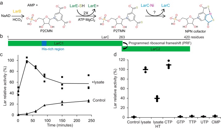
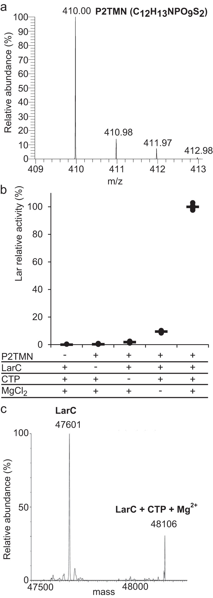
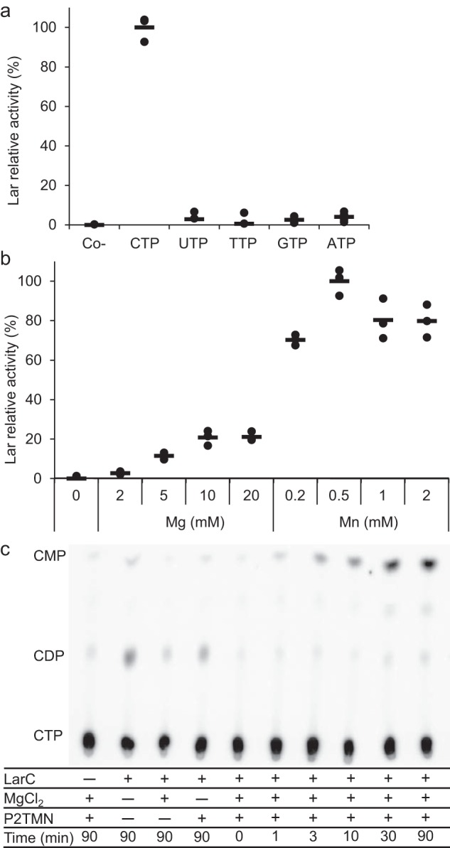
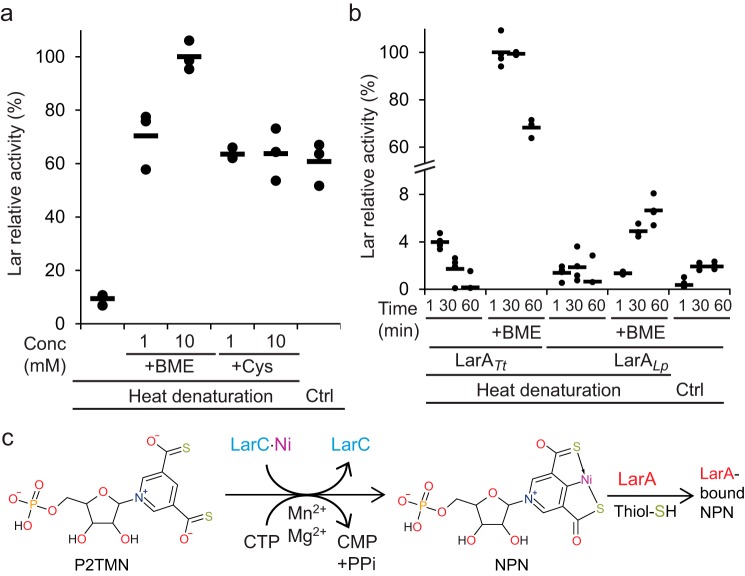
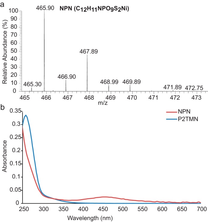
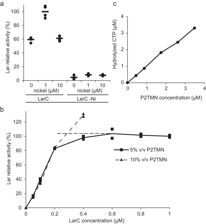
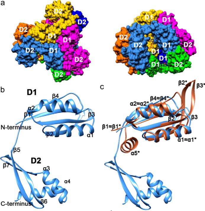
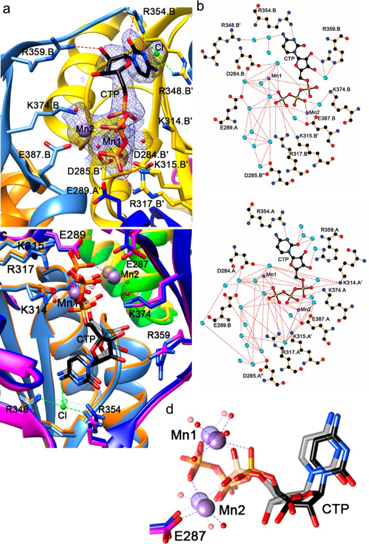
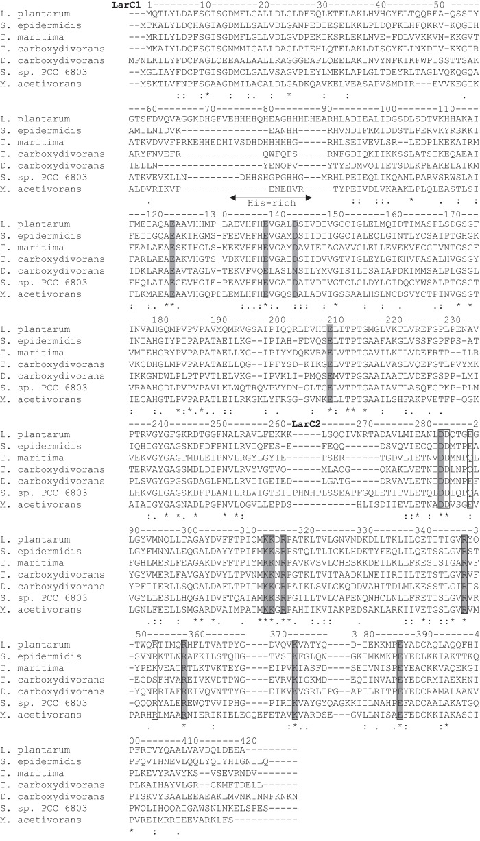
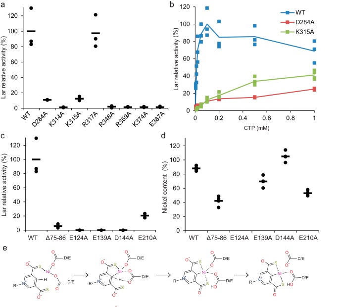
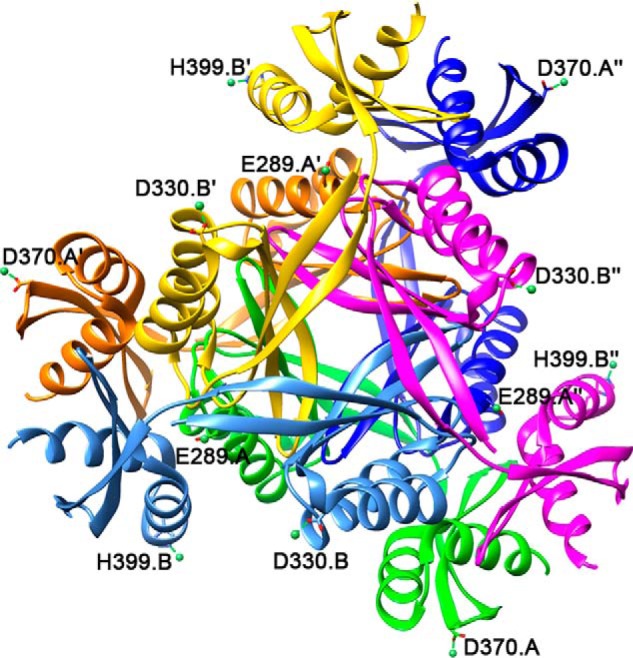
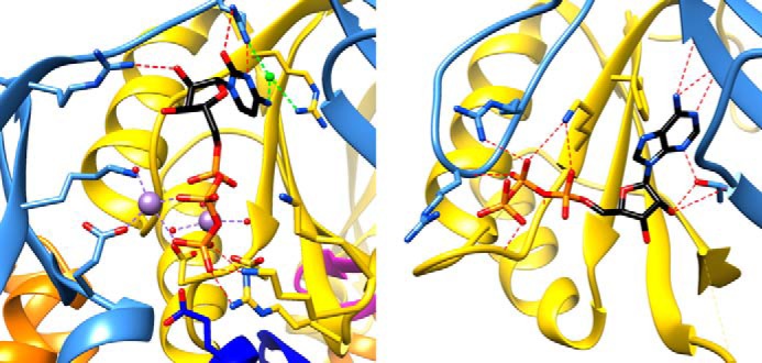
Bacterial lactate racemase is a nickel-dependent enzyme that contains a cofactor, nickel pyridinium-3,5-bisthiocarboxylic acid mononucleotide, hereafter named nickel-pincer nucleotide (NPN). The LarC enzyme from the bacterium Lactobacillus plantarum participates in NPN biosynthesis by inserting nickel ion into pyridinium-3,5-bisthiocarboxylic acid mononucleotide. This reaction, known in organometallic chemistry as a cyclometalation, is characterized by the formation of new metal-carbon and metal-sulfur σ bonds. LarC is therefore the first cyclometallase identified in nature, but the molecular mechanism of LarC-catalyzed cyclometalation is unknown. Here, we show that LarC activity requires Mn2+-dependent CTP hydrolysis. The crystal structure of the C-terminal domain of LarC at 1.85 Å resolution revealed a hexameric ferredoxin-like fold and an unprecedented CTP-binding pocket. The loss-of-function of LarC variants with alanine variants of acidic residues leads us to propose a carboxylate-assisted mechanism for nickel insertion. This work also demonstrates the in vitro synthesis and purification of the NPN cofactor, opening new opportunities for the study of this intriguing cofactor and of NPN-utilizing enzymes.

Keywords: bacterial metabolism; cyclometalation; lactate racemase; lactic acid; natural product biosynthesis; nickel; pincer complex; protein structure.


Conflict of interest statement

The authors declare that they have no conflicts of interest with the contents of this article

Figures

Figure 1.
Figure 1.
Cell lysates and CTP have similar effects on NPN cofactor biosynthesis. a, NPN cofactor biosynthesis starts with NaAD that is carboxylated and hydrolyzed by LarB forming P2CMN. Two LarE proteins each catalyze a sacrificial sulfur transfer reaction to synthesize P2TMN from P2CMN. LarC inserts nickel into P2TMN, forming the NPN cofactor containing a metallacycle (bold blue lines) with Ni-C and Ni-S σ bonds. b, schematic representation of the larC gene. LarC1 and LarC are the two open reading frames that code for LarC. c, effect of 10% (v/v) Lc. lactis cell lysates on in vitro NPN cofactor biosynthesis by LarB, LarE, and LarC. Lar activity with cell lysates after 30 min was set to 100%. d, effect of 10% cell lysates, 10% cell lysates deproteinized with heat treatment (lysate HT), or 1 mm nucleotides on the in vitro NPN cofactor biosynthesis by LarB, LarE, and LarC after 30 min. Lar activity with cell lysates was set to 100%. b and c, NPN cofactor biosynthesis was assessed after addition of LarATt apoprotein and l-lactate by assaying for d-lactate production. The lines indicate the mean values, and the points indicate the individual data points.
Figure 2.
Figure 2.
P2TMN is the substrate of LarC. a, P2TMN identification by MS in negative ionization mode with the molecular formula in parentheses. b, NPN cofactor biosynthesis using purified P2TMN, LarC, CTP, and MgCl2. Lar activity with all compounds was set to 100%. NPN cofactor biosynthesis was assessed after addition of LarATt apoprotein and l-lactate and by assaying for d-lactate production. c, mass of purified LarC. The expected mass of LarC is 47,601.5 Da, and the mass of LarC + CTP + Mg2+ − 2H+ is 48,107 Da.
Figure 3.
Figure 3.
LarC is a CTP-hydrolyzing nickel cyclometallase. a, dependence of LarC reaction on nucleotide identity. Lar activity with CTP was set to 100%. b, dependence of LarC reaction on MgCl2 and MnCl2 concentration. Lar activity with 0.5 mm MnCl2 was set to 100%. Lar activity was assessed after addition of LarATt apoprotein in excess and l-lactate by assaying for d-lactate production. The lines indicate the mean values, and the points indicate the individual data points. c, radiogram from TLC of the LarC reaction using P2TMN, MgCl2, and [α-32P]CTP. The positions of CDP and CMP were confirmed by UV detection.
Figure 4.
Figure 4.
Characterization of the LarA activation by NPN. a, activation of LarATt apoprotein. b, time-dependent activation of LarATt and LarALp apoproteins by in vitro-synthesized NPN, with or without (Ctrl) heat denaturation of LarB, LarC, and LarE and with or without addition of cysteine (+Cys) and β-mercaptoethanol (+BME) (1 or 10 mm in a and 10 mm in b). LarA was incubated with in vitro-synthesized NPN for 1 min in a and for the indicated time in b. Lar activity of LarATt with 10 mm BME was set to 100%. Lar activity was assessed after addition of l-lactate by assaying for d-lactate production. The lines indicate the mean values, and the points indicate the individual data points. c, LarC reaction summary.
Figure 5.
Figure 5.
Spectra of NPN. a, NPN identification by MS in negative ionization mode with the molecular formula in parentheses. b, UV-visible absorbance spectra of purified P2TMN (blue) and NPN (red).
Figure 6.
Figure 6.
Characterization of LarC reaction. a, dependence of LarC reaction on nickel. Lar activity was measured using P2TMN, MgCl2, CTP, and either 1 μm LarC purified from cells cultivated in the presence (LarC) or absence (LarCNi) of 1 mm nickel, with or without further addition of nickel to the assay. NPN biosynthesis was performed for 10 min, in order to allow for several turnovers to take place. The Lar activity of LarC with 1 μm nickel was set to 100%. b, titration of P2TMN by LarC. Lar activity with 1 μm LarC and 5% (v/v) of purified P2TMN was set to 100%. Dashed lines represent the conditions of complete P2TMN consumption (horizontal) or of complete LarC consumption (vertical); the intersection is the point where all LarCs and all substrates are consumed with 5% (v/v) P2TMN. Lar activity was assessed after addition of LarATt apoprotein in excess and l-lactate by assaying for d-lactate production. The lines indicate the mean values, and the points indicate the individual data points. c, amount of CTP hydrolysis versus substrate addition, based on densitometry of the TLC autoradiograph.
Figure 7.
Figure 7.
Masses of LarC2 after cleavage of the purified full-length LarC protein.
Figure 8.
Figure 8.
LarC2 structure. Individual chains and their symmetry-related copies are colored as follows: A, green; A′, orange; A″, dark blue; B, light blue; B′, yellow; B″, magenta. N-terminal domain 1 (D1, residues 272–356) forms the center of the trimer (top view), and C-terminal domain 2 (D2, residues 357–412) protrudes outward. The CTP molecules are shown in stick representation with carbon atoms in black, nitrogen in blue, oxygen in red, and phosphorus in orange. a, hexameric structure of LarC2. Surface representation of the top view (left) and side view (right). b, secondary structure of LarC2 (helices = α, strands = β). c, comparison of LarC2 chain B to the most similar match identified by Dali, a GlnB-like putative CutA protein (brown) from Thermotoga maritima (PDB code 1o5j). CutA helices and sheets are labeled with an asterisk.
Figure 9.
Figure 9.
CTP-bound LarC2 structure. a, CTP-binding pocket in the chain B trimer. Ribbon and protein C atoms are colored by chain as described above. Mn2+ ions and chelation are shown in purple, water molecules in red, and chloride ion and chelation in light green. The 2mFo − DFc electron density map for ligands is shown as blue mesh at 1 σ. b, 2D representation of CTP binding in the LarC2–CTP complex for trimer A (above) and B (below). Atoms are shown with carbon in black, nitrogen in blue, oxygen in red, manganese in purple, and water in cyan; hydrogen bonds/metal chelation are shown as red dashes, protein bonds in brown, and CTP bonds in black. c, comparison of CTP binding in trimer A and trimer B where the ligands of trimer A are transparent. d, metal chelation differences between trimers A and B.
Figure 10.
Figure 10.
Alignment of LarC homologs from Lb. plantarum NCIMB 8826, Staphylococcus epidermidis ATCC 12228, T. maritima MSB8, Thermosinus carboxydivorans NOR1, Desulfotomaculum carboxydivorans CO-1-SRB, Synechocystis sp. PCC 6803, and Methanosarcina acetivorans C2A. The numbering above the sequence corresponds to the residue numbering of Lb. plantarum LarC. The sequence of Lb. plantarum LarC was constructed assuming a PRF (7) at position 263, marking the end of LarC1. The residues conserved in all seven sequences are indicated with an asterisk, and the less conserved residues are indicated with one or two dots (as detailed in the ClustalX program (32)). The residues that were substituted by mutagenesis are highlighted in gray. The residues interacting with CTP in the LarC2 structure are boxed with black lines. The alignment was performed using ClustalX (32).
Figure 11.
Figure 11.
LarC variants affect activity. a–c, analysis of selected substitutions in LarC2 (a and b) and LarC1 (c). Lar activity using P2TMN, CTP (0.1 mm in a and c), MgCl2 and WT LarC (WT), or the indicated LarC variants after addition of LarATt apoprotein plus l-lactate and assaying the d-lactate production was measured. Lar activity of the WT with 0.1 mm CTP was set to 100%. d, nickel content of LarC variants. The lines indicate the mean values, and the points indicate the individual data points. e, hypothetical model for the mechanism of cyclometalation by LarC. Dotted lines indicate broken and newly formed bonds, and straight arrows indicate coordination bonds.
Figure 12.
Figure 12.
Hexameric view of the nickel-bound LarC2 structure (PDB code 6BWR). Nickel atoms and coordination are in green, and chelating side chains are labeled.
Figure 13.
Figure 13.
Comparison of CTP and ATP binding within two members of the GlnB-like family. Left, CTP binding to LarC2. Right, ATP binding to PII of E. coli (PDB code 5L9N (17)), with corresponding chains colored the same way as LarC2. Side chains at the ATP-binding pocket are shown with hydrogen bonds as red dashes, and the manganese ions and chelation are shown in purple, water molecules are in red, and chloride ion and chelation are in light green.

References

    1. Boer J. L., Mulrooney S. B., and Hausinger R. P. (2014) Nickel-dependent metalloenzymes. Arch. Biochem. Biophys. 544, 142–152 10.1016/j.abb.2013.09.002 - DOI - PMC - PubMed
    1. Desguin B., Zhang T., Soumillion P., Hols P., Hu J., and Hausinger R. P. (2015) A tethered niacin-derived pincer complex with a nickel-carbon bond in lactate racemase. Science 349, 66–69 10.1126/science.aab2272 - DOI - PubMed
    1. Desguin B., Goffin P., Viaene E., Kleerebezem M., Martin-Diaconescu V., Maroney M. J., Declercq J. P., Soumillion P., and Hols P. (2014) Lactate racemase is a nickel-dependent enzyme activated by a widespread maturation system. Nat. Commun. 5, 3615 10.1038/ncomms4615 - DOI - PMC - PubMed
    1. Desguin B., Soumillion P., Hols P., and Hausinger R. P. (2016) Nickel-pincer cofactor biosynthesis involves LarB-catalyzed pyridinium carboxylation and LarE-dependent sacrificial sulfur insertion. Proc. Natl. Acad. Sci. U.S.A. 113, 5598–5603 10.1073/pnas.1600486113 - DOI - PMC - PubMed
    1. Fellner M., Desguin B., Hausinger R. P., and Hu J. (2017) Structural insights into the catalytic mechanism of a sacrificial sulfur insertase of the N-type ATP pyrophosphatase family, LarE. Proc. Natl. Acad. Sci. U.S.A. 114, 9074–9079 10.1073/pnas.1704967114 - DOI - PMC - PubMed

Publication types

MeSH terms

LinkOut - more resources