Review
. 2018 Aug 1;25(8):653-664.
doi: 10.5551/jat.RV17022. Epub 2018 Jun 9.

Thrombus Formation and Propagation in the Onset of Cardiovascular Events

Affiliations
Review

Thrombus Formation and Propagation in the Onset of Cardiovascular Events

Yujiro Asada et al. J Atheroscler Thromb. .

Abstract

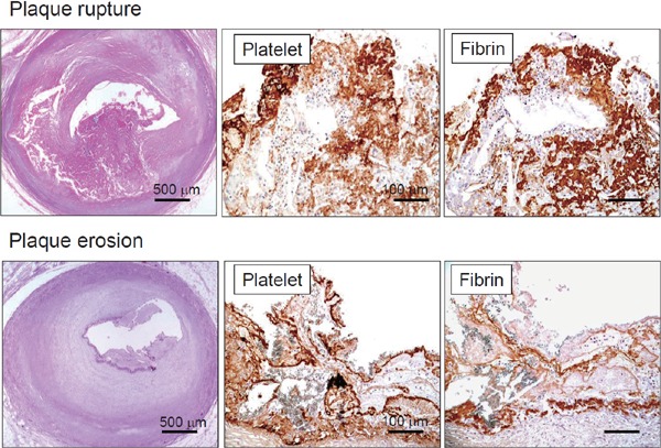
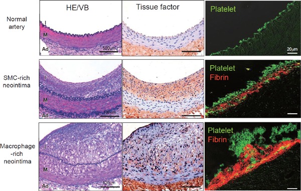
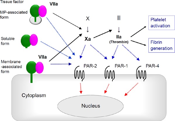
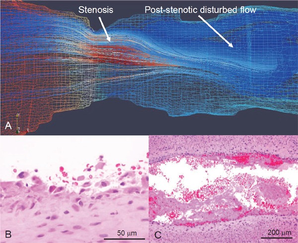
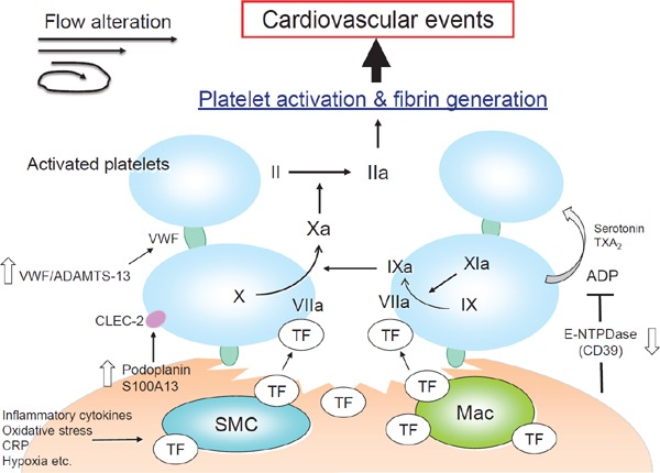
Ischemic cardiovascular disease is a major cause of morbidity and mortality worldwide and thrombus formation on disrupted atherosclerotic plaques is considered to trigger its onset. Although the activation of platelets and coagulation pathways has been investigated intensively, the mechanisms of thrombus formation on disrupted plaques have not been understood in detail. Platelets are thought to play a central role in the formation of arterial thrombus because of rapid flow conditions; however, thrombus that develops on disrupted plaques consistently includes large amounts of fibrin in addition to aggregated platelets. While, thrombus does not always become large enough to completely occlude the vascular lumen, indicating that the propagation of thrombus is also critical for the onset of cardiovascular events. Various factors, such as vascular wall thrombogenicity, altered blood flow and imbalanced blood hemostasis, modulate thrombus formation and propagation on disrupted plaques. Pathological findings derived from humans and experimental animal models of atherothrombosis have identified important factors that affect thrombus formation and propagation, namely platelets, extrinsic and intrinsic coagulation factors, proinflammatory factors, plaque hypoxia and blood flow alteration. These findings might provide insight into the mechanisms of thrombus formation and propagation on disrupted plaques that lead to the onset of cardiovascular events.

Keywords: Atherothrombosis; Blood flow; Factor Ⅺ; Platelet; Tissue factor.


Conflict of interest statement

None declared

Figures

Fig. 1.
Fig. 1.
Microphotographs of coronary plaque rupture and erosion with thrombi Ruptured plaque comprises large necrotic core and disrupted thin fibrous cap accompanied by thrombus formation. Eroded plaque is fibrous and rich in smooth muscle cells, without visible atheromatous component. Both types of thrombi comprise platelets and fibrin (Ref. , with permission).
Fig. 2.
Fig. 2.
Immunohistochemical microphotographs of tissue factor and thrombus in rabbit normal and atherosclerotic femoral arteries (Left column) Representative microphotographs of normal femoral artery and of femoral arteries at 3 weeks after balloon injury of conventional (SMC-rich neotima) or 0.5% cholesterol diet (Macrophage-rich neointima). (Middle column) Tissue factor is expressed in SMC- and macrophage-rich neointima, and in adventitia. (Right column) Thrombus 15 minutes after balloon injury on normal artery comprises only small aggregated platelets, whereas that on neointima comprises a mixture of platelets and fibrin. Thrombus on macrophage-rich neointima is much larger. Abbreviations: Ad, adventitia; HE/VB, hematoxylin and eosin/Victoria blue; I, intima; M, media (modified from Ref. , with permission).
Fig. 3.
Fig. 3.
Tissue factor-dependent coagulation pathway and PARs Membrane- and microparticle (MP)-associated TF binding to factor VIIa triggers coagulation pathway, whereas soluble form of TF with factor VIIa does not. Downstream coagulation factors activate proteinase-activated receptors (PARs) that play other non-coagulative biological roles.
Fig. 4.
Fig. 4.
Computational flow simulation and microphotographs of erosive injury of rabbit stenotic femoral artery with SMC-rich plaque Rabbit femoral arteries 3 weeks after balloon injury are constricted using a vascular occluder. (A) Representative computational reconstructed image and flow simulation in Reynolds-Averaged Navier-Stokes model. Red and blue mesh indicates high and low pressure, respectively, on wall. Flow velocity in this model increases at stenosis and decreases at post-stenotic portion, resulting in disrupted flow. (B, C) Representative microphotographs of erosive injury and thrombus formation. Neointimal endothelial cells and SMCs are broadly detached at post-stenotic portion 15 minute after vascular stenosis (B) (Ref. , with permission). Large mural thrombi are formed at the portion 60 minute after vascular stenosis (C).
Fig. 5.
Fig. 5.
Activation of platelets and coagulation pathway at disrupted atherosclerotic plaque ADAMTS-13, a disintegrin and metalloprotease with a thrombospondin type 1 motif 13; ADP, adenosine diphosphate; CLEC-2, c-type lectin-like receptor 2; CRP, c-reactive protein; E-NTPDase, ecto-nucleoside triphosphate diphosphohydrolase; Mac, macrophage; SMC, smooth muscle cell; TF, tissue factor; TXA2, thromboxane A2; VWF, von Willebrand factor.

References

    1. Falk E, Shah PK, Fuster V. Coronary plaque disruption. Circulation. 1995; 92: 657-671 - PubMed
    1. Virmani R, Kolodgie FD, Burke AP, Farb A, Schwartz SM. Lessons from sudden coronary death: a comprehensive morphological classification scheme for atherosclerotic lesions. Arterioscler Thromb Vasc Biol. 2000; 20: 1262-1275 - PubMed
    1. Shah PK. Mechanisms of plaque vulnerability and rupture. J Am Coll Cardiol. 2003; 41: 15S-22S - PubMed
    1. Komohara Y, Fujiwara Y, Ohnishi K, Shiraishi D, Takeya M. Contribution of macrophage polarization to metabolic diseases. J Atheroscler Thromb. 2016; 23: 10-17 - PubMed
    1. Libby P. Mechanisms of acute coronary syndromes and their implications for therapy. N Engl J Med. 2013; 368: 2004-2013 - PubMed