Review
. 2018;94(6):248-258.
doi: 10.2183/pjab.94.017.

Post-transcriptional regulation of immune responses by RNA binding proteins

Affiliations
Review

Post-transcriptional regulation of immune responses by RNA binding proteins

Takashi Mino et al. Proc Jpn Acad Ser B Phys Biol Sci. 2018.

Abstract

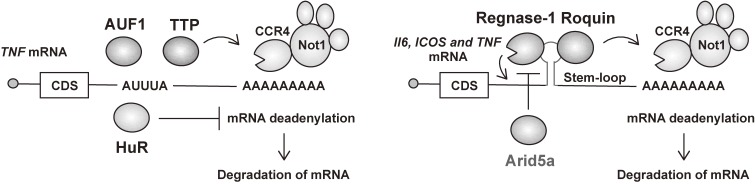
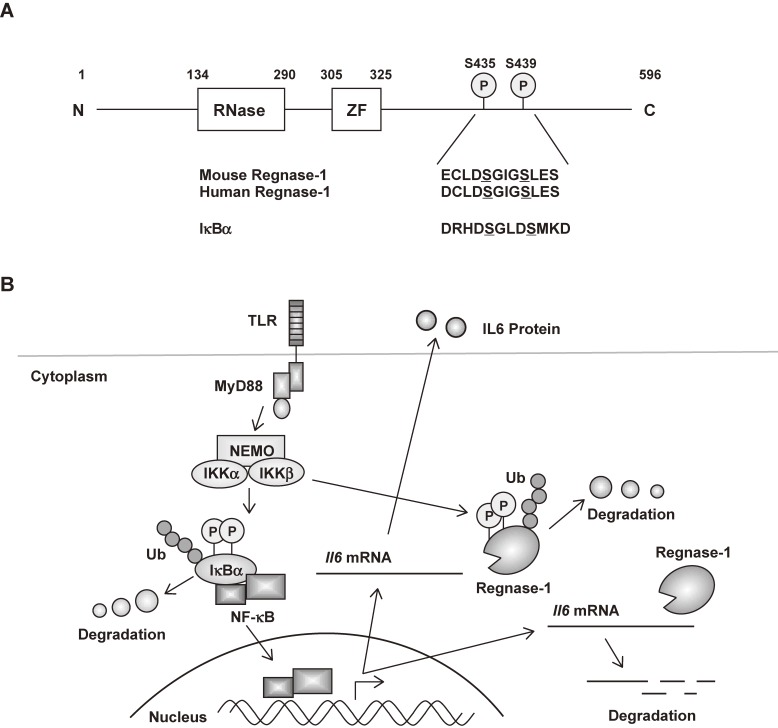
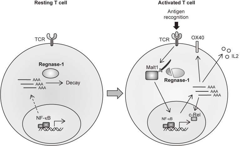
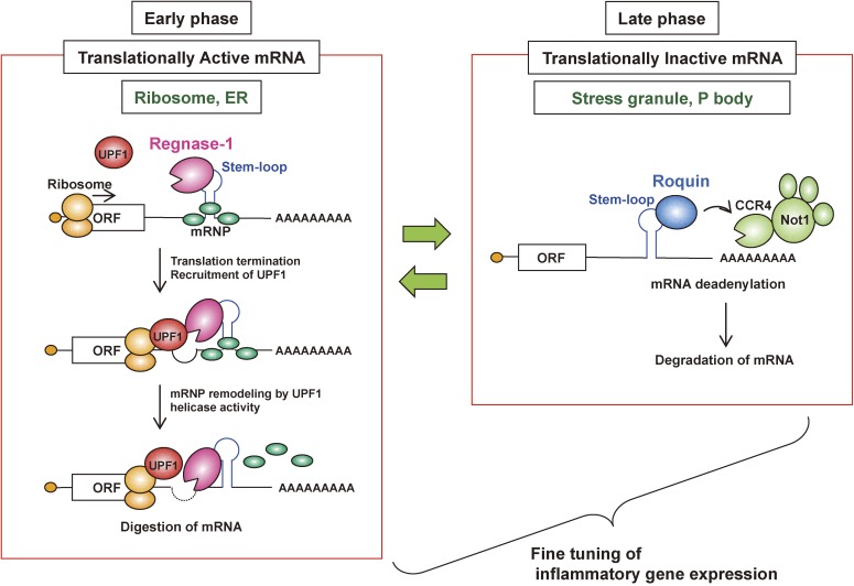
Cytokines are critical mediators of inflammation and host immune defense. Cytokine production is regulated at both transcriptional and post-transcriptional levels. Post-transcriptional damping of inflammatory mRNAs is mediated by a set of RNA binding proteins (RBPs) interacting with cis-elements, such as AU-rich elements (ARE) and stem-loop structures. Whereas ARE-binding proteins such as tristetraprolin and a stem-loop recognizing protein, Roquin, downregulate cytokine mRNA abundance by recruiting a CCR4-NOT deadenylase complex, another stem-loop RBP, Regnase-1, acts as an endoribonuclease, directly degrading target cytokine mRNAs. These RBPs control translation-active or -inactive mRNAs in distinct intracellular locations. The presence of various RBPs regulating mRNAs in distinct locations enables elaborate control of cytokines under inflammatory conditions. Dysregulation of cytokine mRNA decay leads to pathologies such as the development of autoimmune diseases or impaired activation of immune responses. Here we review current knowledge about the post-transcriptional regulation of immune responses by RBPs and the importance of their alteration during inflammatory pathology and autoimmunity.

Keywords: adaptive immunity; cytokines; inflammation; innate immunity; mRNA decay; translation.


Figures

Figure 1.
Figure 1.
Post-transcriptional regulation of inflammation-related mRNAs by RBPs. Regulatory RBPs target the 3′ UTRs of mRNAs to repress expression of the target transcript. AREs and stem-loop structures in 3′ UTRs are well-studied cis-elements recognized by a set of RBPs for controlling the stability of target mRNAs. Whereas TTP, AUF1 and HuR recognize AREs, Regnase-1, Roquin and Arid5a bind to stem-loop structures. CDS, coding sequence.
Figure 2.
Figure 2.
Post-transcriptional regulation of Il-6 expression by Regnase-1 in innate immune cells. (A) Schematic representation of mouse Regnase-1. The sequence of the DSGXXS motif is also shown. ZF, CCCH type zinc finger. (B) In unstimulated cells, Regnase-1 suppresses Il-6 production by degrading Il-6 mRNA through its 3′ UTR. Upon TLR stimulation, IKK complex phosphorylates Regnase-1 at Ser435 and Ser439, which results in the rapid degradation of Regnase-1 protein similar to IκBα. The transcribed Il-6 mRNA is thereby stabilized, which facilitates robust Il-6 production. At a later stage, de novo synthesis of Regnase-1 leads to the degradation of Il-6 mRNA and its own mRNA, promoting the resolution of inflammation.
Figure 3.
Figure 3.
Malt1-induced cleavage of Regnase-1 in T cells regulates immune activation. Regnase-1 regulates the mRNAs of a set of genes, including c-Rel, Ox40 and Il-2, through cleavage of their 3′ UTRs. TCR stimulation leads to cleavage of Regnase-1 at Arg111 by Malt1, resulting in a ‘brake’ of Regnase-1 function and leading to activation of T cells.
Figure 4.
Figure 4.
Regnase-1 and Roquin regulate a common element in inflammatory mRNAs by spatiotemporally distinct mechanisms. Although Regnase-1 and Roquin regulate an overlapping set of mRNAs via a common stem-loop structure located in their 3′ UTRs, they function by spatiotemporally distinct mechanisms. Whereas Roquin destabilizes translationally inactive mRNAs by recruiting the CCR4-NOT deadenylase complex in processing-bodies (PBs) and stress granules (SGs), Regnase-1 localizes to the endoplasmic reticulum (ER) and ribosomes, destabilizes translationally active mRNAs, and requires the RNA helicase activity of UPF1, similar to NMD. Regnase-1 and Roquin tend to control mRNAs at the early and late phases of inflammation, respectively. mRNP complex, mRNA-protein complex.
Figure 5.
Figure 5.
Model of Regnase-1-mediated regulation of iron uptake in the duodenum. Regnase-1 destabilizes PHD3 mRNA, thereby increasing the activity of HIF2α, a critical regulator of iron homeostasis in the duodenum. Accumulated HIF2α then induces the up-regulation of iron transporters and reductases (Dmt1, Ferroportin and Dcytb), leading to adequate iron uptake. Moreover, Regnase-1 itself is transcriptionally up-regulated via HIF2α under iron-deficient conditions. Therefore, the Regnase-1-PHD3-HIF2α axis forms a positive feedback loop for HIF2α amplification to regulate iron uptake in the duodenum.

References

    1. Chovatiya R., Medzhitov R. (2014) Stress, inflammation, and defense of homeostasis. Mol. Cell 54, 281–288. - PMC - PubMed
    1. Moresco E.M., LaVine D., Beutler B. (2011) Toll-like receptors. Curr. Biol. 21, R488–R493. - PubMed
    1. Takeuchi O., Akira S. (2010) Pattern recognition receptors and inflammation. Cell 140, 805–820. - PubMed
    1. Beutler B. (2009) Microbe sensing, positive feedback loops, and the pathogenesis of inflammatory diseases. Immunol. Rev. 227, 248–263. - PMC - PubMed
    1. Medzhitov R., Horng T. (2009) Transcriptional control of the inflammatory response. Nat. Rev. Immunol. 9, 692–703. - PubMed