. 2018 May 25:9:1024.
doi: 10.3389/fmicb.2018.01024. eCollection 2018.

Serodiagnostic Potential of Alpha-Enolase From Sarcoptes scabiei and Its Possible Role in Host-Mite Interactions

Affiliations

Serodiagnostic Potential of Alpha-Enolase From Sarcoptes scabiei and Its Possible Role in Host-Mite Interactions

Jing Xu et al. Front Microbiol. .

Abstract

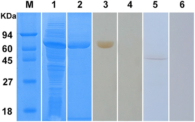
Infestation of the epidermis with the highly contagious ectoparasite, Sarcoptes scabiei, causes scabies, which is characterized by intense itching, pruritus, and secondary infection. This condition affects humans, livestock, and wildlife worldwide, incurring large economic losses and reducing the quality of human life. In the present study, we cloned the alpha-enolase, a key enzyme in the glycolytic and gluconeogenesis pathways, from S. scabiei var. cuniculi, characterized it and produced soluble recombinant enolase protein (rSsc-eno). We determined the localization of Ssc-eno in isolated mites and mites in lesioned skin. The results showed that native enolase was intensely localized in the tegument of the mouthparts, the entire legs, and the whole mites' body, as well as in the gut and reproduction system. Interestingly, we found that native enolase was widely distributed in mites in lesioned skin, with obvious high protein intensity compared with isolated mites. Building on good immunoreactivity, an indirect enzyme-linked immunosorbent assay (ELISA) based on rSsc-eno showed 92% sensitivity and 95.8% specificity, compared with other indirect ELISA in this study, rSsc-eno based ELISA is better in detecting scabies in rabbits. Besides, this method can detect S. scabiei infection as early as 1 week post infection. Compared with other detection methods, such as traditional microscopic examination and recently published universal conventional PCR, rSsc-eno ELISA was more effective to detect early infection in rabbits. Additionally, in vitro incubation experiments demonstrated the concentration-dependent acaricidal activity of rabbit anti-rSsc-eno sera against larval mites, suggested its potential as a vaccine candidate.

Keywords: Sarcoptes scabiei; early diagnosis; embedding; enolase; immunohistochemistry; indirect ELISA.


Figures

Figure 1
Figure 1
Flow chart of the direct paraffin wax embedding method for isolated scabies mites. (A) pooled mites fixed in 4% phosphate-buffered paraformaldehyde, the statue of mites after shaking; (B) dehydration and clearing, statue of pooled mites after treated with either gradient alcohol (75, 85, 95, 100, and 100%) or xylene and then standing for a few minutes; (C) wax immersion, the statue of pooled mites before putting into a drying oven; (D) preparation of paraffin wax blocks; (E) the resulting paraffin wax containing pooled mites after removing from its mold; (F) the resulting paraffin section (5-μm thickness) cut by using a rotary microtome and took photo under microscope. The arrows indicate mites. ep, Eppendorf tube; ce, centrifuge tube; pb, Penicillin Bottle; pt, peeled tip; mbm, metal base mold; eb, embedding box.
Figure 2
Figure 2
Schematic diagram of the in vitro incubation experiment for larval scabies mites. (A) Round shaped gauze. (B) Larval mites under the microscope (40×).
Figure 3
Figure 3
Sequence alignment of S. scabiei enolase (Ssc-eno). Alignments of the deduced amino acid sequence of Ssc-eno (KPM02829.1) with homologous proteins from arthropods (Dermatophagoides farinae: AHV90299.1; Lucilia cuprina: KNC30153.1; Haemaphysalis flava, AIS82610.1; Anopheles darlingi, ETN65833.1) and humans (alpha-enolase from Homo sapiens: AAY43128.1) were performed using Clustal X software version 1.83 and then shaded by BOXshade version 3.21. Predicted secondary structure elements of Ssc-eno, including coils, strands, and helices, are shown above the alignment as dashed lines, straight lines, and loops, respectively. The enolase signature motif and catalytic motif are enclosed in red boxes.
Figure 4
Figure 4
Phylogenetic relationships of S. scabiei enolase (Ssc-eno) with homologous enolases. The tree was constructed from a multiple sequence alignment performed using Clustal W2 and plotted using MEGA 5.10. Numbers indicate bootstrap values (>50%). The protein sequences used in the tree, with their GenBank accession numbers, are listed as follows: Anopheles darling, ETN65833.1; Lucilia cuprina, KNC30153.1; Daphnia pulex, EFX83276.1; Blattella germanica, ABC96322.1; Zootermopsis nevadensis, KDR20985.1; Tribolium castaneum, XP_975266.1; Sarcoptes scabiei, KPM02829.1; Dermatophagoides farinae, AHV90299.1; Euroglyphus maynei p, OTF73661.1; Tetranychus urticae, XP_015782335.1; Haemaphysalis flava, AIS82610.1; Limulus polyphemus, XP_013772811.1; Canis lupus familiaris a, XP_013972892.1; Oryctolagus cuniculus a x2, XP_002716189.2; Homo sapiens a, NP_001419.1; Canis lupus familiaris g, XP_003639985.1; Homo sapiens g, NP_001966.1; Oryctolagus cuniculus g, XP_002712960.1; Homo sapiens b, NP_001967.3; Canis lupus familiaris b x2, XP_005619976.1;Oryctolagus cuniculus b, NP_001075554.1; Trichinella spiralis, AAK50056.1; Ascaris suum, ERG79934.1; Brugia malayi, XP_001896281.1; Clonorchis sinensis, GAA51601.1; Fasciola hepatica, CAK47550.1; Schistosoma japonicum, ACV41761.1; Echinococcus granulosus, ACY30465.1; Taenia pisiformis, AGU16441.1; Taenia multiceps, AFJ44747.1; Taenia solium, AQQ11626.1; Saccharomyces cerevisiae, AAA88713.1; Plasmodium falciparum, AAA18634.1; Eimeria tenella, AAK38886.1; Toxoplasma gondii eno1, AAP24058.1; Toxoplasma gondii eno2, AAP24057.1; Giardia lamblia ATCC 50803, XP_001709336.1; Leishmania donovani, ACE74540.1; Trypanosoma brucei, AAF73201.1; Streptococcus pneumoniae, OBX44201.1. Notes: a (α-enolase), b (β-enolase), g (γ-enolase), x2 (isoform 2).
Figure 5
Figure 5
Sodium dodecyl sulfate-polyacrylamide gel electrophoresis (SDS-PAGE) and western blotting analysis of S. scabiei enolase (Ssc-eno). M, molecular weight markers; Lane 1, recombinant (r)Ssc-eno expressed in E. coli BL21 (DE3) after induction; Lane 2, purified rSsc-eno (6μg); Lane 3, purified rSsc-eno probed with serum from a rabbit naturally infected with S. scabiei (6μg); Lane 4, purified rSsc-eno probed with naïve rabbit serum (6μg); Lane 5, total protein from S. scabiei probed with IgG purified from the anti-rSsc-eno rabbit serum (30μg); Lane 6, total protein from S. scabiei probed with purified IgG from pre-immune rabbit serum (30μg).
Figure 6
Figure 6
Immunolocalization of native S. scabiei enolase (Ssc-eno). Mites or skin samples were fixed in paraformaldehyde and embedded in paraffin. The green fluorescent color shows the location of the native Ssc-eno protein. The sections (5μm) were incubated with either rabbit anti-rSsc-eno IgG at 1:100 (A,B,E,F) or preimmune IgG at 1:100 (C,D), diluted in phosphate buffered saline (PBS). Arrows indicate: a positive signal in the tegument around the mouthparts (A); the entire legs (A); the whole mites' body (A,B); the gut (B); and the reproduction organ (A,B). No staining was observed in mites and host tissues using IgG purified from preimmune serum (C,D), confirming that the detected immunolabeling was specific. T, tegument; M, mouthparts; L, legs; G, gut; R, reproduction organ.
Figure 7
Figure 7
Sensitivity and specificity of the enzyme-linked immunosorbent assay (ELISA) for recombinant S. scabiei enolase (rSsc-eno). The horizontal line represents the cut-off value (0.396). Statistically significant differences were observed between S. scabiei-positive sera and the other positive sera, including C. pisiformis-positive sera (n = 14) and P. ovis var. cuniculi-positive sera (n = 9) (Mann-Whitney U, z = −6.750, p < 0.001), as well as between S. scabiei-positive sera and healthy rabbit sera (Mann–Whitney U, z = −5.843, p < 0.001). No difference was noted between healthy rabbit serum samples and the other positive serum samples (Mann–Whitney U, z = −1.266, p = 0.205).
Figure 8
Figure 8
Serum antibody profiles of rabbits induced by S. scabiei var. cuniculi infection. The horizontal line indicates the cut-off value (0.396). Specific serum IgG antibodies were measured by an enzyme-linked immunosorbent assay (ELISA) for recombinant S. scabiei enolase (rSsc-eno) in the course of the experimental infections. Asterisks indicate statistically significant differences of anti-Ssc-eno antibody level between the infection group and the control group (**P < 0.01), and the error bars represent the standard deviation. “ns” indicates not significant.
Figure 9
Figure 9
Survival curves of S. scabiei var. cuniculi incubated with rabbit anti-recombinant S. scabiei enolase (rSsc-eno) sera. Anti-Ssc-eno sera, anti-His sera and negative sera were diluted (in phosphate buffered saline (PBS)) into 1:10 (A), 1:20 (B), and 1:40 (C), while PBS was used as a blank control for each concentration of sera. For each concentration (n = 10), three repeats were included. The in vitro assay was observed every 12 h for 72 h.

References

    1. Agarwal S., Kulshreshtha P., Bambah Mukku D., Bhatnagar R. (2008). alpha-Enolase binds to human plasminogen on the surface of Bacillus anthracis. Biochim. Biophys. Acta 1784, 986–994. 10.1016/j.bbapap.2008.03.017 - DOI - PubMed
    1. Andriantsoanirina V., Izri A., Botterel F., Foulet F., Chosidow O., Durand R. (2014). Molecular survey of knockdown resistance to pyrethroids in human scabies mites. Clin. Microbiol. Infect. 20, O139–O141. 10.1111/1469-0691.12334 - DOI - PubMed
    1. Angelone-Alasaad S., Min A. M., Pasquetti M., Alagaili A. N., D'Amelio S., Berrilli F., et al. (2015). Universal conventional and real-time PCR diagnosis tools for Sarcoptes scabiei. Parasit. Vectors 8:587 10.1186/s13071-015-1204-8 - DOI - PMC - PubMed
    1. Arlian L. G. (1989). Biology, host relations, and epidemiology of Sarcoptes scabiei. Annu. Rev. Entomol. 34, 139–159. 10.1146/annurev.en.34.010189.001035 - DOI - PubMed
    1. Arlian L. G., Morgan M. S. (2017). A review of Sarcoptes scabiei: past, present and future. Parasit. Vectors 10:297. 10.1186/s13071-017-2234-1 - DOI - PMC - PubMed

LinkOut - more resources