. 2018 Jun 5;4(3):e244.
doi: 10.1212/NXG.0000000000000244. eCollection 2018 Jun.

Absence of NEFL in patient-specific neurons in early-onset Charcot-Marie-Tooth neuropathy

Affiliations

Absence of NEFL in patient-specific neurons in early-onset Charcot-Marie-Tooth neuropathy

Markus T Sainio et al. Neurol Genet. .

Abstract

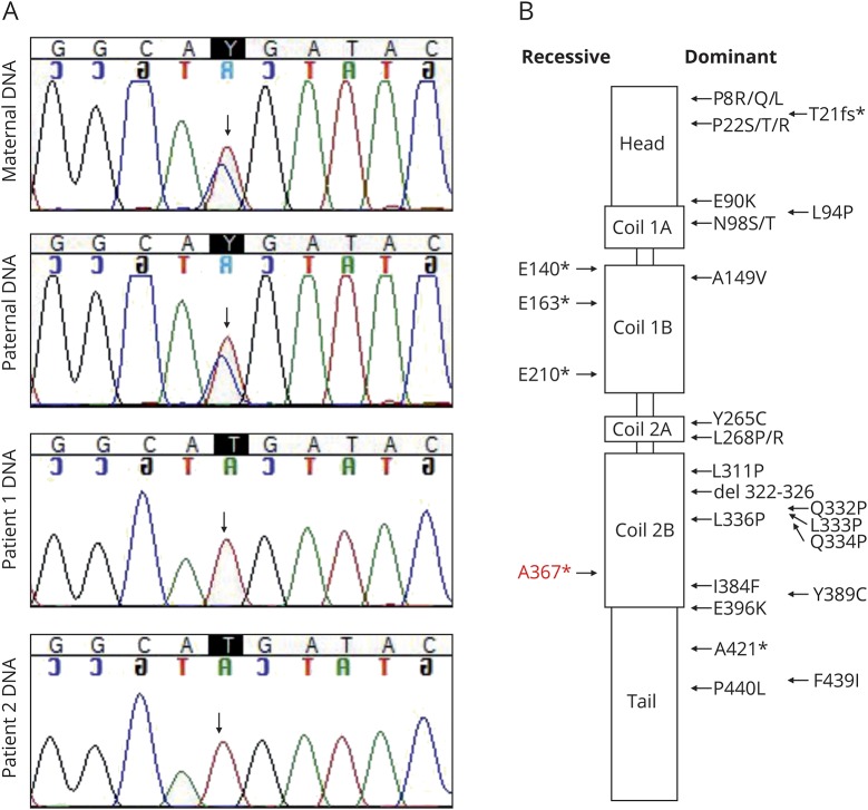
Objective: We used patient-specific neuronal cultures to characterize the molecular genetic mechanism of recessive nonsense mutations in neurofilament light (NEFL) underlying early-onset Charcot-Marie-Tooth (CMT) disease.

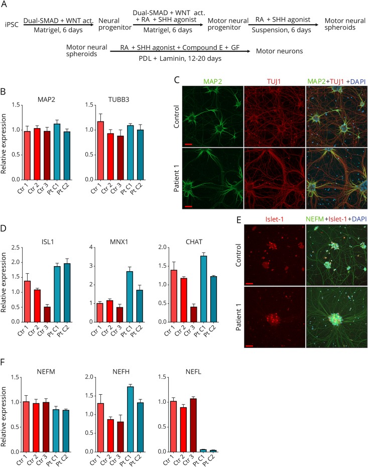
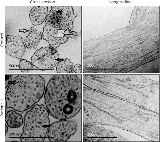
Methods: Motor neurons were differentiated from induced pluripotent stem cells of a patient with early-onset CMT carrying a novel homozygous nonsense mutation in NEFL. Quantitative PCR, protein analytics, immunocytochemistry, electron microscopy, and single-cell transcriptomics were used to investigate patient and control neurons.

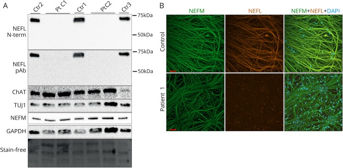
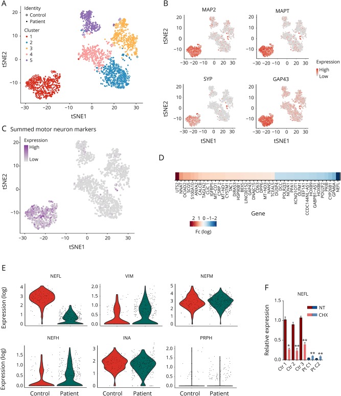
Results: We show that the recessive nonsense mutation causes a nearly total loss of NEFL messenger RNA (mRNA), leading to the complete absence of NEFL protein in patient's cultured neurons. Yet the cultured neurons were able to differentiate and form neuronal networks and neurofilaments. Single-neuron gene expression fingerprinting pinpointed NEFL as the most downregulated gene in the patient neurons and provided data of intermediate filament transcript abundancy and dynamics in cultured neurons. Blocking of nonsense-mediated decay partially rescued the loss of NEFL mRNA.

Conclusions: The strict neuronal specificity of neurofilament has hindered the mechanistic studies of recessive NEFL nonsense mutations. Here, we show that such mutation leads to the absence of NEFL, causing childhood-onset neuropathy through a loss-of-function mechanism. We propose that the neurofilament accumulation, a common feature of many neurodegenerative diseases, mimics the absence of NEFL seen in recessive CMT if aggregation prevents the proper localization of wild-type NEFL in neurons. Our results suggest that the removal of NEFL as a proposed treatment option is harmful in humans.


Figures

Figure 1
Figure 1. Dominant and recessive missense and nonsense variants in neurofilament light (NEFL)
(A) Sequencing traces of the c.1099C>T variant in the family members show that both parents of the patients are heterozygous carriers of the mutation. (B) NEFL protein domains are depicted, and the localization of the reported missense and nonsense variants is indicated (modified from references 17 and 25). The nonsense variant A367* identified in this study is shown in red.
Figure 2
Figure 2. Neuron differentiation and validation
(A) Work-flow of fibroblast-derived induced pluripotent stem cell (iPSC) differentiation into motor neurons; Wnt signaling pathway activator (WNT act), retinoic acid (RA), Sonic hedgehog (SHH), growth factors (GF; BDNF, IGF-1 and CNTF), Poly-d-lysine (PDL). (B) Validation of the expression of neural transcripts MAP2 and TUBB3 against GAPDH by quantitative PCR (qPCR) in total culture RNA of patient 1 clones 1 (Pt C1) and 2 (Pt C2) and controls 1-3 (ctr 1-3) after motor neuron differentiation. (C) Immunocytochemical analysis of MAP2 (green) and TUBB3 (red) proteins in patient 1 and control neuronal cultures. (D) Validation of the expression of motor neural transcripts ISL1, MNX1, and CHAT by qPCR as in B. (E) Immunocytochemical analysis of ISL1 (red) and NEFM (green) protein in patient 1 and control neuronal cultures. ISL1-positive neurons are shown in larger cell clusters in the final differentiation stage (day 14 of in PDL + laminin-coated plates). (F) Expression of intermediate filament subunits neurofilament medium (NEFM), neurofilament heavy (NEFH), and neurofilament light (NEFL) by qPCR as in B. The bars in each graph represent mean levels ± SD, n = 3 for each cell line. All scale bars 50 μm. 4′,6-diamidino-2-phenylindole (DAPI) indicates nuclear staining.
Figure 3
Figure 3. Complete loss of neurofilament light (NEFL) protein in cultured patient neurons
(A) Immunoblotting of whole cell lysates of patient 1 clones 1 and 2 (Pt C1 and C2) and controls 1-3 (ctr 1-3) after motor neuronal differentiation with an N-terminal monoclonal or a polyclonal pan-NEFL antibody. Protein levels of neuronal markers ChAT, TUJ1, and neurofilament medium (NEFM) and the loading control glyceraldehyde 3-phosphate dehydrogenase (GAPDH) as well as the stain-free blot are shown. (B) Immunocytochemical analysis of NEFM (green) and NEFL (orange) of neurite architecture in patient 1 and control neurons after motor neural differentiation. 4′,6-diamidino-2-phenylindole (DAPI) indicates nuclear staining. Scale bars 50 μm.
Figure 4
Figure 4. Transcriptome dynamics of patient and control neurons
(A) Clustering of the single cells derived from patient 1 and control iPSCs in the tSNE-plot based on their gene expression fingerprints. Different clusters are color coded. In the combined single-cell sequencing data, patient cells are shown as filled dots and control cells as diamonds. Neurons are clustered as a clearly separate group of cells (cluster 1, red). (B) MAP2, MAPT, SYP, and GAP43 expression is high in the neuronal cluster cells. Red indicates high expression, and gray indicates low. (C) Cells in the neural cluster express motor neuron lineage-specific transcripts, CHAT, SLC18A3, ISL1, MNX1, LHX1, LHX3, DCC, ONECUT1, and ONECUT2, summed in the tSNE-plot. Purple indicates high expression, and gray indicates low. (D) The most significantly upregulated and downregulated transcripts (adjusted p < 0.001 and absolute fold change ≥1.5) in the neural cluster between patient and control cells. Neurofilament light (NEFL) is the most downregulated transcript in the patient neurons. (E) In the violin plots, each individual cell is shown with its specific transcript level, depicting the most downregulated transcript NEFL in patient neurons, and evenly expressed intermediate filament subunit transcripts INA, NEFM, NEFH, PRPH, and VIM. Expression refers to normalized log(e) expression scale. (F) Expression of NEFL against GAPDH by qPCR from total culture RNA of patient clones 1 (Pt C1) and 2 (Pt C2) and controls 1-3 (Ctr 1-3) after motor neuronal differentiation, nontreated (NT) or treated with 200 μg/mL cycloheximide (CHX) for 18 hours. The comparisons were made individually between each cell line with and without CHX treatment, n = 3 for each cell line and treatment (unpaired 2-tailed t test, **p < 0.001, *p < 0.01). Bars represent mean levels ± SD.
Figure 5
Figure 5. Neurite structure is not disrupted by the lack of neurofilament light (NEFL)
Representative electron microscopy images of neurite architecture in patient 1 and control neurons. Intermediate filaments (outlined arrow) and microtubules (filled arrow) are indicated in cross sections. Normal neurofilament network is seen in longitudinal sections of patient neurites. Scale bars 500 nm.

References

    1. Brown HG, Troncoso JC, Hoh JH. Neurofilament-L homopolymers are less mechanically stable than native neurofilaments. J Microsc 1998;191:229–237. - PubMed
    1. Hirano A, Nakano I, Kurland LT, Mulder DW, Holley PW, Saccomanno G. Fine structural study of neurofibrillary changes in a family with amyotrophic lateral sclerosis. J Neuropathol Exp Neurol 1984;43:471–480. - PubMed
    1. Israeli E, Dryanovski DI, Schumacker PT, et al. . Intermediate filament aggregates cause mitochondrial dysmotility and increase energy demands in giant axonal neuropathy. Hum Mol Genet 2016;25:2143–2157. - PMC - PubMed
    1. Jordanova A, De Jonghe P, Boerkoel CF, et al. . Mutations in the neurofilament light chain gene (NEFL) cause early onset severe Charcot-Marie-Tooth disease. Brain 2003;126:590–597. - PubMed
    1. Yadav P, Selvaraj BT, Bender FL, et al. . Neurofilament depletion improves microtubule dynamics via modulation of Stat3/stathmin signaling. Acta Neuropathol 2016;132:93–110. - PMC - PubMed

LinkOut - more resources