. 2016 Dec;2(4):121-136.
doi: 10.1016/j.bsbt.2016.11.001. Epub 2016 Nov 17.

Mussel-inspired polydopamine for bio-surface functionalization

Affiliations

Mussel-inspired polydopamine for bio-surface functionalization

Y H Ding et al. Biosurf Biotribol. 2016 Dec.

Abstract

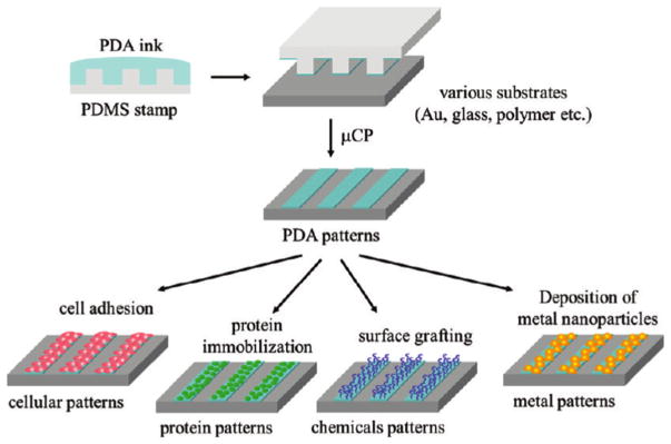
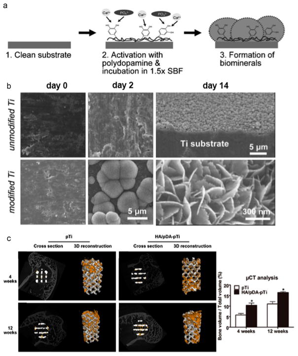
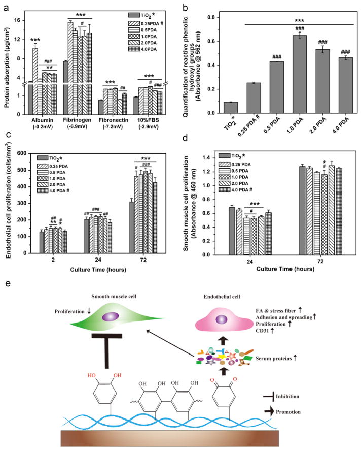
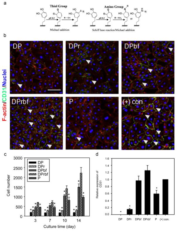
Surface functionalization via molecular design has been a key approach to incorporate new functionalities into existing biomaterials for biomedical application. Mussel-inspired polydopamine (PDA) has aroused great interest as a new route to the functionalization of biomaterials, due to its simplicity and material independency in deposition, favorable interactions with cells, and strong reactivity for secondary functionalization. Herein, this review attempts to highlight the recent findings and progress of PDA in bio-surface functionalization for biomedical applications. The efforts made to elucidate the polymerization mechanism, PDA structure, and the preparation parameters have been discussed. Interactions between PDA coatings and the various cell types involved in different biomedical applications including general cell adhesion, bone regeneration, blood compatibility, and antimicrobial activity have also been highlighted. A brief discussion of post-functionalization of PDA and nanostructured PDA is also provided.

Keywords: Biomedical application; Functionalization; Polydopamine; Polymerization.


Figures

Fig. 1
Fig. 1
Schematic illustration of preparation of tunable micropatterned substrate based on PDA via microcontact printing and secondary reactions. Adapted and reproduced from Ref. [36].
Fig. 2
Fig. 2
The mineralization and bone regeneration facilitated by the PDA coating. a) Scheme for PDA-assisted calcium phosphate (CaP) crystal formation [42]. b) Electron microscopy images showing the effects of the PDA coating on the formation of CaP minerals after incubation the substrates in simulated body fluid (SBF) for 2 and 14 days. Adapted and reproduced from Ref. [42]. c) Microcomputed Tomography (Micro-CT) images of the porous Ti6Al4V scaffolds (pTi) and PDA-assisted hydroxyapatite coating modified scaffolds (HA/PDA-pTi) after implantation for 4 and 12 weeks (the yellow color components was newly formed bone), and the quantified percentages of regenerated bone volume/total volume in these implants. * indicate statistical significance compared to the pTi group, p < 0.05. Adapted and reproduced from Ref. [50].
Fig. 3
Fig. 3
The PDA coating promoted vascular EC growth while inhibited SMC proliferation. a) Quantification of the protein adsorption to PDA and pristin TiO2 sufraces. b) Quantification of reactive phenolic hydroxyl groups determined via the micro-BCA assay. c) EC proliferation determined by cell density after culturing cells for 2 h, 24 h, and 72 h. d) SMC proliferation determined by Cell Counting Kit-8 assay after culturing cells for 24 h and 72 h. e) Schematic diagram of the proposed mechanism by which PDA selectively modulating EC and SMC behavior. 0.25 PDA, 0.5 PDA, 1.0 PDA, 2.0 PDA, and 4.0 PDA represents the PDA coatings on the titanim dioxide (TiO2) surfaces prepared at various initial dopamine concentrations of 0.25, 0.5, 1.0, 2.0, 4.0 g/L in 10 mM Tris buffer, pH 8.5. Statistically significant differences are marked as follows: * vs. TiO2; # vs. 0.25 PDA for a) and b); # vs. 4.0 PDA for c) and d). Adapted and reproduced from Ref. [41].
Fig. 4
Fig. 4
The immobilization of multiple biomelecules via the PDA coating layer. a) Reaction scheme to immobilize biomolecules with thiol or amine groups to the PDA coating layer via Michael additon and/or Schiff base reaction. Adapted and reproduced from Ref. [60]. b) Fluorescent images of human umbilical vein endothelial cells on various substrates. DP: PDA-coated poly(lactic acid-co-ε-caprolactone) film. DPr: RGD immobilized onto DP. DPbf: basic fibroblast growth factor (bFGF) immobilized onto DP. DPrbf: RGD, bFGF immobilized onto DP. P: RGD, bFGF passively adsorbed onto poly(lactic acid-co-ε-caprolactone) film. (+) con.: soluble bFGF treated group. c) Quantification of cell number after culturing human umbilical vein endothelial cells on various substrates for 3, 7, 10, and 14 days. * indicates significance as compared to the DPrbf group at each time point (p < 0.05). d) Expression of CD31 of human umbilical vein endothelial cells cultured on various substrates determined by Western blot analysis. * indicates significance as compared to the (+) con. group (p < 0.05). b), c) and d) were adapted and reproduced from Ref. [62].
Fig. 5
Fig. 5
The stratgies of heparin immobilization via the PDA coating layer. a) Reaction scheme to prepare the heparin-dopamine conjugate. Adapted and reproduced from Ref. [67]. b) Schematics of the PDA-mediated, one-step surface immobilization of multiple biomolecules. Adapted and reproduced from Ref. [68]. c) Schematics of the PDA-assisted, one-step deposition of poly (ethylene imine) (PEI) for further heparin immobilization. Adapted and reproduced from Ref. [71]. d) Fluorescent images of EC/SMC co-culture, cell attachment number, and attachment ratio of EC/SMC showing the immobilized heparin via the PDA-assisted PEI deposition layer with substrate topography synergistically promoted competitive attachment of ECs over SMCs. TiO2: the pristine titanium dioxide substrate. PEI/PDA: PDA-assisted PEI coating on the TiO2 substrate. Hep-PEI/PDA: heparin immobilized onto PEI/PDA. Statistically significant differences are marked as follows: * vs. TiO2; # vs. Hep-PEI/PDA; * or # for p < 0.05; *** or ### for p < 0.001. Adapted and reproduced from Ref. [72].
Fig. 6
Fig. 6
PDA nanoparticles (PDA-NPs) for functionalization of porous scaffolds enhance tissue regeneration. a) SEM images of PDA-NPs immobilized onto β-tricalcium phosphate (TCP) scaffolds after incubation in the PDA-NP suspension for 4 h and 48 h. b) Release profiles of bone morphogenetic protein 2 (BMP-2) from various scaffolds in phosphate-buffered saline (PBS) solution. c) ALP activities of BMSCs cultured on various scaffolds for 7 and 14 days. d) Hematoxylin and eosin (H&E) staining images of various scaffolds retrieved after 12-week implantation and quantitative evaluation of newly formed bone on various scaffolds. m: material; nb: new bone; v- vessel; white asterisk: osteocyte; blue arrow: woven bone formation. Statistically significant differences are marked as follows: * for p ≤ 0.05. Adapted and reproduced from Ref. [80].
Fig. 7
Fig. 7
Self-assembling of PDA microcapsules for biomedical application. a) Schematics of the preparation of the PDA microporous architectures and subsequent loading and release of BMP-2. b) Evaluation of the BMP-2 loading ability and cumulative release from the PDA microporous architectures, i.e. PDA-CPS, and the PDA films. c) Cell proliferation and ALP activity of rat BMSCs after culturing on the pristine Ti, PDA-CPS, and PDA films. * indicates the significant difference (p < 0.05). Adapted and reproduced from Ref. [86].
Scheme 1
Scheme 1
First steps of melanin formation by dopamine oxidation and proposed models of PDA structure.

References

    1. Hodneland CD, Lee YS, Min DH, Mrksich M. Selective immobilization of proteins to self-assembled monolayers presenting active site-directed capture ligands. Proc Natl Acad Sci. 2002;99:5048–5052. - PMC - PubMed
    1. Langer R, Tirrell DA. Designing materials for biology and medicine. Nature. 2004;428:487–492. - PubMed
    1. Wu G, Li P, Feng H, Zhang X, Chu PK. Engineering and functionalization of biomaterials via surface modification. J Mater Chem B. 2015;3:2024–2042. - PubMed
    1. Zhu J. Bioactive modification of poly (ethylene glycol) hydrogels for tissue engineering. Biomaterials. 2010;31:4639–4656. - PMC - PubMed
    1. Nuzzo RG, Fusco FA, Allara DL. Spontaneously organized molecular assemblies. 3. Preparation and properties of solution adsorbed monolayers of organic disulfides on gold surfaces. J Am Chem Soc. 1987;109:2358–2368.