. 2018 Oct;94(3-4):330-338.
doi: 10.1111/cge.13392. Epub 2018 Jul 10.

Genotype-phenotype correlations of low-frequency variants in the complement system in renal disease and age-related macular degeneration

Affiliations

Genotype-phenotype correlations of low-frequency variants in the complement system in renal disease and age-related macular degeneration

M J Geerlings et al. Clin Genet. 2018 Oct.

Abstract

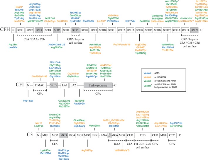
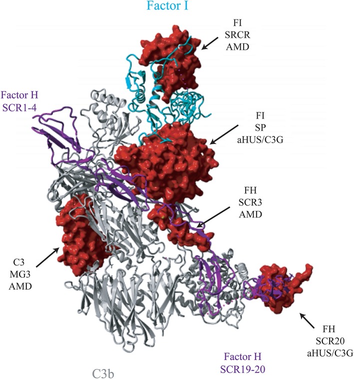
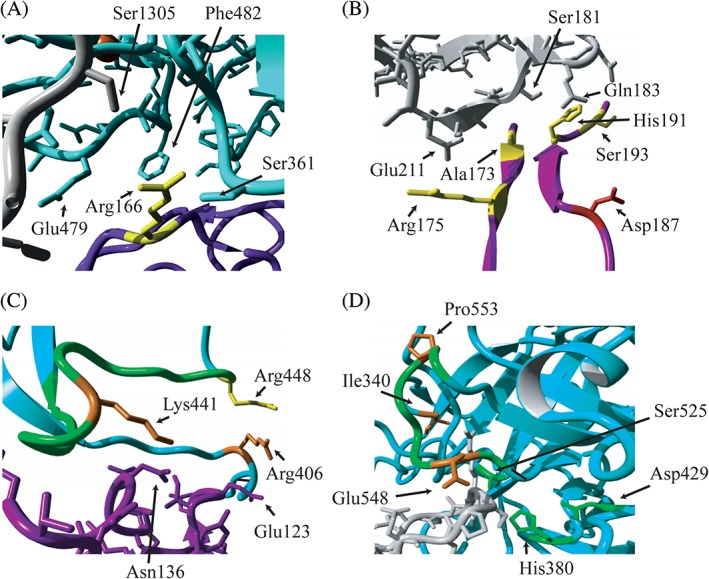
Genetic alterations in the complement system have been linked to a variety of diseases, including atypical hemolytic uremic syndrome (aHUS), C3 glomerulopathy (C3G), and age-related macular degeneration (AMD). We performed sequence analysis of the complement genes complement factor H (CFH), complement factor I (CFI), and complement C3 (C3) in 866 aHUS/C3G and 697 AMD patients. In total, we identified 505 low-frequency alleles, representing 121 unique variants, of which 51 are novel. CFH contained the largest number of unique low-frequency variants (n = 64; 53%), followed by C3 (n = 32; 26%) and CFI (n = 25; 21%). A substantial number of variants were found in both patients groups (n = 48; 40%), while 41 (34%) variants were found only in aHUS/C3G and 32 (26%) variants were AMD specific. Genotype-phenotype correlations between the disease groups identified a higher frequency of protein altering alleles in short consensus repeat 20 (SCR20) of factor H (FH), and in the serine protease domain of factor I (FI) in aHUS/C3G patients. In AMD, a higher frequency of protein-altering alleles was observed in SCR3, SCR5, and SCR7 of FH, the SRCR domain of FI, and in the MG3 domain of C3. In conclusion, we observed a substantial overlap of variants between aHUS/C3G and AMD; however, there is a distinct clustering of variants within specific domains.

Keywords: C3 glomerulopathy; age-related macular degeneration; alternative pathway; atypical hemolytic uremic syndrome; complement system.


Conflict of interest statement

The authors declare no potential conflict of interests.

Figures

Figure 1
Figure 1
Genetic variants identified in 886 aHUS/C3G patients and 697 AMD patients. Colors represent the phenotype in which the variants were identified: green (both in aHUS/C3G and AMD), orange (aHUS/C3G only), blue (AMD only) or green^ (with circumflex) (protective for AMD but found recurrently in aHUS/C3G). Protein domains that carried a significantly higher percentage of alleles in aHUS/C3G or AMD are colored gray. Protein interaction sites are reported underneath the domains with dotted lines. Only low frequency variants (minor allele frequency <5%) based on the ExAC database are shown. CFA, cofactor activity; DAA, decay‐accelerating activity; CRP, C‐reactive protein
Figure 2
Figure 2
Location of protein domains containing a higher percentage of alleles in aHUS/C3G or AMD on the structure of the C3b‐FH‐FI complex. Three‐dimensional structure of C3b (gray) in complex with FI (cyan) and FH construct (purple) containing FH SCR1‐4 and SCR19‐20. Dotted line represents 12‐residue polyglycine linker that connects SCR1‐4 and SCR19‐20. Surfaces of fragments of C3b (233‐348), FI (119‐188, 340‐570) and FH (166‐193, 1169‐1215) carrying missense changes in domains, which contain a significantly higher percentage of alleles in aHUS/C3G or AMD, are shown in red. The figure is generated based on the PDB 5O32,29 using YASARA Version 17.8.1531
Figure 3
Figure 3
Localization of altered residues on the interface of C3b, FH and FI in proteins domains containing a higher percentage of alleles in aHUS/C3G or AMD. Fragments of the 3‐dimensional structure of C3b (gray), FI (cyan) and FH construct (purple) are shown. The residues altered in the SCR3 domain of FH (A and B) and the SP domain of FI (C and D) are shown. Residues that were found mutated in AMD only (yellow), aHUS/C3G (red) or both phenotypes (orange) are indicated, as well as amino acids of interacting partners that are in close proximity of the mutated residues (A, B, C). Important structural elements of FI are indicated in green: the charged loop 435‐448 (C); the activation loop 548‐553 and the catalytic triad (H380, D429, S525) (D). The figure is generated based on the PDB 5O32,29 using YASARA Version 17.8.1531

References

    1. Liszewski MK, Java A, Schramm EC, Atkinson JP. Complement dysregulation and disease: insights from contemporary genetics. Annu Rev Pathol. 2017;12:25‐52. - PMC - PubMed
    1. Klein RJ, Zeiss C, Chew EY, et al. Complement factor H polymorphism in age‐related macular degeneration. Science. 2005;308:385‐389. - PMC - PubMed
    1. Warwicker P, Goodship TH, Donne RL, et al. Genetic studies into inherited and sporadic hemolytic uremic syndrome. Kidney Int. 1998;53:836‐844. - PubMed
    1. Caprioli J, Noris M, Brioschi S, et al. Genetics of HUS: the impact of MCP, CFH, and IF mutations on clinical presentation, response to treatment, and outcome. Blood. 2006;108:1267‐1279. - PMC - PubMed
    1. Malina M, Roumenina LT, Seeman T, et al. Genetics of hemolytic uremic syndromes. Presse Med. 2012;41:e105‐e114. - PubMed

Publication types

MeSH terms