. 2018 Apr 27:2:193-209.
doi: 10.1016/j.isci.2018.03.003.

Tolerance to Morphine-Induced Inhibition of TTX-R Sodium Channels in Dorsal Root Ganglia Neurons Is Modulated by Gut-Derived Mediators

Affiliations

Tolerance to Morphine-Induced Inhibition of TTX-R Sodium Channels in Dorsal Root Ganglia Neurons Is Modulated by Gut-Derived Mediators

Ryan A Mischel et al. iScience. .

Abstract

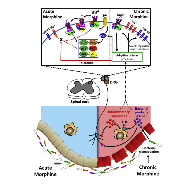
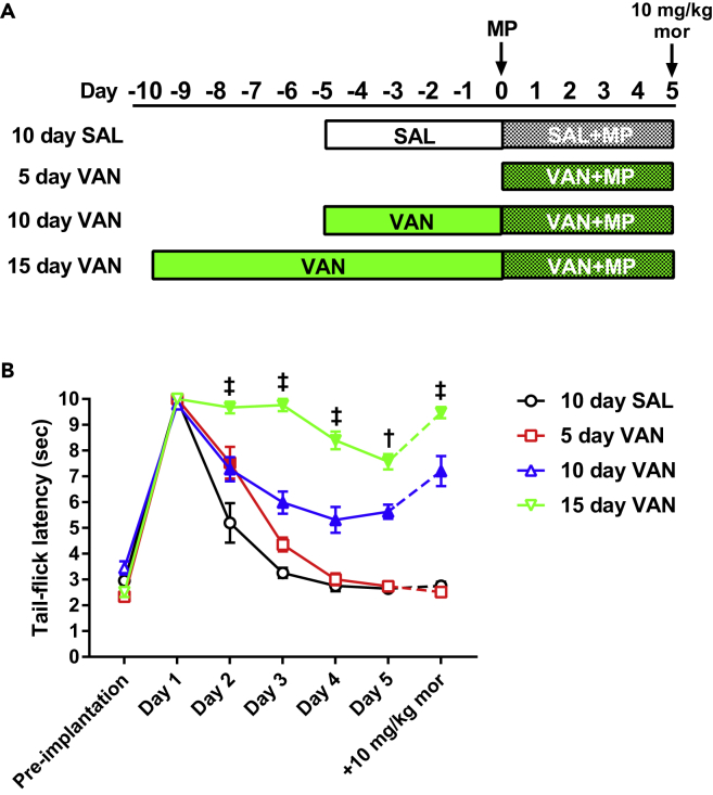
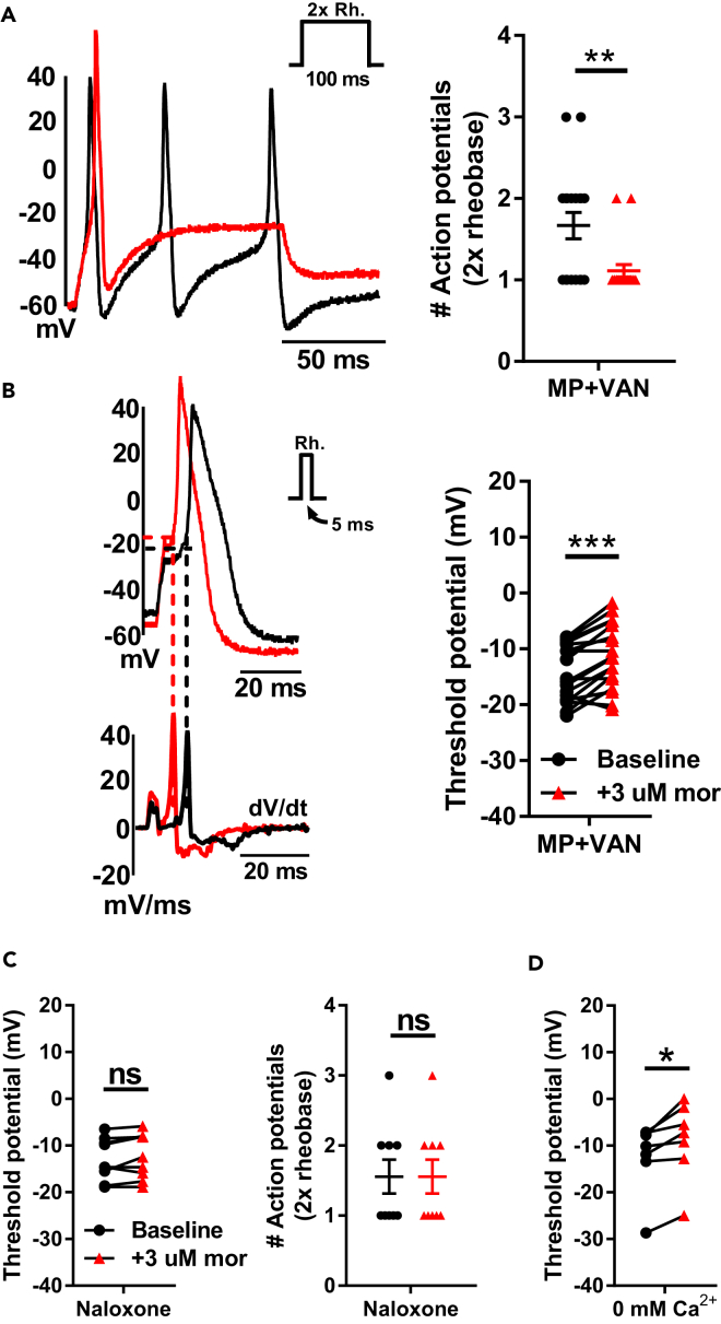
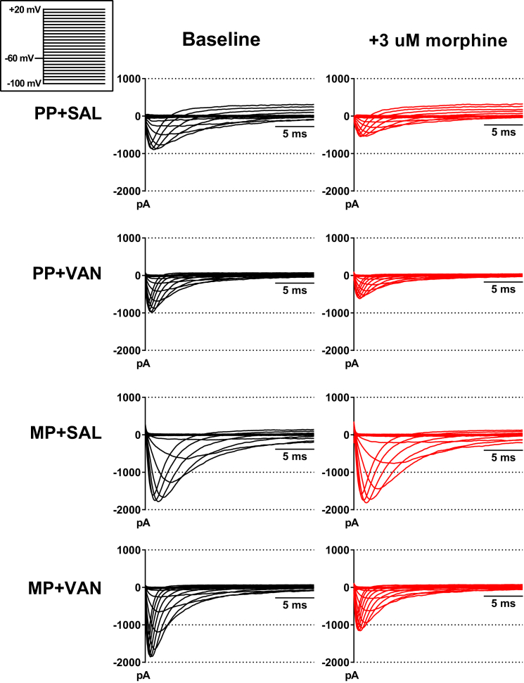
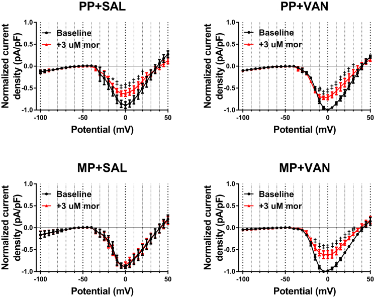
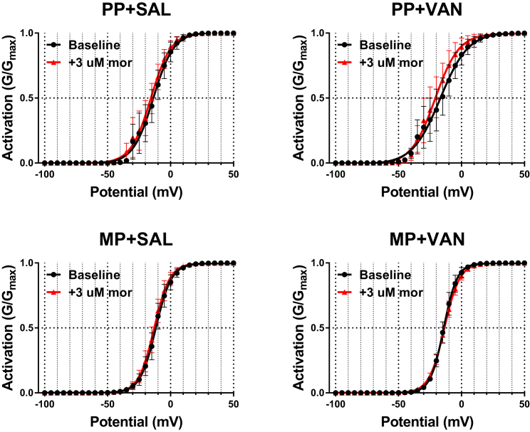
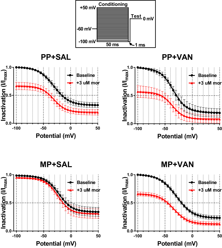
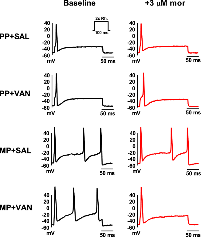
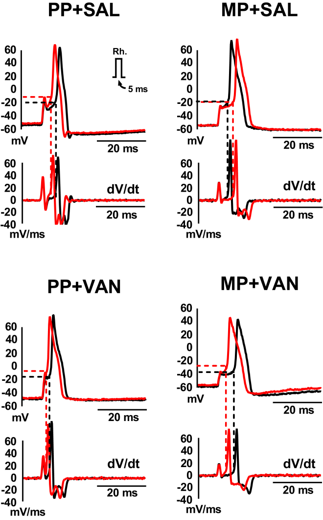
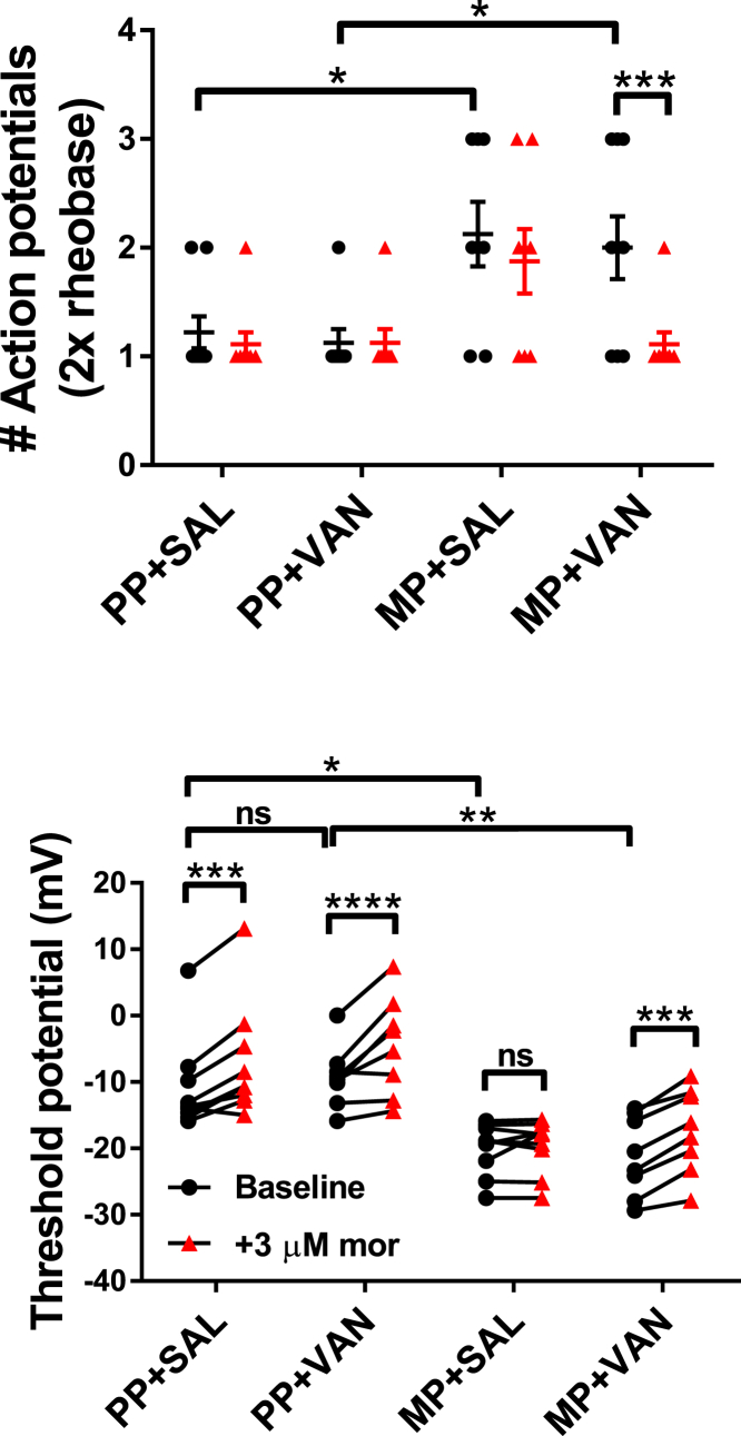
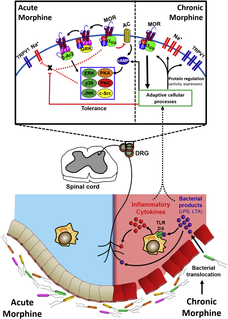
In the clinical setting, analgesic tolerance is a primary driver of diminished pain control and opioid dose escalations. Integral to this process are primary afferent sensory neurons, the first-order components of nociceptive sensation. Here, we characterize the factors modulating morphine action and tolerance in mouse small diameter dorsal root ganglia (DRG) neurons. We demonstrate that acute morphine inactivates tetrodotoxin-resistant (TTX-R) Na+ channels in these cells. Chronic exposure resulted in tolerance to this effect, which was prevented by treatment with oral vancomycin. Using colonic supernatants, we further show that mediators in the gut microenvironment of mice with chronic morphine exposure can induce tolerance and hyperexcitability in naive DRG neurons. Tolerance (but not hyperexcitability) in this paradigm was mitigated by oral vancomycin treatment. These findings collectively suggest that gastrointestinal microbiota modulate the development of morphine tolerance (but not hyperexcitability) in nociceptive primary afferent neurons, through a mechanism involving TTX-R Na+ channels.


Conflict of interest statement

DECLARATION OF INTERESTS All authors declare no conflict of interest or competing financial interests.

Figures

None
Graphical abstract
Figure 1
Figure 1
Antinociceptive Morphine Tolerance in Mice Is Prevented by Oral Vancomycin in a Manner Dependent on the Duration of Treatment (A) Schematic of the treatment regimen utilized for each cohort in this study. (B) Tail-immersion assay for mice receiving 5, 10, or 15 days of oral vancomycin (VAN, 10 mg/kg). Control subjects received 10 days of oral saline (SAL). Following morphine pellet (MP) implantation, the 10 day SAL and 5 day VAN cohorts demonstrated a progressive reduction in tail-flick latency and lacked a response to acute morphine challenge on day 5 (10 mg/kg, subcutaneously), indicating tolerance development. The 10 day VAN treatment resulted in a significant increase in tail-flick latency throughout the testing period (versus 10 day SAL) and an additional increase with acute morphine challenge on day 5, indicating tolerance prevention. This tolerance prevention was significantly enhanced with 15 day VAN treatment (versus 10 day VAN). 10 day SAL (N = 8), 5 day VAN (N = 10), 10 day VAN (N = 14), 15 day VAN (N = 10); significance indicated versus 10 day SAL (filled symbols, p < 0.05) and versus 10 day VAN (p < 0.001, p < 0.0001) by two-way repeated-measures ANOVA with Bonferroni post hoc analysis; data expressed as mean ± SEM (see Table S1 for additional values).
Figure 2
Figure 2
Characterization of Responses to Acute Morphine Challenge in Small Diameter DRG Neurons (A and B) Oral vancomycin treatment (VAN, 10 mg/kg) for 10 days blocks cellular level morphine tolerance in DRG neurons. Representative raw traces (left) and individual observations (right) indicate the number of action potentials at double rheobase (A) and action potential threshold (B) at baseline (black) and following acute morphine challenge (3 μM, red). Morphine perfusion significantly reduced the number of action potentials at double rheobase and depolarized the action potential threshold. (N = 7, n = 18), **p < 0.01, ***p < 0.001 by two-tailed paired Student's t test; data expressed as mean ± SEM. (C) Acute morphine challenge is unable to reduce the excitability of DRG neurons in the presence of naloxone (1 μM). The action potential threshold (left) and number of action potentials at double rheobase (right) are presented at baseline (black) and following acute morphine challenge (3 μM, red). No significant shift in either parameter was noted. N = 4, n = 9; ns, not significant by two-tailed paired Student's t test; data expressed as mean ± SEM. (D) Morphine-induced depolarizations of action potential threshold persist in the complete absence of internal and external Ca2+. The thresholds for each cell are presented at baseline (black) and following acute morphine challenge (3 μM, red) and indicate a significant depolarization with morphine perfusion. N = 5, n = 7; *p < 0.05 by two-tailed paired Student's t test (see Table S2 for additional values).
Figure 3
Figure 3
Representative Voltage-Clamp Traces of TTX-R Na+ Currents in the Absence and Presence of Acute Morphine Challenge Raw traces for voltage steps from −10 to +20 mV are presented at baseline (black) and following acute morphine challenge (3 μM, red). Neurons from PP + SAL and PP + VAN mice demonstrate a significant reduction in the magnitude of inward currents following morphine perfusion. This effect is mitigated in neurons from MP + SAL mice (indicating tolerance development), but preserved in neurons from MP + VAN mice (indicating tolerance prevention). A notable trend toward enhancement of inward current magnitude was noted in cohorts administered morphine pellet (MP) when compared with cohorts given placebo (PP). However, this effect was not statistically significant in this study.
Figure 4
Figure 4
Acute Morphine Challenge Results in Reduction of TTX-R Na+ Current Magnitude That Is Prevented by Chronic Morphine Exposure In Vivo, but Preserved with Concurrent Oral Vancomycin Treatment Current-voltage (I-V) relationships of TTX-R Na+ channels in voltage-clamped DRG neurons are shown at baseline (black) and following acute morphine challenge (3 μM, red). Neurons from PP + SAL and PP + VAN mice demonstrate a significant reduction of current density for steps from −10 to +20 mV. This effect was mitigated in neurons from MP + SAL mice (indicating tolerance development), but preserved in neurons from MP + VAN mice (indicating tolerance prevention). PP + SAL (N = 7, n = 7), PP + VAN (N = 4, n = 6), MP + SAL (N = 7, n = 9), MP + VAN (N = 5, n = 7), *p < 0.05, #p < 0.01, p < 0.001, p < 0.0001 by two-way repeated-measures ANOVA with Bonferroni post hoc analysis; data expressed as mean ± SEM.
Figure 5
Figure 5
Acute Morphine Challenge Does Not Alter the Voltage Dependence of Activation of TTX-R Na+ Channels Relative conductances (G/Gmax) of TTX-R Na+ channels at baseline (black) and following acute morphine challenge (3 μM, red). Values transformed from I-V curves are plotted as a function of the step potential and fit with a Boltzmann function (see Transparent Methods). No significant shifts in the voltage of half-maximum (V1/2) activation, slope factor (k), or Boltzmann fit parameters are noted in any of the treatment cohorts following acute morphine challenge. PP + SAL (N = 7, n = 7), PP + VAN (N = 4, n = 6), MP + SAL (N = 7, n = 9), MP + VAN (N = 5, n = 7), analyzed by two-way repeated-measures ANOVA with Bonferroni post hoc analysis and ordinary least squares non-linear regression; data expressed as mean ± SEM (see Table S3 for additional values).
Figure 6
Figure 6
Acute Morphine Challenge Enhances Steady-State Inactivation of TTX-R Na+ Channels Relative peak current density (I/Imax) of TTX-R Na+ channels at baseline (black) and following acute morphine challenge (3 μM, red). Values obtained during the test pulse are plotted as a function of the conditioning pulse potential and fit with a Boltzmann function (see Transparent Methods). Boltzmann fit comparisons indicate that acute morphine challenge reduced I/Imax (i.e., enhanced inactivation) at all potentials tested in neurons from PP + SAL and PP + VAN mice. This effect was mitigated in neurons from MP + SAL mice (indicating tolerance development), but preserved in neurons from MP + VAN mice (indicating tolerance prevention). No significant shifts in the voltage of half-maximum (V1/2) inactivation and slope factor (k) were noted in any of the treatment cohorts with acute morphine challenge. PP + SAL (N = 7, n = 7), PP + VAN (N = 4, n = 6), MP + SAL (N = 7, n = 9), MP + VAN (N = 5, n = 7), analyzed by two-way repeated-measures ANOVA with Bonferroni post hoc analysis and ordinary least squares non-linear regression; data expressed as mean ± SEM (see Table S4 for additional values).
Figure 7
Figure 7
Representative Current-Clamp Traces for the Number of Action Potentials at Double Rheobase in Colonic Supernatant Experiments Current-clamp traces at double rheobase are presented at baseline (black) and following acute morphine challenge (3 μM, red) for naive DRG neurons exposed to colonic supernatants from each treatment cohort. Neurons in supernatants from PP + SAL and PP + VAN mice demonstrated low excitability at baseline, most often characterized by a single action potential at double rheobase. Owing to a floor effect, no reduction could be detected with morphine perfusion. Neurons in supernatants from MP + SAL mice demonstrated enhanced excitability at baseline, characterized by a greater number of action potentials at double rheobase. No reduction was detected with acute morphine challenge, indicating tolerance development. Neurons in supernatants from MP + VAN mice were also hyperexcitable at baseline, but demonstrated a reduction in the number of action potentials at double rheobase with acute morphine challenge, indicating tolerance prevention (see Figure 9 for individual observations).
Figure 8
Figure 8
Representative Current-Clamp Traces for Action Potential Threshold in Colonic Supernatant Experiments Action potential threshold (Vt) is presented at baseline (black) and following acute morphine challenge (3 μM, red) for naive DRG neurons exposed to colonic supernatants from each treatment cohort. Neurons in supernatants from PP + SAL and PP + VAN mice demonstrated low excitability at baseline, characterized by depolarization of Vt. Morphine perfusion significantly depolarized Vt further, indicating a lack of tolerance development. Neurons in supernatants from MP + SAL mice demonstrated enhanced excitability at baseline, characterized by hyperpolarization of Vt. No depolarization was noted with acute morphine challenge, indicating tolerance development. Neurons in supernatants from MP + VAN mice were also hyperexcitable at baseline, but demonstrated depolarization of Vt with acute morphine challenge, indicating tolerance prevention (see Figure 9 for individual observations).
Figure 9
Figure 9
Mediators in the Gut Wall of Mice with Chronic Morphine Exposure Induce Tolerance in Naive DRG Neurons That Is Prevented by Oral Vancomycin Treatment Individual observations of naive DRG neurons exposed to colonic supernatants from each treatment cohort. Action potentials at double rheobase and action potential threshold (Vt) are presented at baseline (black) and following acute morphine challenge (3 μM, red). Neurons in supernatants from PP + SAL and PP + VAN mice demonstrated low excitability at baseline, characterized by fewer action potentials at double rheobase and depolarization of the action potential threshold (Vt). A significant depolarization of Vt was noted with morphine perfusion; however, no alteration in the number of action potentials at double rheobase could be detected owing to a floor effect. Neurons in supernatants from MP + SAL mice demonstrated enhanced excitability at baseline, characterized by more action potentials at double rheobase and hyperpolarization of Vt. No shift in either parameter was noted with acute morphine challenge, indicating tolerance development. Neurons in supernatants from MP + VAN mice also demonstrated enhanced excitability at baseline, but responded to acute morphine challenge with reductions in the number of action potentials at double rheobase and depolarizations of Vt. PP + SAL (N = 8, n = 9), PP + VAN (N = 6, n = 8), MP + SAL (N = 5, n = 9), MP + VAN (N = 7, n = 8); ns, not significant; *p < 0.05, **p < 0.01, ***p < 0.001, ****p < 0.0001 by two-way repeated-measures ANOVA with Bonferroni post hoc analysis; data expressed as mean ± SEM (see Table S5 for additional values).
Figure 10
Figure 10
General Schematic of the Working Hypothesis Morphine acting acutely on small diameter DRG neurons results in inhibition of TTX-R Na+ channels. This action is MOR dependent, likely occurring via direct Gβγ inhibition or a downstream phosphorylation/dephosphorylation event. Chronic morphine exposure compromises gut epithelial tight junction integrity, allowing translocation of luminal bacteria and the subsequent release of bacterial products and pro-inflammatory cytokines. These act on small diameter DRG neurons, producing tolerance to the inhibitory action of acute morphine on TTX-R Na+ channels. Elimination of Gram-positive gut microbiota by oral vancomycin prevents this tolerance, but does not prevent the upregulation of TTX-R Na+ and TRPV1 channels induced by chronic morphine.

References

    1. Aghajanian G., Wang Y. Common alpha-2 and opiate effector mechanisms in the locus ceruleus: intracellular studies in brain slices. Neuropharmacology. 1987;26:789–800. - PubMed
    1. Akbar A., Yiangou Y., Facer P., Walters J.R., Anand P., Ghosh S. Increased capsaicin receptor TRPV1-expressing sensory fibres in irritable bowel syndrome and their correlation with abdominal pain. Gut. 2008;57:923–929. - PMC - PubMed
    1. Akins P., McCleskey E. Characterization of potassium currents in adult rat sensory neurons and modulation by opioids and cyclic AMP. Neuroscience. 1993;56:759–769. - PubMed
    1. Amaya F., Decosterd I., Samad T.A., Plumpton C., Tate S., Mannion R.J., Costigan M., Woolf C.J. Diversity of expression of the sensory neuron-specific TTX-resistant voltage-gated sodium ion channels SNS and SNS2. Mol. Cell. Neurosci. 2000;15:331–342. - PubMed
    1. Angst M.S., Clark J.D. Opioid-induced hyperalgesia: a qualitative systematic review. Anesthesiology. 2006;104:570–587. - PubMed

LinkOut - more resources