. 2018 Aug 15;526(12):1978-1990.
doi: 10.1002/cne.24468.

Sensitivity and specificity of phospho-Ser129 α-synuclein monoclonal antibodies

Affiliations

Sensitivity and specificity of phospho-Ser129 α-synuclein monoclonal antibodies

Vedad Delic et al. J Comp Neurol. .

Abstract

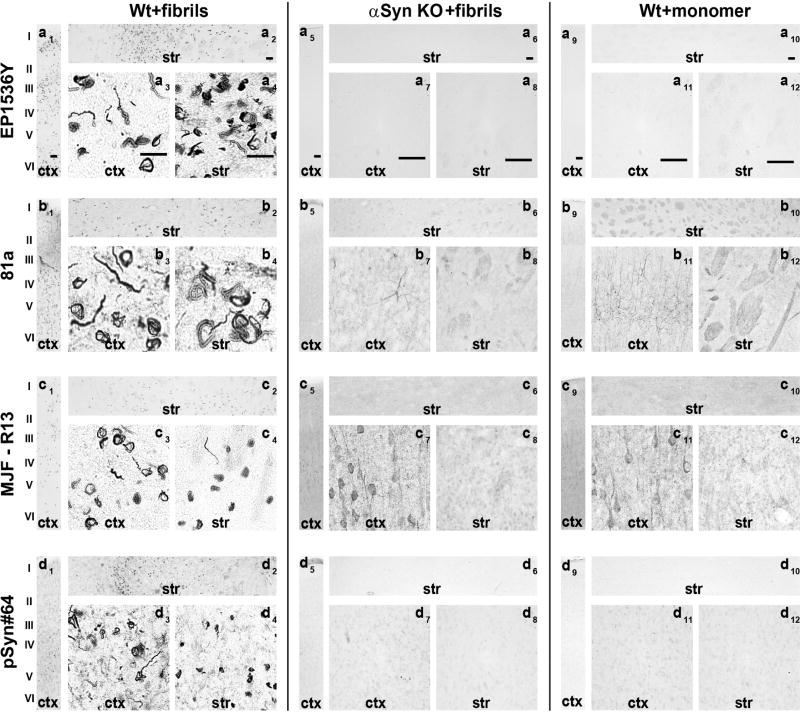
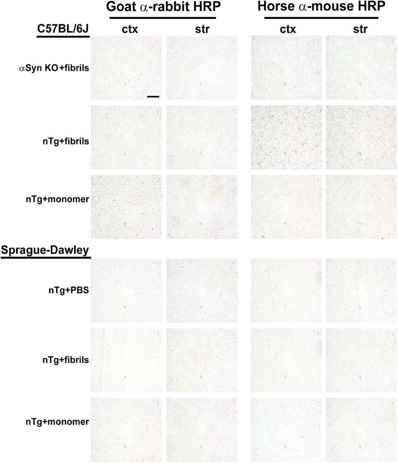
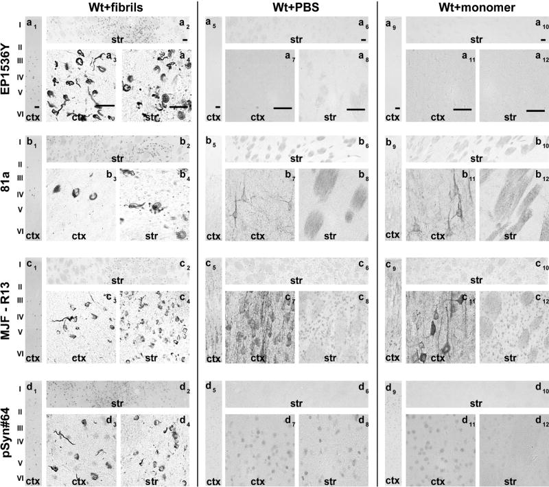
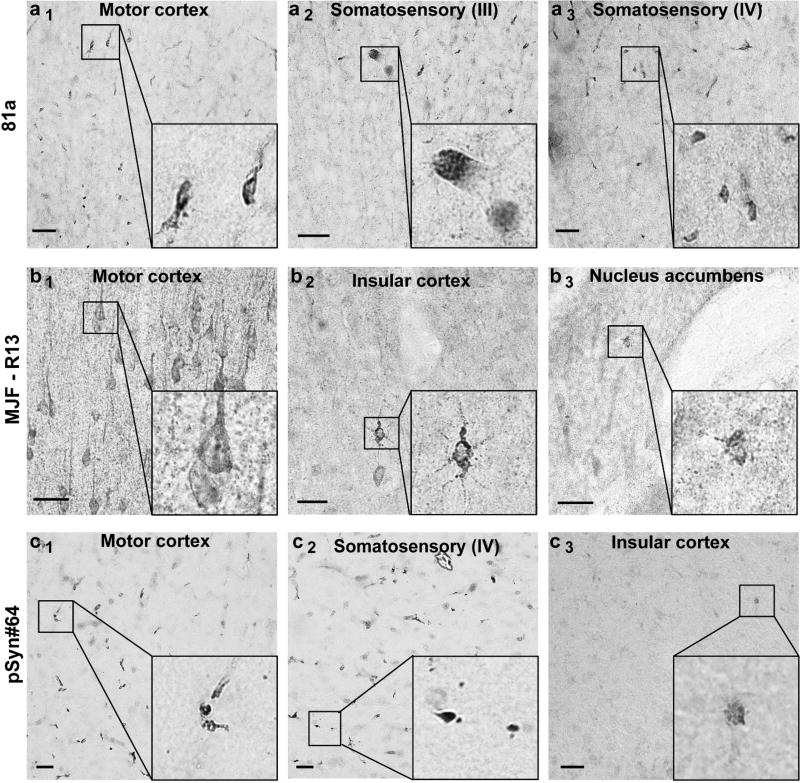
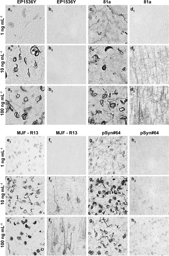
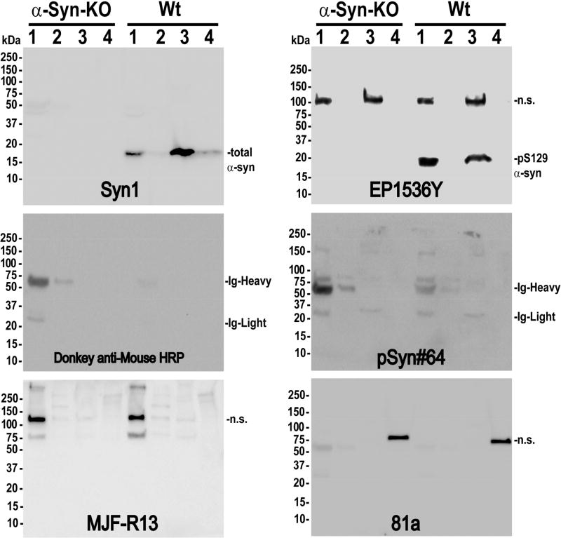
α-Synuclein (α-syn) is an abundant presynaptic protein that is the primary constituent of inclusions that define Lewy body diseases (LBDs). In these inclusions, α-syn is phosphorylated at the serine-129 residue. Antibodies directed to this phosphorylation site are used to measure inclusion abundance and stage disease progression in preclinical models as well as in postmortem tissues in LBDs. While it is critical to reliably identify inclusions, phospho-specific antibodies often cross-react with nonspecific antigens. Four commercially available monoclonal antibodies, two from rabbits (clones EP1536Y and MJF-R13) and two from mice (81a and pSyn#64), have been the most widely used in detecting pS129-α-syn inclusions. Here, we systematically evaluated these antibodies in brain sections and protein lysates from rats and mice. All antibodies detected pS129-α-syn inclusions in the brain that were induced by preformed α-syn fibrils in wild-type rats and mice. Antibody titrations revealed that clones EP1536Y and 81a comparably labeled inclusions in both the perikarya and neuronal processes in contrast to clones MJF-R13 and pSyn#64 that incompletely labeled inclusions at various antibody concentrations. Except for EP1536Y, the clones produced nonspecific diffuse neuropil labeling in α-syn knockout mice as well as mice and rats injected with monomeric α-syn, with some nonspecific staining titrating with pS129-α-syn inclusions. By immunoblot, all the clones cross-reacted with proteins other than α-syn, warranting caution in interpretations of specificity. Clone EP1536Y uniquely and robustly detected endogenous pS129-α-syn in highly soluble protein fractions from the mouse brain. In summary, EP1536Y had the highest sensitivity and specificity for detecting pS129-α-syn.

Keywords: Lewy bodies; Lewy neurites; NACP; SNCA.


Figures

Fig. 1
Fig. 1. Assessment of pSer129-α-Syn and inclusion detection in mouse brain
Wild-type and α-syn knockout mice (C57bl/6J) were injected bilaterally into the striatum with 10 µg of α-syn pre-formed fibrils or monomeric protein as previously described(Abdelmotilib et al., 2017). Brain sections were evaluated from mice six months later. Representative widefield images of DAB-staining (converted to grayscale for contrast) with the indicated monoclonal antibody (left margins) in animals injected with A) fibrils in wild-type mice, B) fibrils in α-Syn knockout mice, or C) monomer protein in wild-type mice. All antibodies were included at 10 ng mL−1 concentration. Coronal sections are shown with 0.4 mm of Bregma, with primary motor cortex (ctx) and dorsal striatum (str) shown at two different magnifications. Scale bars are 0.1 mm.
Fig. 2
Fig. 2. Lack of signal in secondary-only (anti-mouse and anti-rabbit) antibody coronal brain sections
Wild-type and α-syn knockout mice (C57bl/6J) and wild-type Sprague Dawley rats (SDTac) were injected as indicated (left hand margins) bilaterally into the striatum, and processed six months later. Representative widefield images of DAB-staining (converted to grayscale for contrast) with the indicated secondary antibody (top margin). Coronal sections are shown near Bregma from primary motor cortex (ctx) and dorsal striatum (str), with the scale bar set to 0.1 mm.
Fig. 3
Fig. 3. Assessment of pSer129-α-Syn and inclusion detection in rat brain
Wild-type Sprague Dawley rats (SDTac) were injected bilaterally into the striatum with 20 µg of α-syn pre-formed fibrils or monomeric protein as previously described(Abdelmotilib et al., 2017). Brain sections were evaluated from rats six months later. Representative widefield images of DAB-staining (converted to grayscale for contrast) with the indicated monoclonal antibody (left margins) in animals injected with A) fibrils in wild-type rats, B) saline control in wild-type rats, and C) monomer protein in wild-type rats. All antibodies were included at 10 ng mL−1 concentration. Coronal sections are shown within ~1 mm of Bregma, with primary motor cortex (ctx) and dorsal striatum (str) shown at two different magnifications. Scale bars are 0.1 mm.
Fig. 4
Fig. 4. Assessment of pSer129-α-syn monoclonal antibodies in α-syn knockout mice
Coronal brain sections were obtained from adult (3–6 months) α-syn knockout mice and evaluated with the monoclonal antibody indicated in the left margin (a, b, c), all used at 10 ng mL−1. Representative images from DAB staining (grayscale shown for contrast) are shown. Insets black bounding boxes show higher magnifications of features of interest. Scale bars are 0.1 mm.
Fig 5
Fig 5. Off-target pSer129-α-syn monoclonal antibody staining titrates with the labeling of inclusions
Frozen coronal sections of primary motor cortex 0.4mm of Bregma from wild-type C57Bl/6J mice injected with α-syn fibrils in the striatum (a,b) or (c,d,e,g) non-injected wild-type mice were evaluated with the indicated monoclonal pSer129 α-syn antibody (top margin) at the indicated concentration (left margin).
Fig 6
Fig 6. Confocal analysis of pS129 α-syn-positive inclusions in mice and rats
C57Bl/6J wild-type mice and Sprague Dawley wild-type rats were injected bilaterally into the striatum with either 10 µg or 20 µg (respectively) of α-syn fibrils and frozen brain sections analyzed six months later. Confocal images were obtained from a single plane with a 63× objective set for optimal pinhole. Red color indicates pS129-α-syn staining (Cy3) and blue color indicates DAPI. The monoclonal antibody used at 20 ng mL−1 are given in the left margin. Images from both the primary motor cortex (cortex) and dorsal striatum (striatum) were obtained. Scale bar is 50 µm.
Fig 7
Fig 7. pS129 α-syn monoclonal antibodies detect off-target proteins in brain lysates
Cortical tissue was dissected from wild-type and α-syn knockout mice (C57bl/6J), as indicated, and sequentially processed into buffers of increasing stringency, starting with [lane] 1) saline, 2) high-salt supplemented saline, 3) triton-X 100, and 4) SDS. Molecular weight markers are shown in left margins. The Syn1 monoclonal antibody was used to label total α-syn protein as a positive control, with a secondary-only membrane incubated with donkey anti-mouse antibody as a negative control to reveal globulins.

References

    1. Abdelmotilib H, Maltbie T, Delic V, Liu Z, Hu X, Fraser KB, Moehle MS, Stoyka L, Anabtawi N, Krendelchtchikova V, Volpicelli-Daley LA, West A. alpha-Synuclein fibril-induced inclusion spread in rats and mice correlates with dopaminergic Neurodegeneration. Neurobiol Dis. 2017;105:84–98. - PMC - PubMed
    1. Arawaka S, Sato H, Sasaki A, Koyama S, Kato T. Mechanisms underlying extensive Ser129-phosphorylation in alpha-synuclein aggregates. Acta Neuropathol Commun. 2017;5(1):48. - PMC - PubMed
    1. Baba M, Nakajo S, Tu PH, Tomita T, Nakaya K, Lee VM, Trojanowski JQ, Iwatsubo T. Aggregation of alpha-synuclein in Lewy bodies of sporadic Parkinson's disease and dementia with Lewy bodies. Am J Pathol. 1998;152(4):879–884. - PMC - PubMed
    1. Brahmachari S, Ge P, Lee SH, Kim D, Karuppagounder SS, Kumar M, Mao X, Shin JH, Lee Y, Pletnikova O, Troncoso JC, Dawson VL, Dawson TM, Ko HS. Activation of tyrosine kinase c-Abl contributes to alpha-synuclein-induced neurodegeneration. J Clin Invest. 2016;126(8):2970–2988. - PMC - PubMed
    1. Burke RE, Dauer WT, Vonsattel JP. A critical evaluation of the Braak staging scheme for Parkinson's disease. Ann Neurol. 2008;64(5):485–491. - PMC - PubMed

Publication types

LinkOut - more resources