Case Reports
. 2018 Jul 2;128(7):3071-3087.
doi: 10.1172/JCI98164. Epub 2018 Jun 11.

Dominant-negative IKZF1 mutations cause a T, B, and myeloid cell combined immunodeficiency

Affiliations
Case Reports

Dominant-negative IKZF1 mutations cause a T, B, and myeloid cell combined immunodeficiency

David Boutboul et al. J Clin Invest. .

Abstract

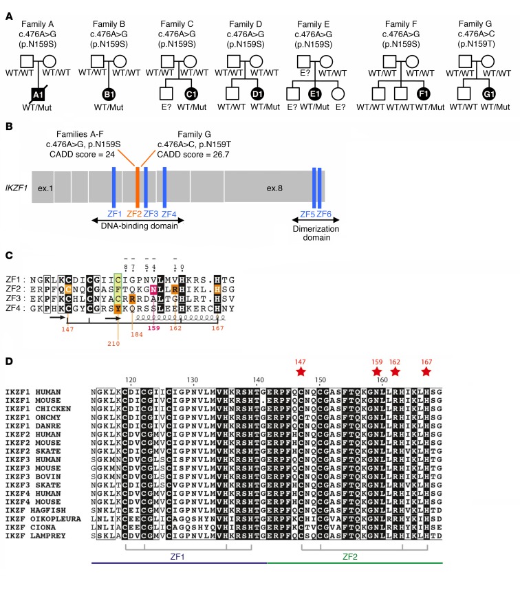
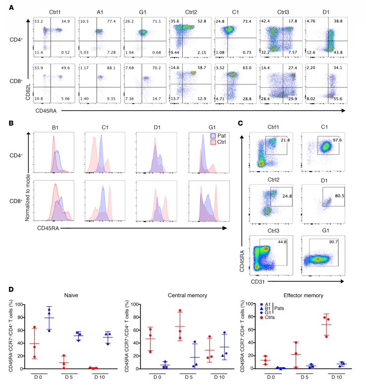
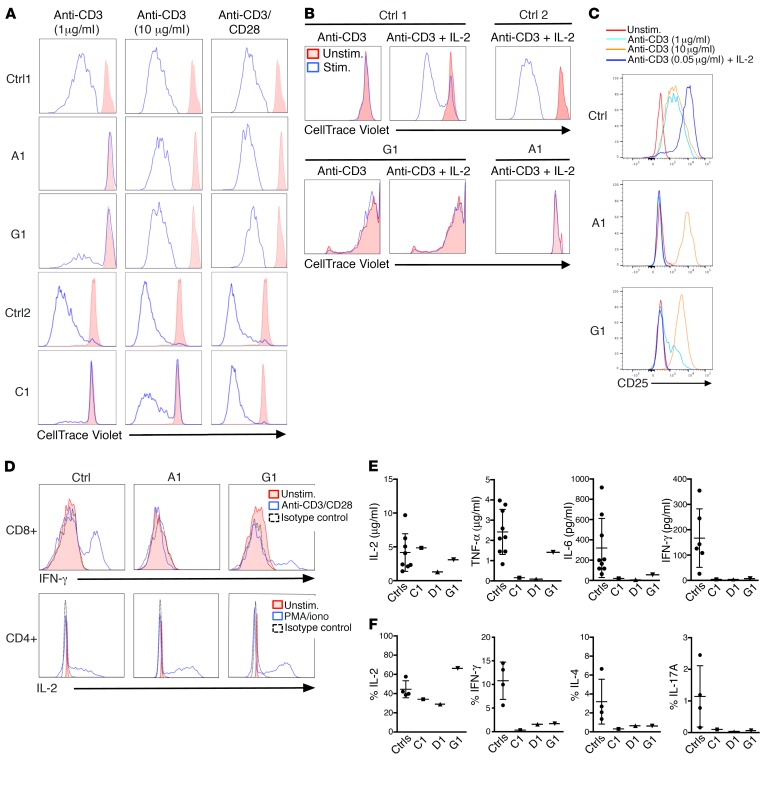
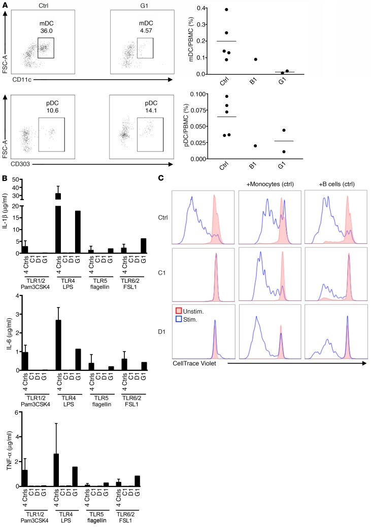
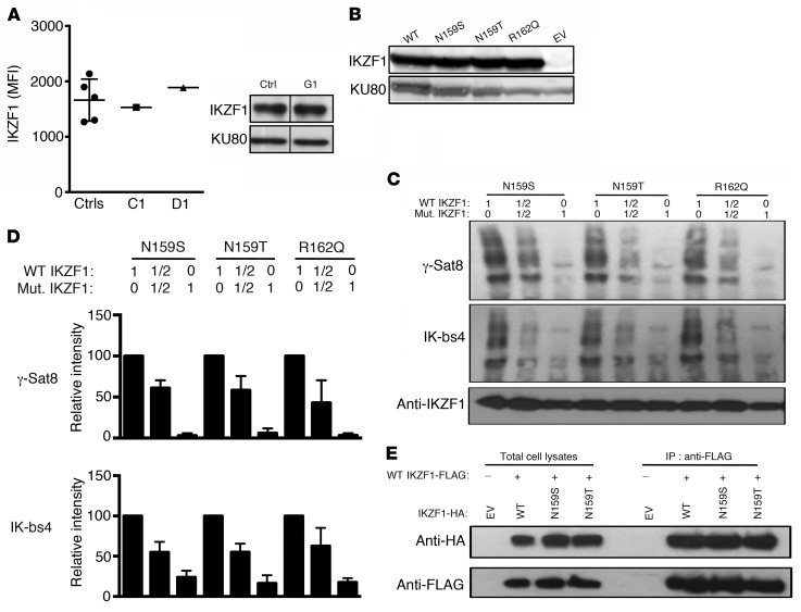
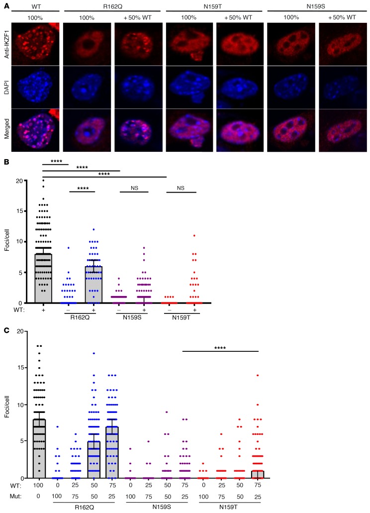
Ikaros/IKZF1 is an essential transcription factor expressed throughout hematopoiesis. IKZF1 is implicated in lymphocyte and myeloid differentiation and negative regulation of cell proliferation. In humans, somatic mutations in IKZF1 have been linked to the development of B cell acute lymphoblastic leukemia (ALL) in children and adults. Recently, heterozygous germline IKZF1 mutations have been identified in patients with a B cell immune deficiency mimicking common variable immunodeficiency. These mutations demonstrated incomplete penetrance and led to haploinsufficiency. Herein, we report 7 unrelated patients with a novel early-onset combined immunodeficiency associated with de novo germline IKZF1 heterozygous mutations affecting amino acid N159 located in the DNA-binding domain of IKZF1. Different bacterial and viral infections were diagnosed, but Pneumocystis jirovecii pneumonia was reported in all patients. One patient developed a T cell ALL. This immunodeficiency was characterized by innate and adaptive immune defects, including low numbers of B cells, neutrophils, eosinophils, and myeloid dendritic cells, as well as T cell and monocyte dysfunctions. Notably, most T cells exhibited a naive phenotype and were unable to evolve into effector memory cells. Functional studies indicated these mutations act as dominant negative. This defect expands the clinical spectrum of human IKZF1-associated diseases from somatic to germline, from haploinsufficient to dominant negative.

Keywords: Genetics; Immunology; Monocytes; Monogenic diseases; T cells.


Conflict of interest statement

Conflict of interest: The authors have declared that no conflict of interest exist.

Figures

Figure 1
Figure 1. Pedigree analysis in patients with IKZF1N159S/T mutations.
(A) Pedigrees of 7 kindreds with IKZF1N159S/T mutations (families A–G). Affected individuals are shown in black. Diagonal lines indicate deceased individuals. WT, wild-type allele; Mut, mutant allele. “E?” indicates unknown genotype. (B) Schematic representation of the IKZF1 coding region, from exon 1 to 8, and corresponding domains, shown in dark gray: the DNA-binding domain containing 4 ZFs and the dimerization domain containing 2 ZFs. Sites of mutations are shown on top with predicted combined annotation dependent depletion (CADD) scores. (C) Alignment of the ZF1-4 DNA-binding domains of IKZF1. Two cysteines and 2 histidines (C2H2) in each finger are responsible for Zn2+ coordination (bottom connecting lines), except for ZF4, in which the last histidine is replaced by a cysteine (strictly conserved in the Ikaros-like family). Amino acids at positions –1, –4, –7, and –8 relative to the first histidine (i.e., 0) are known to be involved in DNA binding (19). Mutated amino acids are colored in pink (this study) or orange (previous studies). The ZF predicted secondary structures (deduced from the model of 3D structure shown in Supplemental Figure 8) are shown below the alignment. (D) Alignment of ZF domains of IKZF1 family members including IKZF1/IKAROS, IKZF2/HELIOS, IKZF3/AIOLOS, and IKZF4/EOS. Sequences of species from the Chordata phylum including marine Urochordata (Ciona and Oikopleura) and primitive fish such as hagfish are aligned. Red stars indicate residues found to be mutated in patients. All these residues are highly conserved through species.
Figure 2
Figure 2. Peripheral T cell phenotypes associated with heterozygous IKZF1N159S/T mutations.
(A) Dot plots from flow cytometric analyses showing the frequency of naive (CD45RA+CD62L+), central memory (CD45RACD62L+), effector memory (CD45RACD62L), and TEMRA cells (CD45RA+CD62L) among CD4+ and CD8+ T cells of patients with IKZF1N159S/T mutations (A1, G1, C1, and D1). Data are shown from independent experiments analyzed with a paired healthy control donor (Ctrl1, Ctrl2, and Ctrl3). (B) Histograms representing CD45RA staining of CD4+ and CD8+ T cells from patients (Pat: B1, C1, D1, and G1 in blue) overlaid with histograms from healthy donor controls (Ctrl in red). (C) Dot plots from flow cytometric analyses showing the frequency of recent thymic emigrants (CD45RA+CD31+) among CD4+ T cells from the indicated patient with a paired healthy donor control (Ctrl). (D) Evolution of naive (CD45RA+CCR7+), central memory (CD45RACCR7+), and effector memory cells (CD45RACCR7) in percentages (%) of PHA-derived CD4+ T cell blasts cultured for 10 days from PBMCs of patients (Pats: A1, B1, and G1) and 3 healthy donor controls (Ctrls). Data represent mean ± SD of 3 independent experiments.
Figure 3
Figure 3. Functional T cell defects associated with heterozygous IKZF1N159S/T mutations.
(AC) PBMCs from patients A1, C1, and G1 and healthy donor controls (Ctrl) were stimulated with indicated agonists for 6–7 days. (A) Cells stimulated (stim.) with plate-coated anti-CD3 at the indicated concentrations or with anti-CD3/CD28–coated beads. (B) Cells stimulated with plate-coated anti-CD3 at 0.05 μg/ml with or without IL-2. unstim., unstimulated. (C) Cells stimulated with plate-coated anti-CD3 at the indicated concentrations with or without IL-2. Proliferation was determined by dilution of CellTrace Violet dye analyzed by flow cytometry. Histograms showing cell divisions by dilution of the CellTrace Violet dye (A and B) and CD25 expression (C). Data are representative of 3 experiments. (D) Histograms from PHA-derived T cell blasts from patients A1, G1, and a healthy donor control (Ctrl) stimulated with (blue) or without (red) anti-CD3/CD28–coated beads (upper panels) or with PMA/ionomycin (iono; lower panels) for 24 hours. IL-2 (lower panels) and IFN-γ (upper panels) were measured with intracellular flow cytometry assays. Black dashed line indicates isotype control. Data are representative of 2 experiments. (E) PBMCs from patients C1, D1, and G1 and healthy donor controls (Ctrls) were stimulated with anti-CD3/CD28–coated beads for 24 hours. Cell-free supernatants were harvested, and cytokines (IL-2, TNF-α, IL-6, and IFN-γ) were measured with the Luminex 200 System. Data indicate the mean of replicate sample for C1 (n = 2), D1 (n = 1), and G1 (n = 1) compared with 6–9 different healthy donor controls (mean ± SD). The n values represent the number of replicates. (F) PBMCs from patients C1, D1, G1, and 2 paired healthy donor controls (Ctrls) were stimulated with PMA/ionomycin for 6 hours. IL-2, IFN-γ, and IL-4 (for Th1/2 evaluation) and IL-17A (for Th17 evaluation) were measured with intracellular flow cytometry assays. Data represent the percentage of the cells positive for the indicated cytokine among CD4+ T cells. Data are issued from 2 experiments performed in patient C1 and 1 experiment from D1 and G1.
Figure 4
Figure 4. Myeloid abnormalities associated with heterozygous IKZF1N159S/T mutations.
(A) PBMCs from G1 and a healthy donor control (Ctrl) were analyzed for the presence of DCs. Dot plots (left panels) of lineage-negative (CD3, CD19, and CD56), HLA-DR–positive, and CD14 or CD16 negative population corresponding to DCs. The CD11c and CD303 markers enabled the delineation of mDCs (CD11c+CD303) and pDCs (CD11cCD303+). Right panels show the results expressed as percentage of PBMCs from patients B1, G1, and 5 healthy donor controls. (B) PBMCs from C1, D1, and G1 and healthy donor controls (Ctrl) were stimulated with the indicated TLR agonist for 24 hours. Cell-free supernatants were harvested, and cytokines (IL-6, IL-1β, TNF-α) were measured with the Luminex 200 System. Data are from one experiment performed in patients C1, D1, and G1 and compared with 4 healthy donor controls (mean ± SD). (C) PBMCs from C1 and D1 were stimulated with (blue line) or without (solid red) soluble anti-CD3 and anti-CD28 antibodies (1 μg/ml each) in the presence or absence of the healthy donor’s monocytes or B cells for 5 days. The proliferation was determined by dilution of CellTrace Violet dye analyzed by flow cytometry. Data are representative of 3 experiments.
Figure 5
Figure 5. Expression, dimerization, and DNA binding of IKZF1N159S/T mutants.
(A) IKZF1 expression was evaluated in 3 patients (C1, D1, and G1). Permeabilized CD3+ T cells of C1 and D1 were stained with PE-conjugated anti-IKZF1 antibodies and results expressed as mean fluorescence intensity (MFI) from FACS analysis (left panel). Data are mean ± SD of 5 healthy donor controls paired with the indicated patients. Cell extracts of PHA-derived T cell blasts of G1 were immunoblotted with anti-IKZF1 or anti-KU80 antibodies as a loading control (right panel). (B) Expression of IKZF1 in HEK293T cells transiently transfected with WT IKZF1, IKZF1 mutants (N159S, N159T, R162Q), or empty vector (EV). IKZF1R162Q haploinsufficient mutant was used for comparison. Cell lysates were immunoblotted with anti-IKZF1 or anti-KU80 antibodies as a loading control. (C) EMSAs were performed with nuclear extracts from HEK293T cells transiently transfected with WT IKZF1 and/or the indicated IKZF1 mutants (FLAG-tagged IKZF1). IKZF1R162Q haploinsufficient mutant was used for comparison. The nuclear extracts were tested by gel mobility shift assay for binding to γ-Sat8 (upper panel) and IK-bs4 (middle panel), 2 sequences from pericentromeric regions known to be IKZF1 targets (upper panels). Cell lysates were tested by Western blot for WT and/or mutant IKZF1-FLAG expression with an anti-IKZF1 antibody (lower panel). Data shown are representative of 3 experiments. (D) EMSA bands were quantified by the Bio-Rad Image Lab program. Data were normalized to WT binding defined as 100%. Error bars represent SD from 3 independent experiments. (E) Cell lysates of HEK293T cells transiently transfected with WT IKZF1-FLAG and WT or mutant IKZF1-HA were immunoprecipitated (IP) with anti-FLAG antibody. Data represent Western blot of whole cell lysates and IP samples (right panel) with anti-HA (upper right panels) and anti-FLAG antibodies (lower right panels). Whole cell lysates correspond to 5% of proteins used in IP (left panel). Data shown are representative of 3 experiments.
Figure 6
Figure 6. Interference of IKZF1N159S/T mutants with pericentromeric targeting of WT IKZF1.
(A) NIH3T3 cells were transfected with WT IKZF1 or the indicated IKZF1 mutants (Mut) (R162Q, N159S, N159T) or with 1:1 WT/Mut ratio. After 36 hours, cells were labeled with a monoclonal anti-IKZF1 primary antibody and an Alexa Fluor 546-conjugated (red) secondary antibody plus DAPI. IKZF1R162Q haploinsufficient mutant was used for comparison. Cells were visualized by confocal microscopy, and images shown are representative of 5 experiments. Original magnification, ×190. (B) Focus counts from the experiment in A. IKZF1 foci were counted in 100 transfected NIH 3T3 cells (n = 100) for each condition. Observer bias was eliminated by coding the slides prior to inspection. Statistical analysis was performed between the different groups with a 1-way ANOVA test. Horizontal lines represent the median ± 95% confidence interval (CI). ****P < 0.0001. (C) The experiment in A was repeated twice adding 3:1 and 1:3 WT/Mut ratio, and foci were enumerated as described in B. For each condition, 90–121 transfected cells were counted, and statistical analysis was performed between the different groups with a 1-way ANOVA test. Horizontal lines represent the median ± 95% CI. ****P < 0.0001.

References

    1. Molnár A, et al. The Ikaros gene encodes a family of lymphocyte-restricted zinc finger DNA binding proteins, highly conserved in human and mouse. J Immunol. 1996;156(2):585–592. - PubMed
    1. Yoshida N, et al. Germline IKAROS mutation associated with primary immunodeficiency that progressed to T-cell acute lymphoblastic leukemia. Leukemia. 2017;31(5):1221–1223. doi: 10.1038/leu.2017.25. - DOI - PubMed
    1. John LB, Ward AC. The Ikaros gene family: transcriptional regulators of hematopoiesis and immunity. Mol Immunol. 2011;48(9-10):1272–1278. doi: 10.1016/j.molimm.2011.03.006. - DOI - PubMed
    1. Thomas RM, et al. Ikaros silences T-bet expression and interferon-gamma production during T helper 2 differentiation. J Biol Chem. 2010;285(4):2545–2553. doi: 10.1074/jbc.M109.038794. - DOI - PMC - PubMed
    1. Heizmann B, Sellars M, Macias-Garcia A, Chan S, Kastner P. Ikaros limits follicular B cell activation by regulating B cell receptor signaling pathways. Biochem Biophys Res Commun. 2016;470(3):714–720. doi: 10.1016/j.bbrc.2016.01.060. - DOI - PubMed

Publication types

MeSH terms

Substances