. 2018 Aug;109(8):2435-2445.
doi: 10.1111/cas.13666. Epub 2018 Aug 7.

High-affinity PD-1 molecules deliver improved interaction with PD-L1 and PD-L2

Affiliations

High-affinity PD-1 molecules deliver improved interaction with PD-L1 and PD-L2

Yanyan Li et al. Cancer Sci. 2018 Aug.

Abstract

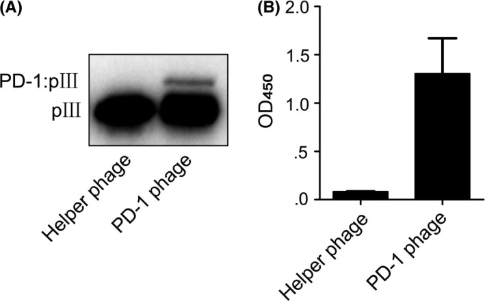
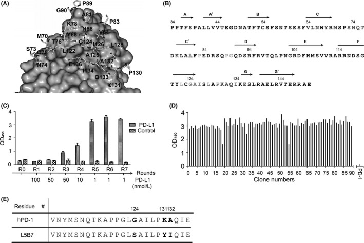
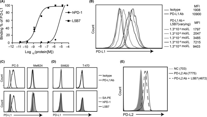
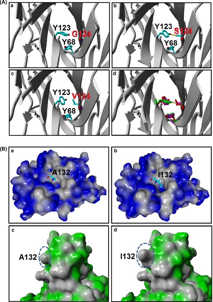
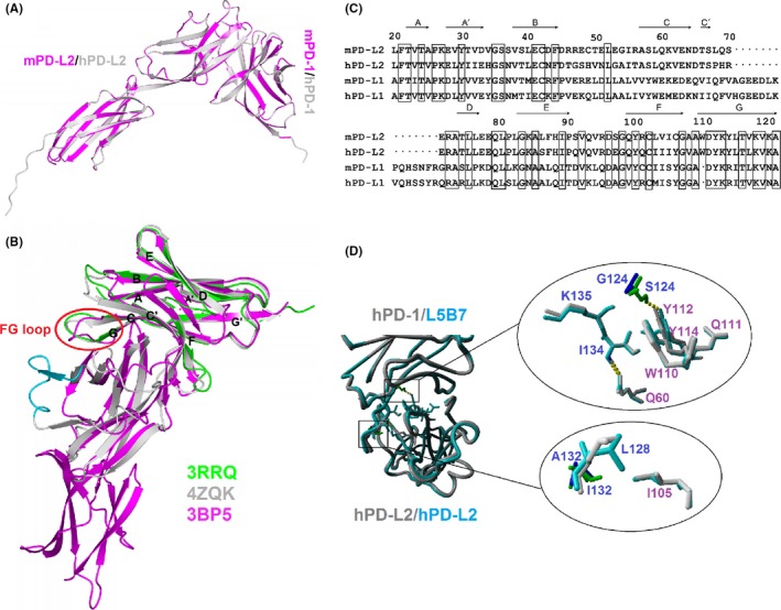
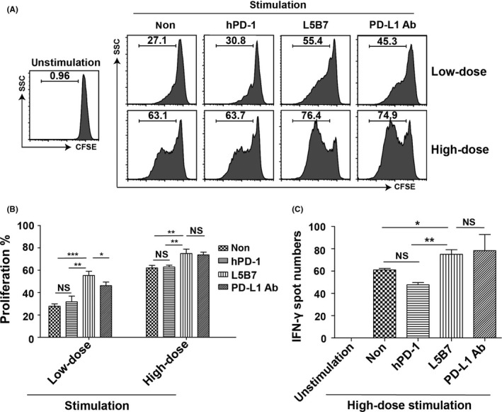
The inhibitory checkpoint molecule programmed death (PD)-1 plays a vital role in maintaining immune homeostasis upon binding to its ligands, PD-L1 and PD-L2. Several recent studies have demonstrated that soluble PD-1 (sPD-1) can block the interaction between membrane PD-1 and PD-L1 to enhance the antitumor capability of T cells. However, the affinity of natural sPD-1 binding to PD-L1 is too low to permit therapeutic applications. Here, a PD-1 variant with approximately 3000-fold and 70-fold affinity increase to bind PD-L1 and PD-L2, respectively, was generated through directed molecular evolution and phage display technology. Structural analysis showed that mutations at amino acid positions 124 and 132 of PD-1 played major roles in enhancing the affinity of PD-1 binding to its ligands. The high-affinity PD-1 mutant could compete with the binding of antibodies specific to PD-L1 or PD-L2 on cancer cells or dendritic cells, and it could enhance the proliferation and IFN-γ release of activated lymphocytes. These features potentially qualify the high-affinity PD-1 variant as a unique candidate for the development of a new class of PD-1 immune-checkpoint blockade therapeutics.

Keywords: PD-1; PD-L1; PD-L2; inhibitory receptor; phage display.


Figures

Figure 1
Figure 1
Display of hPD‐1 extracellular region on M13 phage. A, Western blot of PD‐1 phage. B, The binding of PD‐1 phage to hPD‐L1 detected by ELISA. Helper phage was run as a negative control. Error bars indicate SD (n = 3)
Figure 2
Figure 2
Design of hPD‐1 phage displays libraries and isolation of hPD‐L1 (hPD‐L1)‐binding phage by biopanning. A, The 24 selected residues for construction of phage display libraries. The structure of mPD‐1/hPD‐L1 (PDB number: 3BIK) was used as the template to build the hPD‐1/hPD‐L1 complex model. Residues of hPD‐1 are shown as stick models, whereas hPD‐L1 is represented by gray surface. B, Amino acid division of hPD‐1 for the library construction is shown in colors: Library 1, red; Library 2, green; Library 3, orange; Library 4, cyan; Library 5, pink. The Ala81 was assigned to Library 2 and Library 3. Residue Ser93 (blue) was a mutation from Cys. Secondary structural elements of hPD‐1 extracellular region were shown on top of the sequence. C, Polyclonal phage ELISA. The polyclonal phage particles from each round of biopannings were tested for their binding to 0.5 μg/mL immobilized hPD‐L1. An irrelevant protein (pHLA) was used as the negative control. The result of Library 5 was shown as a representative. D, Monoclonal phage ELISA. 400 individual colonies of each library from the 7th round of biopanning were tested for their binding to hPD‐L1 by monoclonal phage ELISA. The result of Library 5 is shown as a representative. E, Partial sequence alignment of hPD‐1 and L5B7. The 3 mutated amino acids are shown as G124S, K131Y and A132I
Figure 3
Figure 3
Interaction of hPD‐1 and L5B7 with hPD‐L1 or hPD‐L2 on the cell surface. A, Kinetic analysis of soluble hPD‐1 and L5B7 binding to hPD‐L1 on MDAMB‐231 cells. B, The soluble L5B7 competing with anti‐PD‐L1 Ab (29E.1A3) binding to hPD‐L1 on MDAMB‐231 cells. C,D, The soluble hPD‐1 and L5B7 binding to hPD‐L1 on various cells, (C) PD‐L1+ tumor cells PC‐3 and Mel624, (D) PD‐L1 tumor cells SW620 and T‐47D. Top panel, isotype control and anti‐PD‐L1 Ab. Bottom panel, SAPE, hPD‐1 and L5B7. E, The soluble L5B7 competing with anti‐PD‐L2 Ab (M1H18) binding to hPD‐L2 on mature dendritic cells
Figure 4
Figure 4
Close‐up views of π‐π interaction in Gly124 and hydrophobic interaction in Ala132. A, The enhancement of π‐π interaction in G124S and G124V PD‐1 variants. The hPD‐1/hPD‐L1 (PDB: 4ZQK) served as the template to build G124S and G124V mutation models. The complex was shown in ribbon representation, and the Tyr123 of hPD‐L1, Tyr68, Gly/Ser/Val124 of hPD‐1 are shown in a stick model. Hydrogen bonds are depicted as yellow dashed lines: (a) Gly124; (b) Ser124; (c) Val124; and (d) superposition of Gly124 (green), Ser124 (magenta) and Val124 (red) in hPD‐1 variants. B, Hydrophobic interaction was enhanced in the hPD‐1 variants. Molecular surfaces of hPD‐L1 (a,b) and hPD‐1 (c,d) are represented with blue or green color, respectively, whereas the hydrophobic part is colored gray. Residues Ala/Ile132 of hPD‐1/variant are shown as stick models (a,b), and the corresponding molecular surfaces are indicated as dashed circles (c,d)
Figure 5
Figure 5
Structural analysis of interaction between L5B7 and hPD‐L2. A, Structure of hPD‐1/hPD‐L2 superimposed on mPD‐1/mPD‐L2 (PDB number: 3BP5) represented as ribbon models. mPD‐1/mPD‐L2 are colored magenta. B, Structure‐based sequence alignment of apo‐hPD‐1 (green, PDB number: 3RRQ), hPD‐1/hPD‐L1 (gray, PDB number: 4ZQK), mPD‐1/mPD‐L2 (magenta, PDB number: 3BP5). The black letters label the secondary structural elements of hPD‐1. The red circle represents the orientation discrepancy of the FG loop of PD‐1. The cyan represented redundant hPD‐L1 sequence on structure compared to mPD‐L2. C, The sequence alignment of extracellular IgV domains of mouse/human PD‐L2 and PD‐L1. The black dot shows the missing residues in PD‐L2 compared to PD‐L1. The black box represents a conservative sequence in PD‐L1 and PD‐L2. The secondary structural elements of the extracellular IgV region of mPD‐L2 are shown on top of the sequence. D, The role of G124S and A132I in enhancing L5B7 binding to hPD‐L2. The mutations are located at the key position related to interaction of hPD‐1 and hPD‐L2 (left). The physical impact of G124S and A132I in enhancing L5B7 binding to hPD‐L2 (right). Top, G124S (blue/green); bottom, A132I (blue/green)
Figure 6
Figure 6
The enhancement of proliferation and IFN‐γ release of activated PBMC. A, The flow cytometry data of proliferation of activated PBMC with anti‐CD3 Ab (aCD3) and anti‐CD28 Ab (aCD28) in the presence or absence of soluble hPD‐1, L5B7 and anti‐PD‐L1 Ab (5 μg/mL). PBMCwere stimulated with low‐dose antibodies of 15 ng/mL aCD3 and 7.5 ng/mL aCD28 or high‐dose antibodies of 30 ng/mL aCD3 and 15 ng/mL aCD28. B, The statistical data of proliferating cells in (A). Error bars indicated SD (n = 3). C, ELISpot assays showing IFN‐γ release of PBMC activated with 30 ng/mL aCD3 and 15 ng/mL aCD28 in the presence or absence of soluble hPD‐1, L5B7 and anti‐PD‐L1 Ab (5 μg/mL). Error bars indicated SD (n = 3). Unpaired Student's t test, NS,> .05; *, < .05; **, < .01; ***, < .001

Similar articles

Cited by

References

    1. Lee S, Margolin K. Tumor‐infiltrating lymphocytes in melanoma. Curr Oncol Rep. 2012;14:468‐474. - PMC - PubMed
    1. Page DB, Postow MA, Callahan MK, Allison JP, Wolchok JD. Immune modulation in cancer with antibodies. Annu Rev Med. 2014;65:185‐202. - PubMed
    1. Carosella ED, Ploussard G, LeMaoult J, Desgrandchamps F. A systematic review of immunotherapy in urologic cancer: evolving roles for targeting of CTLA‐4, PD‐1/PD‐L1, and HLA‐G. Eur Urol. 2015;68:267‐279. - PubMed
    1. Brentjens RJ, Davila ML, Riviere I, et al. CD19‐targeted T cells rapidly induce molecular remissions in adults with chemotherapy‐refractory acute lymphoblastic leukemia. Sci Transl Med. 2013;5:177ra38. - PMC - PubMed
    1. Robbins PF, Kassim SH, Tran TL, et al. A pilot trial using lymphocytes genetically engineered with an NY‐ESO‐1‐reactive T‐cell receptor: long‐term follow‐up and correlates with response. Clin Cancer Res. 2015;21:1019‐1027. - PMC - PubMed

MeSH terms