

Confirmation of Luteinizing Hormone (LH) in Living Human Vitreous and the Effect of LH Receptor Reduction on Murine Electroretinogram
- PMID: 29890291
- PMCID: PMC6063313
- DOI: 10.1016/j.neuroscience.2018.05.049
Confirmation of Luteinizing Hormone (LH) in Living Human Vitreous and the Effect of LH Receptor Reduction on Murine Electroretinogram
Abstract
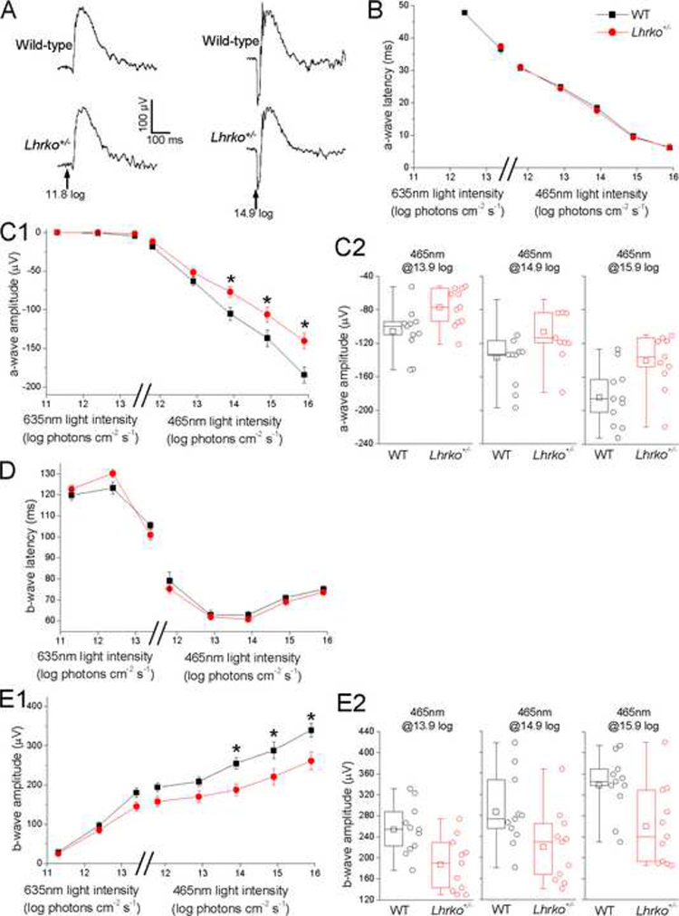
Luteinizing hormone (LH), produced in the anterior pituitary, has been detected in cadaver eyes and LH receptors (LHRs) have been identified in the retina, with the highest density in cone photoreceptors. Our aim was to confirm the presence of LH in the living, human eye as well as to examine the potential impact of a reduction in LHR signaling on visual processing. Vitreous samples were collected from 40 patients (23 diabetics, 17 non-diabetics) who were undergoing vitrectomies for various indications. LH concentration was quantified in each sample via an electro-chemiluminescence immunoassay and Meso Scale Discovery platform and normalized to total protein. In addition, full-field electroretinography (ERG) was performed on 11 adult LHR knockout heterozygous mice (B6;129X1-Lhcgrtm1Zmlei/J) and 11 wild types using the Celeris-Diagnosys system. The median LH values (pg/mg total protein) for non-diabetics, diabetics without proliferative diabetic retinopathy (PDR) and diabetics with PDR were 40.7, 41.9 and 167.8 respectively. LH levels were significantly higher in diabetics with PDR. In our ERG investigation, heterozygous LHRKOs were found to have significantly reduced amplitudes of a-wave and b-waves at high stimulus intensities with no significant change in a-wave or b-wave amplitudes at lower intensities; this is consistent with a selective impairment of cone-mediated responses. Our findings confirm LH is present in the adult human eye. Our findings also suggest that a reduction in LH receptor signaling negatively impacts visual processing of the cone photoreceptors. Overall, our study results support the theory that LH likely plays a physiologic role in the eye.
Keywords: ERG; cone cells; electroretinography; luteinizing hormone; photoreceptors; visual processing.
Copyright © 2018 IBRO. Published by Elsevier Ltd. All rights reserved.
Conflict of interest statement
Potential Conflicts of Interest:
Dr. Tammy Z Movsas is the Director and Principal Investigator at Zietchick Research Institute (ZRI), a private research institute, and has a pending patent application for the use of gonadotropin antagonists for the treatment of ocular disorders. Dr. Arivalagan Muthusamy is a salaried employee at Zietchick Research Institute. The other co-authors are not employees of Zietchick Research Institute nor do they have any potential conflicts of interest.
Figures
Similar articles
-
Vitreous Levels of Luteinizing Hormone and VEGF are Strongly Correlated in Healthy Mammalian Eyes.Curr Eye Res. 2018 Aug;43(8):1041-1044. doi: 10.1080/02713683.2018.1467932. Epub 2018 May 3. Curr Eye Res. 2018. PMID: 29677452 Free PMC article.
-
[Multimodal Approaches for the Analysis of Retinal Functional Disorders―Focusing on Retinal Detachment].Nippon Ganka Gakkai Zasshi. 2017 Mar;121(3):185-231. Nippon Ganka Gakkai Zasshi. 2017. PMID: 30088405 Japanese.
-
Localization of receptors for luteinizing hormone/chorionic gonadotropin in neural retina.Life Sci. 1998;63(12):1057-64. doi: 10.1016/s0024-3205(98)00367-1. Life Sci. 1998. PMID: 9749828
-
Structural and functional plasticity of the luteinizing hormone/choriogonadotrophin receptor.Hum Reprod Update. 2013 Sep-Oct;19(5):583-602. doi: 10.1093/humupd/dmt023. Epub 2013 May 17. Hum Reprod Update. 2013. PMID: 23686864 Review.
-
Inactivating mutations of luteinizing hormone beta-subunit or luteinizing hormone receptor cause oligo-amenorrhea and infertility in women.Horm Res. 2009;71(2):75-82. doi: 10.1159/000183895. Epub 2009 Jan 8. Horm Res. 2009. PMID: 19129711 Review.
Cited by
-
The association between high levels of luteinizing hormone and proliferative retinopathy of prematurity in female preterm infants.J AAPOS. 2020 Jun;24(3):145.e1-145.e5. doi: 10.1016/j.jaapos.2020.02.011. Epub 2020 Jun 6. J AAPOS. 2020. PMID: 32522707 Free PMC article.
-
The potential effect of human chorionic gonadotropin on vasoproliferative disorders of the immature retina.Neuroreport. 2018 Dec 12;29(18):1525-1529. doi: 10.1097/WNR.0000000000001140. Neuroreport. 2018. PMID: 30300333 Free PMC article.
-
Atypical pituitary hormone-target tissue axis.Front Med. 2023 Feb;17(1):1-17. doi: 10.1007/s11684-022-0973-7. Epub 2023 Feb 27. Front Med. 2023. PMID: 36849623 Review.
-
Elimination of Signaling by the Luteinizing Hormone Receptor Reduces Ocular VEGF and Retinal Vascularization during Mouse Eye Development.Curr Eye Res. 2018 Oct;43(10):1286-1289. doi: 10.1080/02713683.2018.1495740. Epub 2018 Jul 18. Curr Eye Res. 2018. PMID: 29966451 Free PMC article.
-
Accumulation of systematic TPM1 mediates inflammation and neuronal remodeling by phosphorylating PKA and regulating the FABP5/NF-κB signaling pathway in the retina of aged mice.Aging Cell. 2022 Mar;21(3):e13566. doi: 10.1111/acel.13566. Epub 2022 Feb 11. Aging Cell. 2022. PMID: 35148456 Free PMC article.
References
-
- Aiello LP, Avery RL, Arrigg PG, Keyt BA, Jampel HD, Shah ST, Pasquale LR, Thieme H, et al. (1994) Vascular endothelial growth factor in ocular fluid of patients with diabetic retinopathy and other retinal disorders. N Engl J Med 331:1480–1487. - PubMed
-
- Babitha V, Yadav VP, Chouhan VS, Hyder I, Dangi SS, Gupta M, Khan FA, Taru Sharma G, et al. (2014) Luteinizing hormone, insulin like growth factor-1, and epidermal growth factor stimulate vascular endothelial growth factor production in cultured bubaline granulosa cells. Gen Comp Endocrinol 198:112. - PubMed
-
- Bringmann A, Wiedemann P (2009) Involvement of Muller glial cells in epiretinal membrane formation. Graefes Arch Clin Exp Ophthalmol 247:865–883. - PubMed
-
- Caldwell RB, Bartoli M, Behzadian MA, El-Remessy AE, Al-Shabrawey M, Platt DH, Caldwell RW (2003) Vascular endothelial growth factor and diabetic retinopathy: pathophysiological mechanisms and treatment perspectives. Diabetes Metab Res Rev 19:442–455. - PubMed
Publication types
MeSH terms
Substances
Grants and funding
LinkOut - more resources
Full Text Sources
Other Literature Sources
Molecular Biology Databases
Miscellaneous
