

Hepatic Mitochondrial Dysfunction and Immune Response in a Murine Model of Peanut Allergy
- PMID: 29890625
- PMCID: PMC6024519
- DOI: 10.3390/nu10060744
Hepatic Mitochondrial Dysfunction and Immune Response in a Murine Model of Peanut Allergy
Abstract
Background: Evidence suggests a relevant role for liver and mitochondrial dysfunction in allergic disease. However, the role of hepatic mitochondrial function in food allergy is largely unknown. We aimed to investigate hepatic mitochondrial dysfunction in a murine model of peanut allergy.
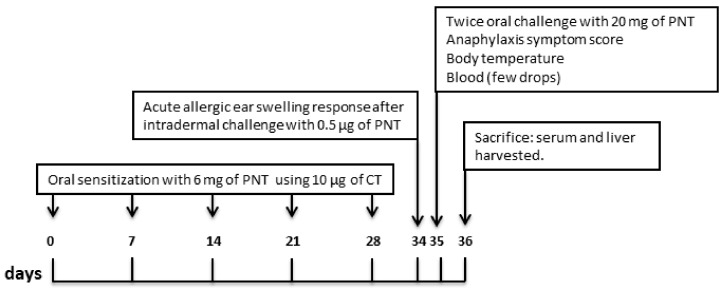
Methods: Three-week-old C3H/HeOuJ mice were sensitized by the oral route with peanut-extract (PNT). We investigated: 1. the occurrence of effective sensitization to PNT by analysing acute allergic skin response, anaphylactic symptoms score, body temperature, serum mucosal mast cell protease-1 (mMCP-1) and anti-PNT immunoglobulin E (IgE) levels; 2. hepatic involvement by analysing interleukin (IL)-4, IL-5, IL-13, IL-10 and IFN-γ mRNA expression; 3. hepatic mitochondrial oxidation rates and efficiency by polarography, and hydrogen peroxide (H₂O₂) yield, aconitase and superoxide dysmutase activities by spectrophotometry.
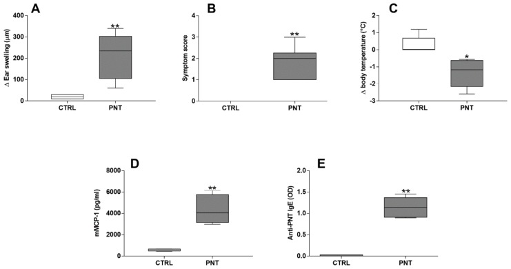
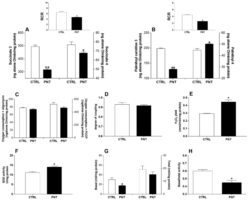
Results: Sensitization to PNT was demonstrated by acute allergic skin response, anaphylactic symptoms score, body temperature decrease, serum mMCP-1 and anti-peanut IgE levels. Liver involvement was demonstrated by a significant increase of hepatic Th2 cytokines (IL-4, IL-5 and IL-13) mRNA expression. Mitochondrial dysfunction was demonstrated by lower state 3 respiration rate in the presence of succinate, decreased fatty acid oxidation in the presence of palmitoyl-carnitine, increased yield of ROS proven by the inactivation of aconitase enzyme and higher H₂O₂ mitochondrial release.
Conclusions: We provide evidence of hepatic mitochondrial dysfunction in a murine model of peanut allergy. These data could open the way to the identification of new mitochondrial targets for innovative preventive and therapeutic strategies against food allergy.
Keywords: Th2 cytokines; food allergy; mitochondrial function; oxidative stress.
Conflict of interest statement
The authors declare no conflict of interest.
Figures

Similar articles
-
Experimental food allergy to peanut enhances the immune response to house dust mite in the airways of mice.Clin Exp Allergy. 2017 Jan;47(1):121-128. doi: 10.1111/cea.12799. Epub 2016 Sep 20. Clin Exp Allergy. 2017. PMID: 27533916
-
Mixed antibody and T cell responses to peanut and the peanut allergens Ara h 1, Ara h 2, Ara h 3 and Ara h 6 in an oral sensitization model.Clin Exp Allergy. 2004 Sep;34(9):1422-8. doi: 10.1111/j.1365-2222.2004.02062.x. Clin Exp Allergy. 2004. PMID: 15347376
-
Anti-hIgE gene therapy of peanut-induced anaphylaxis in a humanized murine model of peanut allergy.J Allergy Clin Immunol. 2016 Dec;138(6):1652-1662.e7. doi: 10.1016/j.jaci.2016.03.053. Epub 2016 Jun 29. J Allergy Clin Immunol. 2016. PMID: 27372563
-
Towards immunotherapy for peanut allergy.Curr Opin Allergy Clin Immunol. 2005 Dec;5(6):558-62. doi: 10.1097/01.all.0000191233.90136.21. Curr Opin Allergy Clin Immunol. 2005. PMID: 16264338 Review.
-
Allergy to peanut oil--clinically relevant?J Eur Acad Dermatol Venereol. 2007 Apr;21(4):452-5. doi: 10.1111/j.1468-3083.2006.02133.x. J Eur Acad Dermatol Venereol. 2007. PMID: 17373969 Review.
Cited by
-
Anti-Allergic Effect of Dietary Polyphenols Curcumin and Epigallocatechin Gallate via Anti-Degranulation in IgE/Antigen-Stimulated Mast Cell Model: A Lipidomics Perspective.Metabolites. 2023 May 5;13(5):628. doi: 10.3390/metabo13050628. Metabolites. 2023. PMID: 37233669 Free PMC article.
-
Butyrate as a bioactive human milk protective component against food allergy.Allergy. 2021 May;76(5):1398-1415. doi: 10.1111/all.14625. Epub 2020 Nov 16. Allergy. 2021. PMID: 33043467 Free PMC article.
-
The Difference of Gut Microbiota and Their Correlations With Urinary Organic Acids Between Autistic Children With and Without Atopic Dermatitis.Front Cell Infect Microbiol. 2022 Jun 21;12:886196. doi: 10.3389/fcimb.2022.886196. eCollection 2022. Front Cell Infect Microbiol. 2022. PMID: 35800387 Free PMC article.
-
Mitochondrial and Nuclear Mitochondrial Variants in Allergic Diseases.Allergy Asthma Immunol Res. 2020 Sep;12(5):877-884. doi: 10.4168/aair.2020.12.5.877. Allergy Asthma Immunol Res. 2020. PMID: 32638566 Free PMC article.
-
Oxidative Stress and Mitochondria Are Involved in Anaphylaxis and Mast Cell Degranulation: A Systematic Review.Antioxidants (Basel). 2024 Jul 29;13(8):920. doi: 10.3390/antiox13080920. Antioxidants (Basel). 2024. PMID: 39199166 Free PMC article. Review.
References
MeSH terms
Substances
LinkOut - more resources
Full Text Sources
Other Literature Sources
