Review
. 2018 Jun 8;23(6):1386.
doi: 10.3390/molecules23061386.

14-3-3: A Case Study in PPI Modulation

Affiliations
Review

14-3-3: A Case Study in PPI Modulation

Alice Ballone et al. Molecules. .

Abstract

In recent years, targeting the complex network of protein⁻protein interactions (PPIs) has been identified as a promising drug-discovery approach to develop new therapeutic strategies. 14-3-3 is a family of eukaryotic conserved regulatory proteins which are of high interest as potential targets for pharmacological intervention in human diseases, such as cancer and neurodegenerative and metabolic disorders. This viewpoint is built on the “hub” nature of the 14-3-3 proteins, binding to several hundred identified partners, consequently implicating them in a multitude of different cellular mechanisms. In this review, we provide an overview of the structural and biological features of 14-3-3 and the modulation of 14-3-3 PPIs for discovering small molecular inhibitors and stabilizers of 14-3-3 PPIs.

Keywords: 14-3-3 PPIs; PPI modulation; small molecules.


Conflict of interest statement

The authors declare no conflict of interest.

Figures

Figure 1
Figure 1
View of the monomer of the 14-3-3ζ isoform (PDB ID: 5EXA) [11] (A). The molecular assembly of the structural elements is shown in cartoon and surface representation. Hydrophobic, basic and acidic residues are depicted in green, blue, and red, respectively (B,C). View of the monomer turned by 90 degrees around the y-axis; helices from 1 to 9 constituting the classical 14-3-3 monomer are represented, as well as residues that belong to the phospho-acceptor side and residues that establish the hydrophobic interactions.
Figure 2
Figure 2
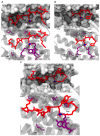
Common recognition motif for 14-3-3 proteins that contain a phosphorylated serine or threonine; mode II for the complex Son of sevenless homolog 1 SOS1/14-3-3ζ (PDB ID: 6F08) [20] (A). Higher side: 14-3-3ζ (white surface) and the peptide SOS1 (red sticks); lower side: Polar contacts (black dashed lines) and hydrophobic interactions (wireframe white spheres) between the residues of 14-3-3ζ (red sticks and white-transparent cartoon) and the pSer binding site of SOS1 (violet sticks). Mode III for the complex ERα/14-3-3σ (PDB ID: 4JC3) [21] (B). Higher side: 14-3-3σ (white surface) and the peptide ERα (red sticks); lower side: Polar contacts (black dashed lines) and hydrophobic interactions (wireframe white spheres) between the residues of 14-3-3σ (red sticks and white-transparent cartoon) and the pSer binding site of ERα (violet sticks). Mode I for the complex ubiquitin specific protease 8 (USP8)/14-3-3ζ (PDB ID: 6F09) [22] (C). Higher side: 14-3-3ζ (white surface) and the peptide USP8 (red sticks); lower side: Polar contacts (black dashed lines) and hydrophobic interactions (wireframe white spheres) between the residues of 14-3-3ζ (red sticks and white-transparent cartoon) and the pSer binding site of USP8 (violet sticks).
Figure 3
Figure 3
Example of a stabilizer that binds to the interface of 14-3-3 (white surface) and a peptide motif (red sticks) binding within the groove. When this peptide motif binds within the 14-3-3 groove, a druggable pocket is formed. A semisynthetic fusicoccin derivative (16-O-Me-FC-H) (cyan sticks) binds to this pocket and stabilizes the potassium channel subfamily K member 9 (TASK-3) and 14-3-3 motif complex. The information was obtained analyzing the PDB entry 4FR3 [39].
Figure 4
Figure 4
Examples of the different natural 14-3-3 protein–protein interaction (PPI) stabilizers. (A) Left side: crystal structure of 14-3-3ζ (white surface) in complex with peptide cystic fibrosis transmembrane conductance regulator (CFTR) R-domain pS753-pS768 (red sticks) and stabilizer fusicoccin-A (cyan sticks) [35]; right side: chemical structure of fusicoccin-A; (B) left side: crystal structure of 14-3-3ζ (white surface) in complex with a dephosphorylated C-RAF peptide (red sticks) and cotylenin A (cyan sticks) [43]; right side: chemical structure of cotylenin A; (C) right side: chemical structure of mizoribine.
Figure 5
Figure 5
Examples of the different semisynthetic 14-3-3 PPI stabilizers. (A) Left side: crystal structure of 14-3-3σ (white surface) in complex with TASK-3 peptide (red sticks) and stabilizer fusicoccin A-THF (cyan sticks) [39]; right side: chemical structure of fusicoccin A-THF; (B) left side: crystal structure of 14-3-3ζ (white surface) in complex with Gab2 peptide (red sticks) and ISIR-005 (cyan sticks) [11]; right side: chemical structure of ISIR-005.
Figure 6
Figure 6
Examples of different synthetic 14-3-3 PPI stabilizers. (A) Left side: crystal structure of TASK-3 (white surface) in complex with plasma membrane H+-ATPase (PMA2) peptide (red spheres) and stabilizer pyrrolidone1 (cyan sticks) [47]; right side: chemical structure of pyrrolidone1; (B) left side: crystal structure of 14-3-3-like protein E (isoform of Nicotiana tabacum) (white surface) in complex with PMA2 peptide (ice blue spheres) and stabilizer pyrazole 34 (cyan sticks) [48]; right side: chemical structure of pyrazole 34; (C) left side: crystal structure of 14-3-3β (white surface) in complex with carbohydrate-response element-binding protein (ChREBP) peptide (purple spheres) and stabilizer AMP (cyan sticks) [49]; right side: chemical structure of AMP; (D) left side: crystal structure of 14-3-3ζ (white surface) in complex with Cdc25C peptide (red sticks) and CLR01 (cyan sticks) [51]; right side: chemical structure of CLR01.
Figure 7
Figure 7
Examples of the different classes of 14-3-3 PPI inhibitors. (A) Crystal structure of peptide R18 (cyan sticks) in the 14-3-3ζ binding groove (white surface) (PDB ID: 1A38) [55]; (B) crystal structure of ExoS-derived alkyne cross-linked cyclic peptide (cyan sticks) in the 14-3-3ζ binding groove (white surface) (PDB ID: 5J31) [57]; (C) crystal structure of modified Tau peptide hybrid 3b (cyan sticks) in the 14-3-3σ binding groove (white surface) (PDB ID: 5HF3) [58]; (D) chemical structures of BV02 and BV101.

Similar articles

Cited by

References

    1. Moore B.W., Perez V.J., Carlson F.D., editors. Physiological and Biochemical Aspects of Nervous Integration. Prentice-Hall Inc.; The Marine Biological Laboratory; Woods Hole, MA, USA: 1967. pp. 343–359.
    1. Aitken A. 14-3-3 proteins: A historic overview. Semin. Cancer Biol. 2006;16:162–172. doi: 10.1016/j.semcancer.2006.03.005. - DOI - PubMed
    1. Ichimura T., Isobe T., Okuyama T., Takahashi N., Araki K., Kuwano R., Takahashi Y. Molecular cloning of cDNA coding for brain-specific 14-3-3 protein, a protein kinase-dependent activator of tyrosine and tryptophan hydroxylases. Proc. Natl. Acad. Sci. USA. 1988;85:7084–7088. doi: 10.1073/pnas.85.19.7084. - DOI - PMC - PubMed
    1. Wang W., Shakes D.C. Molecular evolution of the 14-3-3 protein family. J. Mol. Evol. 1996;43:384–398. doi: 10.1007/BF02339012. - DOI - PubMed
    1. Cau Y., Valensin D., Mori M., Draghi S., Botta M. Structure, function, involvement in diseases and targeting of 14-3-3 proteins: An update. Curr. Med. Chem. 2017;25:5–21. doi: 10.2174/0929867324666170426095015. - DOI - PubMed