. 2018 Jun 8;10(6):749.
doi: 10.3390/nu10060749.

Early Life Vitamin C Deficiency Does Not Alter Morphology of Hippocampal CA1 Pyramidal Neurons or Markers of Synaptic Plasticity in a Guinea Pig Model

Affiliations

Early Life Vitamin C Deficiency Does Not Alter Morphology of Hippocampal CA1 Pyramidal Neurons or Markers of Synaptic Plasticity in a Guinea Pig Model

Stine N Hansen et al. Nutrients. .

Abstract

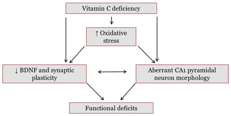
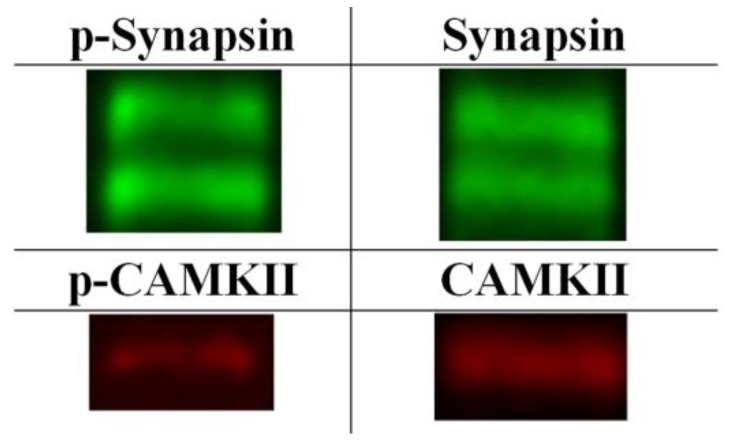
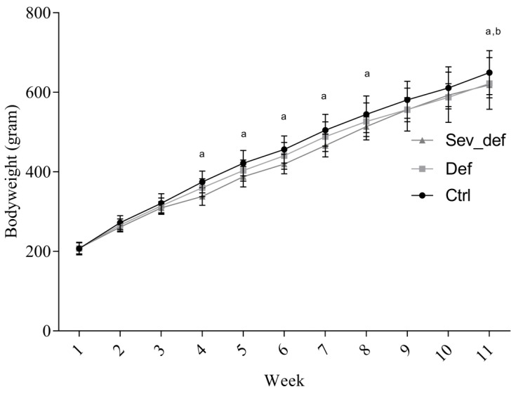
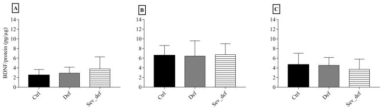
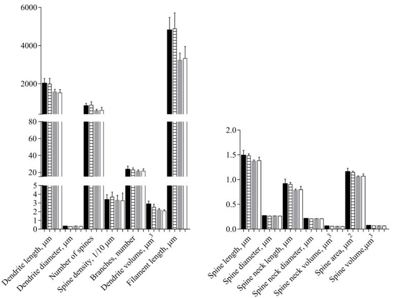
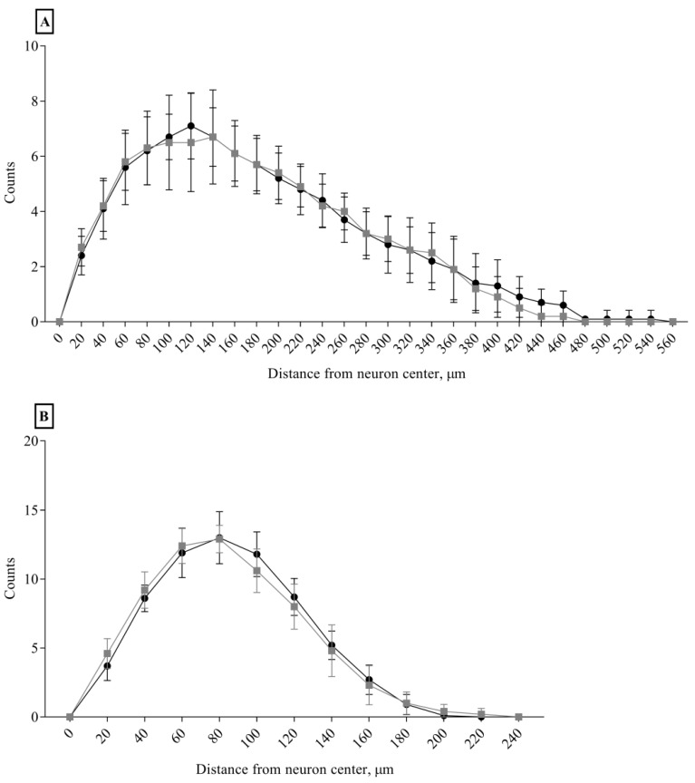
Approximately 15% of the Western world population, including pregnant women and their children, is characterized as vitamin C (vitC) deficient. In guinea pigs, early life vitC deficiency causes spatial memory deficits, decreased hippocampal volume and neuron numbers, in otherwise clinically healthy animals. We hypothesized that vitC deficiency leads to decreased brain-derived neurotrophic factor and synaptic plasticity markers in selected brain areas (frontal cortex, hippocampus and striatum) and cause morphological changes in cornu ammonis 1 pyramidal neurons of the hippocampus either through a direct effect or indirectly by increased oxidative stress. Fifty-seven female guinea pigs were allocated to three groups receiving either 1390, 100 or 0⁻50 mg vitC/kg feed for 11 weeks. Dietary vitC levels were reflected in the plasma, cortical and adrenal gland levels, however, redox imbalance was only present in the adrenal glands allowing for the investigation of a direct influence of vitC deficiency on the chosen parameters in the brain. Synaptic plasticity markers were not affected in the investigated brain areas and no differences in isolated pyramidal neuron morphology was recorded. Based on our findings, it appears that vitC deficiency may primarily elicit impaired neuronal function through increased levels of oxidative stress.

Keywords: cavia porcellus; neuronal morphology; synaptic plasticity; vitamin C deficiency.


Conflict of interest statement

The authors declare no conflict of interest.

Figures

Figure 1
Figure 1
Hypothesized pathways through which vitamin C (vitC) deficiency may cause functional deficits in the brain. Reduced levels of vitC during deficiency may disturb redox balance and subsequently promote oxidative stress in the brain. In addition, vitC deficiency may exert a direct effect on brain function. Both vitC deficiency and increases in oxidative stress have been found to lower levels of brain-derived neurotrophic factor (BDNF) in the brain. A decrease in BDNF, and subsequent negative effects on downstream pathways, may reduce synaptic plasticity. Synaptic plasticity is crucial in memory formation and cognitive functions; hence, dysfunction may lower signal transmission and induce functional deficits. BDNF is also involved in neuronal arbor growth and a reduction may further induce aberrant dendrite development of hippocampal cornu ammonis (CA) 1 pyramidal neurons. These neurons are crucial in memory functions and have been found to be susceptible to increased oxidative stress and states of malnutrition. Collectively, vitC deficiency is hypothesized to cause changes in synaptic plasticity and neuronal morphology, potentially disrupting brain functionality.
Figure 2
Figure 2
Examples of Western blots of p-/synapsin 1 and p-/Ca2+-calmodulin-dependent kinase II. The figure depicts examples of the Western blots of the four synaptic plasticity markers investigated. The two bands seen in the p-synapsin and synapsin Western blots (70 and 74 kDa respectively) are consistent with splice variants as confirmed by manufacturer. P-CAMKII and CAMKII were detected around 50 kDa in accordance with previous findings.
Figure 3
Figure 3
Changes in guinea pig bodyweight during the study. The Sev_def group showed mild growth retardation from week 4, but caught up with Def animals by week 9. At termination, there was a significant difference between Ctrl and Def and Sev_def. a Ctrl vs. Sev_def, p < 0.05; b Ctrl vs. Def, p < 0.05. Two-way repeated-measures ANOVA with Tukey’s multiple comparison correction, n = 19, mean ± SD. Ctrl: Control animals, Def: Vitamin C deficient animals, Sev_def: Severely vitamin C deficient animals.
Figure 4
Figure 4
Brain-derived neurotrophic factor levels in the frontal cortex, hippocampus and striatum. Frontal cortex (A), the hippocampus (B) and the striatum (C) as measured by ELISA. No differences between groups were detected in any of the brain areas. One-wayANOVA with Tukey’s multiple comparison test, n = 6–10, mean ± SD. Ctrl: Control animals, Def: Vitamin C deficient animals, Sev_def: Severely vitamin C deficient animals.
Figure 5
Figure 5
Morphological analyses of apical and basal dendrites of pyramidal neurons in the cornu ammonis 1 of the hippocampus. Control animals (apical—black; basal—gray) and severely vitamin C deficient animals (apical—horisontal stripes; basal—white). No influence of vitamin C deficiency was detected in the morphological parameters, except for a slight decrease in apical dendrite volume (p = 0.0488). t-Test or Welch’s t-test, mean ± SD. Each experimental group consisted of 10 animals and from each animal 5–7 pyramidal neurons situated within the stratum pyramidale of cornu ammonis 1 were analyzed for morphological changes and the data averaged.
Figure 6
Figure 6
Sholl analysis of apical and basal dendrites of cornu ammonis 1 pyramidal neurons. Apical (A) and basal (B) cornu ammonis 1 pyramidal neuron from control animals (black dots) and severely vitamin C deficient animals (gray squares). No effect of vitC deficiency was detected on the dendrite complexity measured as number of dendrite crossings on concentric circles radiating from the neuronal soma with a continuous increase in the radius of 20 μm. Two-way repeated measures ANOVA, mean ± SD. Each experimental group consisted of 10 animals and from each animal 5–7 pyramidal neurons within the stratum pyramidale of cornu ammonis 1 were analyzed for morphological changes and the data averaged.

Similar articles

Cited by

References

    1. Dobbing J. Later growth of the brain and its vulnerability. Pediatrics. 1974;53:2–6. doi: 10.1097/00006254-197407000-00012. - DOI - PubMed
    1. Ikonomidou C., Kaindl A.M. Neuronal death and oxidative stress in the developing brain. Antioxid. Redox Signal. 2011;14:1535–1550. doi: 10.1089/ars.2010.3581. - DOI - PubMed
    1. Valtorta F., Pozzi D., Benfenati F., Fornasiero E.F. The synapsins: Multitask modulators of neuronal development. Semin. Cell Dev. Biol. 2011;22:378–386. doi: 10.1016/j.semcdb.2011.07.008. - DOI - PubMed
    1. Tolsa C.B., Zimine S., Warfield S.K., Freschi M., Rossignol A.S., Lazeyras F., Hanquinet S., Pfizenmaier M., Hüppi P.S. Early alteration of structural and functional brain development in premature infants born with intrauterine growth restriction. Pediatr. Res. 2004;56:132–138. doi: 10.1203/01.PDR.0000128983.54614.7E. - DOI - PubMed
    1. Morgane P.J., Mokler D.J., Galler J.R. Effects of prenatal protein malnutrition on the hippocampal formation. Neurosci. Biobehav. Rev. 2002;26:471–483. doi: 10.1016/S0149-7634(02)00012-X. - DOI - PubMed

MeSH terms

Substances

LinkOut - more resources