. 2018 Jun 9;23(6):1401.
doi: 10.3390/molecules23061401.

New Tetrahydroisoquinoline Derivatives Overcome Pgp Activity in Brain-Blood Barrier and Glioblastoma Multiforme in Vitro

Affiliations

New Tetrahydroisoquinoline Derivatives Overcome Pgp Activity in Brain-Blood Barrier and Glioblastoma Multiforme in Vitro

Iris Chiara Salaroglio et al. Molecules. .

Abstract

P-glycoprotein (Pgp) determines resistance to a broad spectrum of drugs used against glioblastoma multiforme (GB). Indeed, Pgp is highly expressed in GB stem cells and in the brain-blood barrier (BBB), the peculiar endothelium surrounding the brain. Inhibiting Pgp activity in the BBB and GB is still an open challenge. Here, we tested the efficacy of a small library of tetrahydroisoquinoline derivatives with an EC50 for Pgp ≤ 50 nM, in primary human BBB cells and in patient-derived GB samples, from which we isolated differentiated/adherent cells (AC, i.e., Pgp-negative/doxorubicin-sensitive cells) and stem cells (neurospheres, NS, i.e., Pgp-positive/doxorubicin-resistant cells). Three compounds used at 1 nM increased the delivery of doxorubicin, a typical substrate of Pgp, across BBB monolayer, without altering the expression and activity of other transporters. The compounds increased the drug accumulation within NS, restoring doxorubicin-induced necrosis and apoptosis, and reducing cell viability. In co-culture systems, the compounds added to the luminal face of BBB increased the delivery of doxorubicin to NS growing under BBB and rescued the drug’s cytotoxicity. Our work identified new ligands of Pgp active at low nanomolar concentrations. These compounds reduce Pgp activity in BBB and GB and improve in vitro chemotherapy efficacy in this tumor.

Keywords: P-glycoprotein; brain-blood barrier; doxorubicin; glioblastoma multiforme.


Conflict of interest statement

The authors declare there are no conflict of interest.

Figures

Scheme 1
Scheme 1
Synthesis of MC70. Reagents and conditions. (a) CH3OH, catatlytic concentrated H2SO4, reflux, 90 min; (b) LiAlH4, tetrahydrofuran, room temperature 45 min; (c) HCl 37%, 90 °C, 2 h; (d) 6,7-dimethoxy-1,2,3,4-tetrahydroisoquinoline hydrochloride, 4-methylmorpholine, CH3CN, reflux, 6 h.
Figure 1
Figure 1
Effects of Pgp ligands of BBB viability and integrity. (A) Human cellular microvascular endothelial cells/D3 clone hCMEC/D3 cells were grown in the upper insert of Transwell devices for 7 days. The medium was then replaced with fresh medium (0) or with medium containing compounds 16 at the indicated concentrations for 24 h. Cell viability was measured by a chemiluminescence-based assay, in triplicates. Data are presented as means ± SD (n = 4). Versus untreated (0) cells: * p < 0.05. (B) hCMEC/D3 cells were grown in the upper insert of Transwell devices for 7 days. The medium was then replaced with fresh medium (ctrl) or with medium containing 1 nM of compounds 17 for 24 h. Cells were lysed and immunoblotted with the indicated antibodies. β-Tubulin level was used as control of equal protein loading. The figure is representative of one out of three experiments with similar results. (C) Cells were grown in the upper insert of Transwell devices and incubated as indicated in B. 5 μM doxorubicin (doxo) was added during the last 3 h. The amount of doxorubicin in the medium of the lower chamber was measured spectrofluorimetrically, in duplicates. Data are presented as means ± SD (n = 4). Versus dox: * p < 0.005.
Figure 2
Figure 2
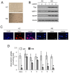
Effects of Pgp ligands on doxorubicin retention in glioblastoma cells. (A) Representative bright field microscope images of glioblastoma cells, cultured as adherent cells (AC) or neurospheres (NS). Magnification: 60 × objective (0.52 numerical aperture); 10 × ocular lens. Bar: 20 µm. The micrographs are representative of patient 2. No significant differences in cell morphology were detected for patient 1 and 3. (B) AC or NS from each patient were lysed and immunoblotted with the indicated antibodies. β-Tubulin level was used as control of equal protein loading. The figure is representative of one out of three experiments with similar results. (C) AC and NS from patient 2 were seeded on sterile glass coverslips, incubated 3 h with 5 μM doxorubicin (doxo), then stained with 4′,6-diamidin-2-phenylindole (DAPI) and analyzed by fluorescence microscopy to detect the intracellular accumulation of the drug. Magnification: 63 × objective (1.4 numerical aperture); 10 × ocular lens. The micrographs are representative of three experiments with similar results. No significant differences were detected for patient 1 and 3. Bar: 5 µm. (D) AC and NS were incubated for 3 h with 5 µM doxorubicin (doxo), in the absence or presence of 1 nM of compounds 16. The intracellular doxorubicin was quantified fluorimetrically, in duplicates. Pooled data of patients 1–3 are presented as means ± SD (n = 3). Vs AC doxo: * p < 0.001; vs. NS doxo: ° p < 0.001.
Figure 3
Figure 3
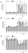
Effects of Pgp ligands on doxorubicin cytotoxicity in glioblastoma cells. Adherent cells (AC) or neurospheres (NS) from glioblastoma samples were grown for 24 h (panel AB) or 48 h (panel C) in fresh medium (ctrl) or in the presence of 1 nM of compounds 16. When indicated, 5 µM doxorubicin (doxo) was co-incubated. (A) The cell culture supernatant was checked spectrophotometrically for the extracellular activity of LDH, in duplicates. Pooled data of patients 1-3 are presented as means ± SD (n = 3). Vs. AC ctrl: * p < 0.001; vs. NS ctrl: ° p < 0.001; vs. NS doxo: # p < 0.001. (B) The activity of caspase 3 was measured fluorimetrically, in duplicates. Data are means ± SD (n = 3). Pooled data of patients 1–3 are presented as means ± SD (n = 3). Vs. AC ctrl: * p < 0.001; vs. NS ctrl: ° p < 0.01; vs. NS doxo: # p < 0.05. (C) Cell viability was measured by a chemiluminescence-based assay, in quadruplicates. Pooled data of patients 1-3 are presented as means ± SD (n = 3). Vs. AC ctrl: * p < 0.001; vs. NS ctrl: ° p < 0.001; vs. NS doxo: # p < 0.005.
Figure 4
Figure 4
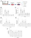
Effects of Pgp ligands on doxorubicin delivery and cytotoxicity in BBB-glioblastoma cells co-cultures. hCMEC/D3 cells were grown for 7 days up to confluence in Transwell inserts; neurospheres (NS) were seeded at day 4 in the lower chamber. After 3 days of co-culture, supernatant in the upper chamber was replaced with fresh medium (ctrl) or with medium containing 5 µM doxorubicin (doxo), in the absence (ctrl) or presence of 1 nM of compounds 16. (A) Fluorimetric quantification of intracellular doxorubicin in NS after 6 h. Pooled data of patients 1-3 are presented as means ± SD (n = 3). Vs. doxo: * p < 0.01. (B) The culture supernatant of NS was checked spectrophotometrically for the extracellular activity of LDH after 24 h. Pooled data of patients 1–3 are presented as means ± SD (n = 3). Vs. untreated cells (ctrl, either “−doxo” or “+doxo”): * p < 0.001. (C) The activity of caspase 3 was measured fluorimetrically in NS lysates after 24 h, in duplicates. Pooled data of patients 1–3 are presented as means ± SD (n = 3). Vs. untreated cells (ctrl, either “−doxo” or “+doxo”): * p < 0.002. (D) Cell viability of NS was measured after 48 h by a chemiluminescence-based assay, in quadruplicates. Pooled data of patients 1–3 are presented as means ± SD (n = 3). Vs. untreated cells (ctrl, either “−doxo” or “+doxo”): * p < 0.02.
Figure 5
Figure 5
Possible dual effects of Pgp ligands on Pgp in BBB and glioblastoma cells. (A) hCMEC/D3 cells were grown for 7 days up to confluence in Transwell inserts, then the supernatant in the upper chamber was replaced with fresh medium (ctrl) or with medium containing 5 µM doxorubicin, in the absence (ctrl) or presence of 1 nM of compounds 16. After 3 h the medium of the lower chamber was removed and added to neuropsheres (NS) for the measure of intracellular doxorubicin uptake, LDH release, caspase-3 activity and cell viability (panels CF). In all these panels, a standard solution of 1 µM doxorubicin (doxo) was used as internal control. (B) Fluorimetric quantification of doxorubicin in the medium of the lower chamber incubated as reported in A. Data are means ± SD (n = 3). Vs ctrl: * p < 0.001. (C) Fluorimetric quantification of intracellular doxorubicin in NS after 6 h, treated with 1 µM doxorubicin (doxo) or with media of the lower chamber containing 5 µM doxorubicin+compounds 16. Pooled data of patients 1–3 are presented as means ± SD (n = 3). Vs. doxo: * p < 0.001. (D) The culture supernatant of NS, treated as in C was checked spectrophotometrically for the extracellular activity of LDH after 24 h. Pooled data of patients 1-3 are presented as means ± SD (n = 3). Vs. untreated cells (ctrl): * p < 0.005; vs. cells treated with 1 µM doxorubicin (doxo): * p < 0.02. (E) The activity of caspase 3 was measured fluorimetrically in NS lysates after 24 h of treatment as in C in duplicates. Pooled data of patients 1–3 are presented as means ± SD (n = 3). Vs. untreated cells (ctrl): * p < 0.02; vs. cells treated with 1 µM doxorubicin (doxo): * p < 0.02. (F) Cell viability of NS, treated as in C, was measured after 48 h by a chemiluminescence-based assay, in quadruplicates. Pooled data of patients 1–3 are presented as means ± SD (n = 3). Vs. untreated cells (ctrl): * p < 0.05; vs. cells treated with 1 µM doxorubicin (doxo): * p < 0.05.

Similar articles

Cited by

References

    1. Ellor S.V., Pagano-Young T.A., Avgeropoulos N.G. Glioblastoma: Background, standard treatment paradigms, and supportive care considerations. J. Law Med. Ethics. 2014;42:171–182. doi: 10.1111/jlme.12133. - DOI - PubMed
    1. Bai R.Y., Staedke V., Riggins G.J. Molecular targeting of GBM: Drug discovery and therapies. Trends Mol. Med. 2011;17:301–312. doi: 10.1016/j.molmed.2011.01.011. - DOI - PMC - PubMed
    1. Beier D., Schulz J.B., Beier C.P. Chemoresistance of glioblastoma cancer stem cells—Much more complex than expected. Mol. Cancer. 2011;10:e128. doi: 10.1186/1476-4598-10-128. - DOI - PMC - PubMed
    1. Corso C.D., Bindra R.S. Success and Failures of Combined Modalities in Glioblastoma Multiforme: Old Problems and New Directions. Semin. Radiat. Oncol. 2016;26:281–298. doi: 10.1016/j.semradonc.2016.06.003. - DOI - PubMed
    1. Salmaggi A., Boiardi A., Gelati M., Russo A., Calatozzolo C., Ciusani E., Sciacca F.L., Ottolina A., Parati E.A., La Porta C., et al. GBM-Derived Tumorospheres Identify a Population of Tumor Stem-Like Cells with Angiogenic Potential and Enhanced Multidrug Resistance Phenotype. Glia. 2006;54:850–860. doi: 10.1002/glia.20414. - DOI - PubMed

Substances