. 2018 Jun 10;19(6):1719.
doi: 10.3390/ijms19061719.

Effect of Different Preconditioning Regimens on the Expression Profile of Murine Adipose-Derived Stromal/Stem Cells

Affiliations

Effect of Different Preconditioning Regimens on the Expression Profile of Murine Adipose-Derived Stromal/Stem Cells

Patrick C Baer et al. Int J Mol Sci. .

Abstract

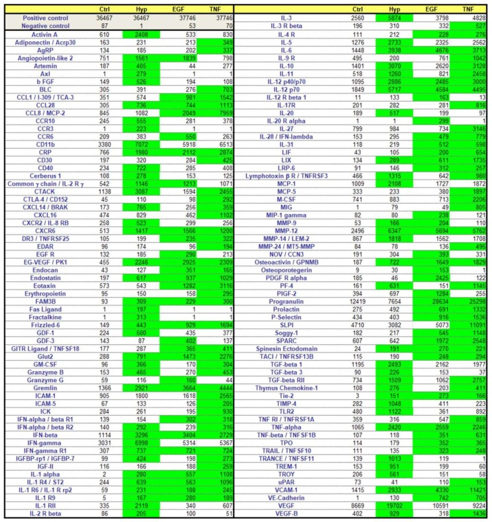
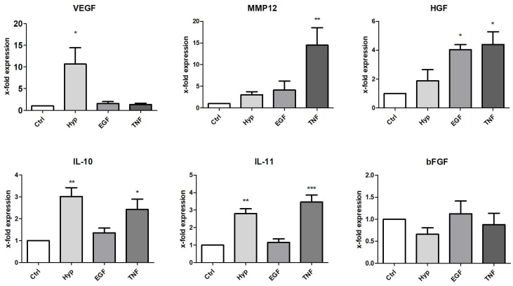
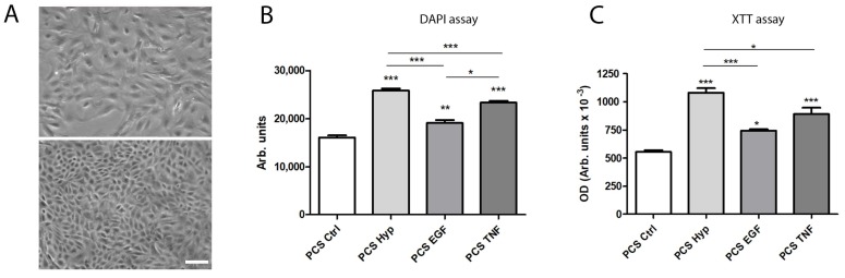
Stem cell-based therapies require cells with a maximum regenerative capacity in order to support regeneration after tissue injury and organ failure. Optimization of this regenerative potential of mesenchymal stromal/stem cells (MSC) or their conditioned medium by in vitro preconditioning regimens are considered to be a promising strategy to improve the release of regenerative factors. In the present study, MSC were isolated from inguinal adipose tissue (mASC) from C57BL/6 mice, cultured, and characterized. Then, mASC were either preconditioned by incubation in a hypoxic environment (0.5% O₂), or in normoxia in the presence of murine epidermal growth factor (EGF) or tumor necrosis factor α (TNFα) for 48 h. Protein expression was measured by a commercially available array. Selected factors were verified by PCR analysis. The expression of 83 out of 308 proteins (26.9%) assayed was found to be increased after preconditioning with TNFα, whereas the expression of 61 (19.8%) and 70 (22.7%) proteins was increased after incubation with EGF or in hypoxia, respectively. Furthermore, we showed the proliferation-promoting effects of the preconditioned culture supernatants on injured epithelial cells in vitro. Our findings indicate that each preconditioning regimen tested induced an individual expression profile with a wide variety of factors, including several growth factors and cytokines, and therefore may enhance the regenerative potential of mASC for cell-based therapies.

Keywords: cytokines; mesenchymal stromal/stem cells; preconditioning; pretreatment; regeneration; secretion; stem cells.


Conflict of interest statement

The authors declare no conflict of interest.

Figures

Figure 1
Figure 1
Cell characterization. (A) Characteristic phase contrast microscopy of murine adipose-derived stromal/stem cells (mASC) cultured in standard cell culture (bar: 100 µm); (B) representative flow cytometric histograms (red) of the expression of characteristic markers. Black histograms show isotype controls.
Figure 2
Figure 2
Cell viability after preconditioning for 48 h. Murine ASC were cultured in 96-well plates and preconditioned for 48 h. The XTT assay was performed and optical density (OD) was measured in a microplate reader at 490 nm vs. 650 nm (arbitrary units, mean ± SD, n = 5). No significant effects of the different pretreatments on the cell viability could be detected.
Figure 3
Figure 3
Color map of increased expressed proteins after the preconditioning regimens. Cells were either cultured under standard conditions (Ctrl) or preconditioned by incubation in a hypoxic environment (Hyp; 0.5% O2) or in the presence of murine epidermal growth factor (EGF) (10 ng/mL) or murine tumor necrosis factor α (TNFα) (10 ng/mL) for 48 h. The expression of 308 proteins was measured in the cell supernatant after preconditioning by a commercially available protein array. The heatmap displays proteins enhanced at least >2-fold versus the control and >150 arbitrary units after pretreatment (green).
Figure 4
Figure 4
Effect of preconditioning on mRNA expression of selected factors. Expression was measured in total RNA from mASC after preconditioning by culture under hypoxia (Hyp, 0.5% O2), or in medium containing EGF (10 ng/mL) or TNFα (10 ng/mL). The expression levels in each experiment were normalized to a housekeeping gene (β-actin) and are expressed relative to the control using the ∆∆CT method. * p < 0.05, ** p < 0.01, *** p < 0.001 versus control, n = 4–6.
Figure 5
Figure 5
Effect of processed culture supernatant (PCS) after preconditioning (by Hyp, EGF, or TNF) or control culture (Ctrl) on the proliferation and viability of epithelial cells. (A) Characteristic phase contrast microscopy of subconfluent (above) and confluent (below) murine renal tubular epithelial cells in culture (bar: 100 µm); (B) DAPI assay: cell proliferation was determined by a fluorometric assay using 4,6-diamino-2-phenylindole (DAPI), measuring the DNA content as an indirect determination of proliferation, after incubation with PCS for 72 h. Fluorescence was measured in a fluorescence reader (355 nm ex/460 nm em), and expressed as arbitrary units (mean ± SD, n = 6); (C) XTT assay: cell viability of mTEC was measured after incubation with PCS for 72 h. The XTT assay was performed and optical density (OD) was measured in a microplate reader at 490 nm vs. 650 nm (arbitrary units, mean ± SD, n = 6). * p < 0.05, ** p < 0.01, *** p < 0.001 versus control and among each preconditioning regimen.

Similar articles

Cited by

References

    1. Caplan A.I. All MSCs are pericytes? Cell Stem Cell. 2008;3:229–230. doi: 10.1016/j.stem.2008.08.008. - DOI - PubMed
    1. Da Silva Meirelles L., Chagastelles P.C., Nardi N.B. Mesenchymal stem cells reside in virtually all post-natal organs and tissues. J. Cell Sci. 2006;119:2204–2213. doi: 10.1242/jcs.02932. - DOI - PubMed
    1. Baer P.C., Geiger H. Adipose-derived mesenchymal stromal/stem cells: Tissue localization, characterization, and heterogeneity. Stem Cells Int. 2012;2012:812693. doi: 10.1155/2012/812693. - DOI - PMC - PubMed
    1. Hsiao S.T.-F., Asgari A., Lokmic Z., Sinclair R., Dusting G.J., Lim S.Y., Dilley R.J. Comparative Analysis of Paracrine Factor Expression in Human Adult Mesenchymal Stem Cells Derived from Bone Marrow, Adipose, and Dermal Tissue. Stem Cells Dev. 2012;21:2189–2203. doi: 10.1089/scd.2011.0674. - DOI - PMC - PubMed
    1. Cruz F.F., Rocco P.R.M. Stem-cell extracellular vesicles and lung repair. Stem Cell Investig. 2017;4 doi: 10.21037/sci.2017.09.02. - DOI - PMC - PubMed

MeSH terms

LinkOut - more resources