. 2018 Jun;41(6):582-590.
doi: 10.14348/molcells.2018.0011. Epub 2018 Jun 12.

Long-Term Priming by Three Small Molecules Is a Promising Strategy for Enhancing Late Endothelial Progenitor Cell Bioactivities

Affiliations

Long-Term Priming by Three Small Molecules Is a Promising Strategy for Enhancing Late Endothelial Progenitor Cell Bioactivities

Yeon-Ju Kim et al. Mol Cells. 2018 Jun.

Abstract

Endothelial progenitor cells (EPCs) and outgrowth endothelial cells (OECs) play a pivotal role in vascular regeneration in ischemic tissues; however, their therapeutic application in clinical settings is limited due to the low quality and quantity of patient-derived circulating EPCs. To solve this problem, we evaluated whether three priming small molecules (tauroursodeoxycholic acid, fucoidan, and oleuropein) could enhance the angiogenic potential of EPCs. Such enhancement would promote the cellular bioactivities and help to develop functionally improved EPC therapeutics for ischemic diseases by accelerating the priming effect of the defined physiological molecules. We found that preconditioning of each of the three small molecules significantly induced the differentiation potential of CD34+ stem cells into EPC lineage cells. Notably, long-term priming of OECs with the three chemical cocktail (OEC-3C) increased the proliferation potential of EPCs via ERK activation. The migration, invasion, and tube-forming capacities were also significantly enhanced in OEC-3Cs compared with unprimed OECs. Further, the cell survival ratio was dramatically increased in OEC-3Cs against H2O2-induced oxidative stress via the augmented expression of Bcl-2, a prosurvival protein. In conclusion, we identified three small molecules for enhancing the bioactivities of ex vivo-expanded OECs for vascular repair. Long-term 3C priming might be a promising methodology for EPC-based therapy against ischemic diseases.

Keywords: cell priming; endothelial progenitor cells; ischemic diseases; vascular repair.


Figures

Fig. 1
Fig. 1. Effects of each factor on the differentiation of CD34+ HSCs into the EPC lineage
(A) Morphology of small and large EPC-CFUs derived from HUCB CD34+ cells. (B–G) After ex vivo expansion of CD34+ HSCs with various concentrations of each factor, the cells were cultured in methylcellulose-containing medium for 14–21 days. Small and large EPC-CFUs were counted. The results are shown as mean ± SEM (*P < 0.05 and **P < 0.01 vs. control).
Fig. 2
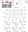
Fig. 2. Characterization of OECs and OEC-3Cs
(A) OEC culture protocol of long-term preconditioning by the 3 chemical cocktail. (B) Scatter plot presenting the log2 (FPKM) values for each gene in OECs (X-axis) versus the OEC-3Cs (Y-axis). (C) Multi-dimensional scale plot representing the biological replicates of OECs, OEC-3Cs, and AD-MSCs. (D) The expression of OEC surface markers (CD34, c-Kit, CXCR4, Tie2, VEGFR2, CD31) was analyzed by flow cytometry. (E–J) Quantification of the flow cytometry results of OEC surface markers. Means ± SEM (n = 4 OEC lines from four separate HUCB samples).
Fig. 3
Fig. 3. Enhanced cell proliferation via ERK signaling in OEC-3Cs
(A–C) Activation of ERK and Akt was evaluated by measuring the phosphorylation levels by Western blotting; relative expression levels were determined by densitometry normalized to β-actin expression (D) Cell proliferation was examined using the BrdU incorporation assay. (E) OECs and OEC-3Cs were exposed to PD98059 (10 μM) for 24 h, and cell proliferation was analyzed. The results are shown as mean ± SEM (*P < 0.05 and **P < 0.01 vs. OEC).
Fig. 4
Fig. 4. Enhanced angiogenic function in OEC-3Cs
(A, B) Cell migration was evaluated by scratch wound-healing assays and migration capacity is displayed as the migration area (%). (C, D) Cell migration and invasion were assessed by Transwell migration and invasion assays. The migration and invasion capacity was determined by the numbers of migrating cells in OECs and OEC-3Cs. (E, F) OECs and OEC-3Cs were seeded into Matrigel-coated wells and the angiogenic function of cells was evaluated in a tube formation assay. Representative images of tube formation (magnification 40×) and quantification of the number of tube branches. Data represent the mean ± SEM of three independent experiments (*P < 0.05 and **P < 0.01 vs. OEC).
Fig. 5
Fig. 5. Cytoprotective effect by the 3 chemical cocktail against H2O2-induced oxidative stress
(A) The apoptosis of OECs was analyzed by flow cytometry using AnnexinV/propidium iodide (PI) staining after exposure to H2O2 (800 μM, 30 min) as follows: live cells (AnnexinV/PI), apoptotic cells (AnnexinV+/PI), dead cells (AnnexinV+/PI+). (B) Protein expression of Bax (pro-apoptotic) and Bcl-2 (pro-survival) was assessed by western blotting. (C, D) Quantification of expression levels of Bax and Bcl-2 relative to β-actin expression. Data represent the mean ± SEM of three independent experiments (**P < 0.01 vs. OEC).

References

    1. Annex B.H. Therapeutic angiogenesis for critical limb ischaemia. Nat Rev Cardiol. 2013;10:387–396. - PubMed
    1. Asahara T., Murohara T., Sullivan A., Silver M., van der Zee R., Li T., Witzenbichler B., Schatteman G., Isner J.M. Isolation of putative progenitor endothelial cells for angiogenesis. Science. 1997;275:964–967. - PubMed
    1. Asahara T., Kawamoto A., Masuda H. Concise review: Circulating endothelial progenitor cells for vascular medicine. Stem Cells. 2011;29:1650–1655. - PubMed
    1. Beck L., Jr, D’Amore P.A. Vascular development: cellular and molecular regulation. FASEB J. 1997;11:365–373. - PubMed
    1. Cai H., Harrison D.G. Endothelial dysfunction in cardiovascular diseases: the role of oxidant stress. Circ Res. 2000;87:840–844. - PubMed