

Dynamic Hyaluronan drives liver endothelial cells towards angiogenesis
- PMID: 29890947
- PMCID: PMC5996548
- DOI: 10.1186/s12885-018-4532-1
Dynamic Hyaluronan drives liver endothelial cells towards angiogenesis
Abstract
Background: Angiogenesis, the formation of new blood vessels from pre-existing vasculature is essential in a number of physiological processes such as embryonic development, wound healing as well as pathological conditions like, tumor growth and metastasis. Hyaluronic acid (HA), a high molecular weight polysaccharide, major component of extracellular matrix is known to associate with malignant phenotypes in melanomas and various other carcinomas. Hyaluronic acid binding protein 1 (HABP1) has been previously reported to trigger enhanced cellular proliferation in human liver cancer cells upon its over-expression. In the present study, we have identified the HA mediated cellular behaviour of liver endothelial cells during angiogenesis.
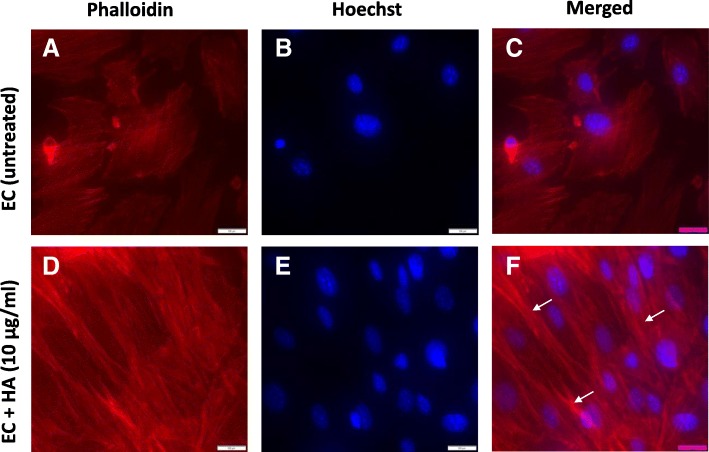
Methods: Endothelial cells have been isolated from perfused liver of mice. Cell proliferation was studied using microwell plates with tetrazole dye. Cell migration was evaluated by measuring endothelial monolayer wound repair as well as through transwell migration assay. Alterations in proteins and mRNA expression were estimated by immunobloting and quantitative real time PCR using Applied Biosystems. The paraformaldehyde fixed endothelial cells were used for immuno- florescence staining and F-actin detection with conjugated antibodies. The images were captured by using Olympus florescence microscope (IX71).
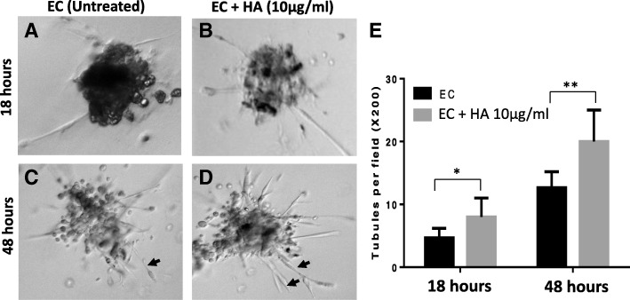
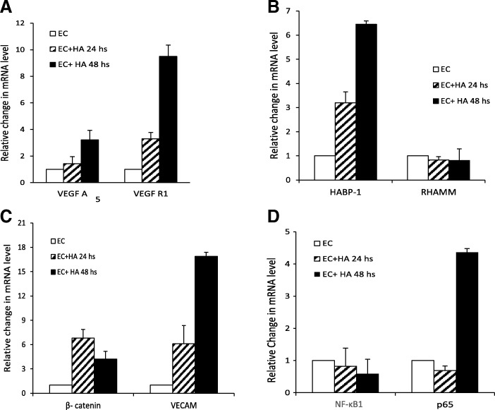
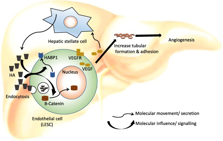
Results: We observed that administration of HA enhanced cell proliferation, adhesion, tubular sprout formation as well as migration of liver endothelial cells (ECs). The effect of HA in the rearrangement of the actins confirmed HA -mediated cytoskeleton re-organization and cell migration. Further, we confirmed enhanced expression of angiogenic factors like VEGF-A and VEGFR1 in endothelial cells upon HA treatment. HA supplementation led to elevated expression of HABP1 in murine endothelial cells. It was interesting to note that, although protein levels of β- catenin remained unaltered, but translocation of this protein from membrane to nucleus was observed upon HA treatment, suggesting its role not only in vessel formation but also its involvement in angiogenesis signalling.
Conclusions: The elucidation of molecular mechanism (s) responsible for HA mediated regulation of endothelial cells and angiogenesis contributes not only to our understanding the mechanism of disease progression but also offer new avenues for therapeutic intervention.
Keywords: Angiogenesis; Hyaluronic acid or Hyaluronan; Liver endothelial cells.
Conflict of interest statement
Ethics approval and consent to participate
Our study (IEC-244/05.05.2017) is carried out as per direction by Institute Ethics committee, All India Institute of Medical Sciences, New Delhi, India.
Consent for publication
Authors certify that no portion of this manuscript has been previously published.
Competing interests
The authors declare that they have no competing interests.
Publisher’s Note
Springer Nature remains neutral with regard to jurisdictional claims in published maps and institutional affiliations.
Figures


Similar articles
-
Oligosaccharides of hyaluronan induce angiogenesis through distinct CD44 and RHAMM-mediated signalling pathways involving Cdc2 and gamma-adducin.Int J Oncol. 2009 Oct;35(4):761-73. doi: 10.3892/ijo_00000389. Int J Oncol. 2009. PMID: 19724912
-
Hyaluronan-mediated angiogenesis in vascular disease: uncovering RHAMM and CD44 receptor signaling pathways.Matrix Biol. 2007 Jan;26(1):58-68. doi: 10.1016/j.matbio.2006.08.261. Epub 2006 Sep 19. Matrix Biol. 2007. PMID: 17055233 Review.
-
Vascular endothelial growth factor-C promotes vasculogenesis, angiogenesis, and collagen constriction in three-dimensional collagen gels.J Vasc Surg. 2005 Apr;41(4):699-707. doi: 10.1016/j.jvs.2005.01.015. J Vasc Surg. 2005. PMID: 15874936
-
Hyaluronan and human endothelial cell behavior.Connect Tissue Res. 2008;49(3):120-3. doi: 10.1080/03008200802148462. Connect Tissue Res. 2008. PMID: 18661325 Review.
-
Hyaluronan oligosaccharides promote excisional wound healing through enhanced angiogenesis.Matrix Biol. 2010 Mar;29(2):107-16. doi: 10.1016/j.matbio.2009.11.002. Epub 2009 Nov 12. Matrix Biol. 2010. PMID: 19913615
Cited by
-
Attenuated Replication-Competent Herpes Simplex Virus Expressing an ECM-Modifying Transgene Hyaluronan Synthase 2 of Naked Mole Rat in Oncolytic Gene Therapy.Microorganisms. 2023 Oct 29;11(11):2657. doi: 10.3390/microorganisms11112657. Microorganisms. 2023. PMID: 38004669 Free PMC article.
-
Identification of crucial genes that induce coronary atherosclerosis through endothelial cell dysfunction in AMI-identifying hub genes by WGCNA.Am J Transl Res. 2022 Nov 15;14(11):8166-8174. eCollection 2022. Am J Transl Res. 2022. PMID: 36505315 Free PMC article.
-
hapln1a+ cells guide coronary growth during heart morphogenesis and regeneration.Nat Commun. 2023 Jun 13;14(1):3505. doi: 10.1038/s41467-023-39323-6. Nat Commun. 2023. PMID: 37311876 Free PMC article.
-
The mechanisms and effects of lactylation modification in different kinds of cancers.Discov Oncol. 2025 Apr 18;16(1):560. doi: 10.1007/s12672-025-02359-9. Discov Oncol. 2025. PMID: 40249419 Free PMC article. Review.
-
Update on the Role of the Endothelial Glycocalyx in Angiogenesis and Vascular Inflammation.Front Cell Dev Biol. 2021 Aug 31;9:734276. doi: 10.3389/fcell.2021.734276. eCollection 2021. Front Cell Dev Biol. 2021. PMID: 34532323 Free PMC article. Review.
References
-
- Rousseau S, Houle F, Kotanides H, Witte L, Waltenberger J, Landry J, Huot J. Vascular endothelial growth factor (VEGF)-driven actin-based motility is mediated by VEGFR2 and requires concerted activation of stress-activated protein kinase 2 (SAPK2/p38) and geldanamycin-sensitive phosphorylation of focal adhesion kinase. J Biol Chem. 2000;275(14):10661–10672. doi: 10.1074/jbc.275.14.10661. - DOI - PubMed
MeSH terms
Substances
Grants and funding
LinkOut - more resources
Full Text Sources
Other Literature Sources
Molecular Biology Databases
Research Materials
Miscellaneous
