. 2018 Jun 11;6(1):51.
doi: 10.1186/s40425-018-0371-5.

Dexamethasone-induced immunosuppression: mechanisms and implications for immunotherapy

Affiliations

Dexamethasone-induced immunosuppression: mechanisms and implications for immunotherapy

Amber J Giles et al. J Immunother Cancer. .

Abstract

Background: Corticosteroids are routinely utilized to alleviate edema in patients with intracranial lesions and are first-line agents to combat immune-related adverse events (irAEs) that arise with immune checkpoint blockade treatment. However, it is not known if or when corticosteroids can be administered without abrogating the efforts of immunotherapy. The purpose of this study was to evaluate the impact of dexamethasone on lymphocyte activation and proliferation during checkpoint blockade to provide guidance for corticosteroid use while immunotherapy is being implemented as a cancer treatment.

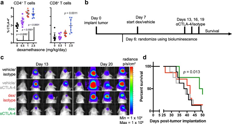
Methods: Lymphocyte proliferation, differentiation, and cytokine production were evaluated during dexamethasone exposure. Human T cells were stimulated through CD3 ligation and co-stimulated either directly by CD28 ligation or by providing CD80, a shared ligand for CD28 and CTLA-4. CTLA-4 signaling was inhibited by antibody blockade using ipilimumab which has been approved for the treatment of several solid tumors. The in vivo effects of dexamethasone during checkpoint blockade were evaluated using the GL261 syngeneic mouse intracranial model, and immune populations were profiled by flow cytometry.

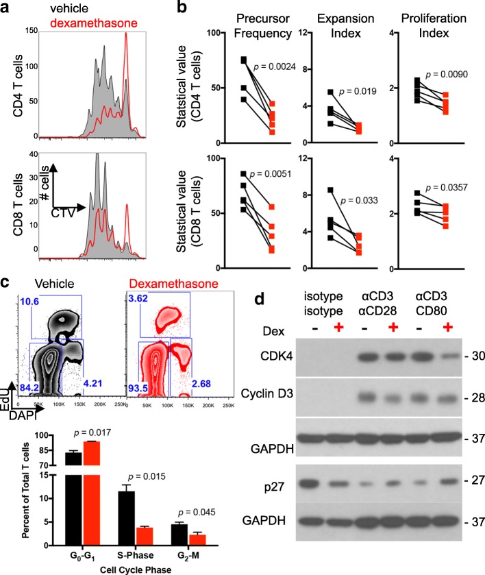
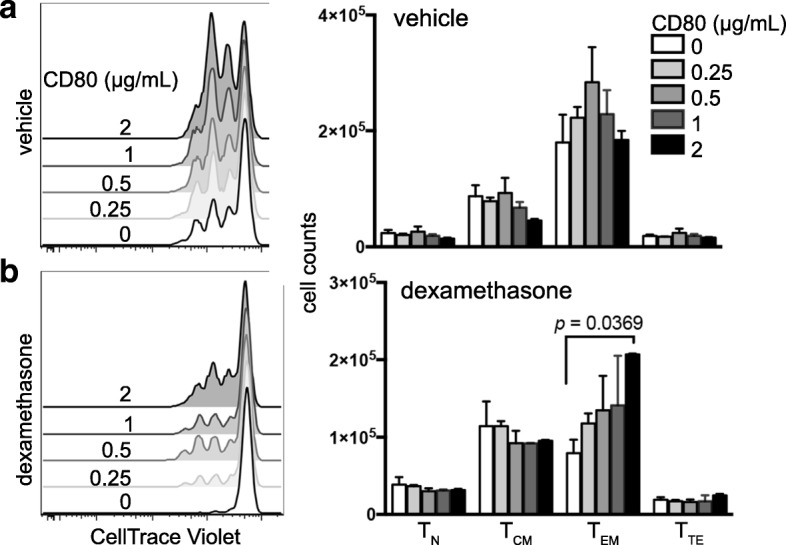
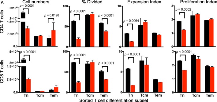
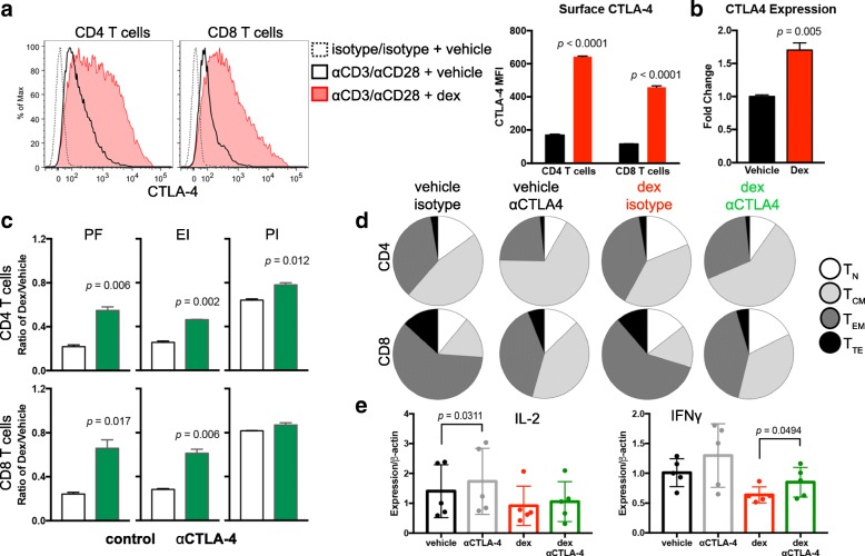
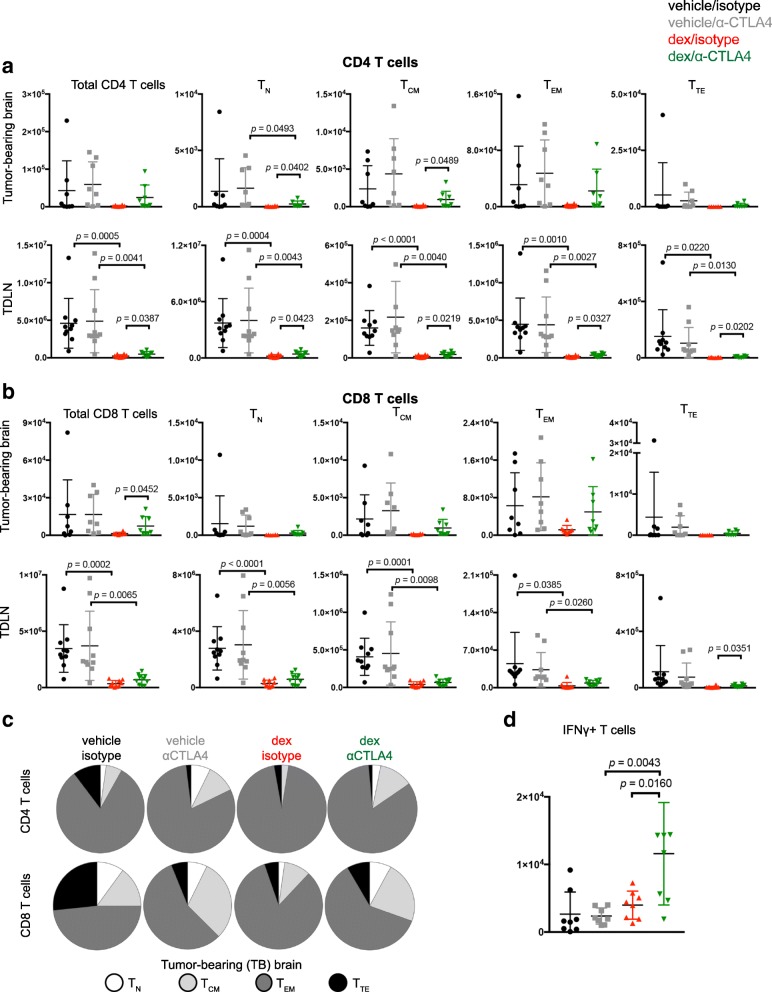
Results: Dexamethasone upregulated CTLA-4 mRNA and protein in CD4 and CD8 T cells and blocked CD28-mediated cell cycle entry and differentiation. Naïve T cells were most sensitive, leading to a decrease of the development of more differentiated subsets. Resistance to dexamethasone was conferred by blocking CTLA-4 or providing strong CD28 co-stimulation prior to dexamethasone exposure. CTLA-4 blockade increased IFNγ expression, but not IL-2, in stimulated human peripheral blood T cells exposed to dexamethasone. Finally, we found that CTLA-4 blockade partially rescued T cell numbers in mice bearing intracranial gliomas. CTLA-4 blockade was associated with increased IFNγ-producing tumor-infiltrating T cells and extended survival of dexamethasone-treated mice.

Conclusions: Dexamethasone-mediated T cell suppression diminishes naïve T cell proliferation and differentiation by attenuating the CD28 co-stimulatory pathway. However, CTLA-4, but not PD-1 blockade can partially prevent some of the inhibitory effects of dexamethasone on the immune response.

Keywords: Checkpoint blockade; Corticosteroids; Dexamethasone; Glioma; Immunotherapy.


Conflict of interest statement

Ethics approval and consent to participate

Not applicable; not a clinical trial.

Competing interests

The authors declare that they have no competing interests.

Publisher’s Note

Springer Nature remains neutral with regard to jurisdictional claims in published maps and institutional affiliations.

Figures

Fig. 1
Fig. 1
T cell proliferation is impaired by dexamethasone. Healthy donor T cells were cultured for four days with αCD3/CD80 microbeads in the presence of vehicle or dexamethasone. a Representative flow cytometry plots of CellTrace violet dilution. Plots were derived from gated CD4 (top row) or CD8 (bottom row) T cells. b Negatively-selected healthy donor T cells were stained and proliferation analyses determined by flow cytometry following four days of culture under the indicated conditions. Precursor Frequency, Expansion Index, and Proliferation Index are shown. Each symbol is the average of duplicate wells, and each paired symbol represents a different donor (n = 5 donors). Statistical significance was determined with a paired two-tailed T test. c Cell cycle analysis was performed on healthy donor T cells cultured with vehicle or dexamethasone and stimulated with αCD3/CD80 microbeads. EdU uptake and DNA content were used to identify G0/G1, S, and G2/M phases. Representative flow images (top) and quantification of duplicate wells are shown (bottom) from two independent experiments. d Lysates from healthy donor T cells incubated with the indicated microbeads and vehicle or dexamethasone were probed for the indicated proteins. GAPDH was used as a loading control and is shown for each individual blot. Data are representative of three independent experiments
Fig. 2
Fig. 2
Increased co-stimulation ameliorates the inhibitory effects of dexamethasone. Negatively-selected healthy donor T cells were cultured with 5 μg/mL αCD3 and increasing concentrations of CD80 in the presence of vehicle or dexamethasone. a-b CD4 T cells cultured with vehicle (a) or dexamethasone (b). Flow cytometry plots showing proliferation of cells cultured with the indicated concentration of CD80 (left) and total numbers of naïve (TN), central memory (TCM), effector memory (TEM), and terminal effector (TTE) T cells following four days of culture (right) are shown. Differentiation subsets were assessed by CD45RO and CCR7 staining. Each condition was plated in duplicate, and data are representative of three independent experiments. Data were analyzed with an unpaired, two-tailed T Test
Fig. 3
Fig. 3
Naïve and effector memory T cells show sensitivity to dexamethasone. a Healthy donor T cells were sorted into TN, TCM, and TEM subsets by flow cytometry. Sorted subsets were cultured with αCD3/CD80 microbeads in the presence of dexamethasone (red) or vehicle control (black). Total cell numbers, Precursor Frequency, Expansion Index, and Proliferation Index of CD4 T cells (top) and CD8 T cells (bottom) are shown. All samples were plated in duplicate and analyzed with a paired, two-tailed T test. Data are representative of three independent experiments
Fig. 4
Fig. 4
CTLA-4 blockade partially restores T cell proliferation in the presence of dexamethasone. a Flow cytometry analysis of CTLA-4 surface expression on CD4 (left) or CD8 (right) T cells stimulated with αCD3/αCD28 microbeads. Unstimulated (dashed line), stimulated in presence of vehicle (solid line), and stimulated in presence of dexamethasone (filled red line) are shown (left) and median fluorescence intensity (MFI) of CTLA-4-expressing T cells is quantified (right). Data are representative of four independent experiments. b Expression of CTLA-4 by qPCR of T cells stimulated in the presence of vehicle or dexamethasone. Data are representative of four independent experiments. c Healthy donor T cells stimulated for four days in the presence of vehicle or dexamethasone and with or without ipilimumab F(ab’)2 antibody. Proliferation analysis of CD4 T cells (top) and CD8 T cells (bottom) was performed, and the ratio of cells stimulated with dexamethasone relative to vehicle control are shown for Precursor Frequency (PF), Expansion Index (EI), and Proliferation Index (PI). All samples were plated in duplicate and the ratios were analyzed with an unpaired, two-tailed T test. Data are representative of 7 healthy donors. d Cells were cultured as in (c). The number of T cells in each differentiation group were quantified by flow cytometry and analyzed by SPICE. e Expression of the indicated cytokines was determined by qPCR. Five healthy donors were assayed for each condition. Each data point represents and average of triplicate wells. Data were analysed with a paired, two-tailed T test
Fig. 5
Fig. 5
CTLA-4 blockade enhances survival of dexamethasone-treated mice. a CTLA-4 was measured on circulating CD4 (left) and CD8 (right) T cells 1 h following oral gavage of vehicle or the indicated concentration of dexamethasone. Each cohort contained eight mice with intracranial GL261 tumors. Vehicle and dexamethasone-treated cohorts were statistically analyzed with an unpaired two-tailed student’s T-test. b Schema of treatment cohorts for (c-d). GL261 ffluc-mCherry glioma cells were orthotopically implanted into C57BL/6 mice one week before treatment initiation. Luminescence readings were acquired 6 days following tumor implantation and weekly thereafter. Mice were treated with vehicle or dexamethasone as indicated. CTLA-4 blocking antibody or isotype control were administered on days 13, 16, and 19 following tumor implantation. c Luminescence of tumor-bearing mice at days 13 and 20 following tumor implantation. d Kaplan Meier survival curves of mice receiving the indicated treatments. n = 7 to 8 mice per cohort. Data are representative of two independent experiments
Fig. 6
Fig. 6
CTLA-4 blockade rescues lymphocyte defects induced by dexamethasone. GL261 ffluc-mCherry tumor-bearing mice were randomized into the indicated cohorts. Vehicle or dexamethasone treatment was initiated on day 7, and isotype or CTLA-4 blocking antibody were administered on days 13, 16, and 19 following tumor implantation. Mice were euthanized on day 23 and tissues were harvested for flow cytometry analysis. a-b CD4 (a) and CD8 (b) T cells were quantified along with the indicated differentiation subsets using CD44 and CD62L expression. Brains (n = 8) and cervical lymph nodes (n = 10) were collected. Data are analyzed using a unpaired students T test. c The relative contribution of each differentiation subset is shown for CD4 (top) and CD8 (bottom) TILs. d The total number of IFNγ-producing T cells were quantified from the tumor-bearing hemispheres of mice from the indicated cohorts. Data are analyzed using an unpaired students T test. N = 8 mice/group

Similar articles

Cited by

References

    1. Hoos A. Development of immuno-oncology drugs - from CTLA4 to PD1 to the next generations. Nat Rev Drug Discov. 2016;15(4):235–247. doi: 10.1038/nrd.2015.35. - DOI - PubMed
    1. Farber SH, Tsvankin V, Narloch JL, Kim GJ, Salama AK, Vlahovic G, et al. Embracing rejection: immunologic trends in brain metastasis. Oncoimmunology. 2016;5(7):e1172153. doi: 10.1080/2162402X.2016.1172153. - DOI - PMC - PubMed
    1. Batich KA, Reap EA, Archer GE, Sanchez-Perez L, Nair SK, Schmittling RJ, et al. Long-term survival in glioblastoma with cytomegalovirus pp65-targeted vaccination. Clin Cancer Res. 2017;23(8):1898–1909. doi: 10.1158/1078-0432.CCR-16-2057. - DOI - PMC - PubMed
    1. Reardon DA, Gokhale PC, Klein SR, Ligon KL, Rodig SJ, Ramkissoon SH, et al. Glioblastoma eradication following immune checkpoint blockade in an Orthotopic. Immunocompetent Model Cancer Immunol Res. 2016;4(2):124–135. doi: 10.1158/2326-6066.CIR-15-0151. - DOI - PubMed
    1. Fecci PE, Ochiai H, Mitchell DA, Grossi PM, Sweeney AE, Archer GE, et al. Systemic CTLA-4 blockade ameliorates glioma-induced changes to the CD4+ T cell compartment without affecting regulatory T-cell function. Clin Cancer Res. 2007;13(7):2158–2167. doi: 10.1158/1078-0432.CCR-06-2070. - DOI - PubMed

Publication types