. 2018 Jun 11;6(1):50.
doi: 10.1186/s40478-018-0552-y.

Sequential stages and distribution patterns of aging-related tau astrogliopathy (ARTAG) in the human brain

Affiliations

Sequential stages and distribution patterns of aging-related tau astrogliopathy (ARTAG) in the human brain

Gabor G Kovacs et al. Acta Neuropathol Commun. .

Abstract

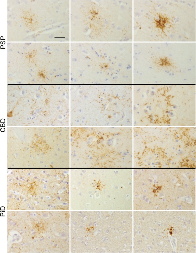
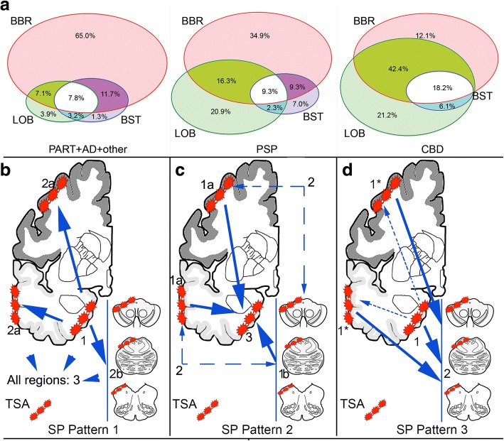
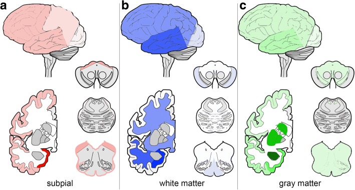
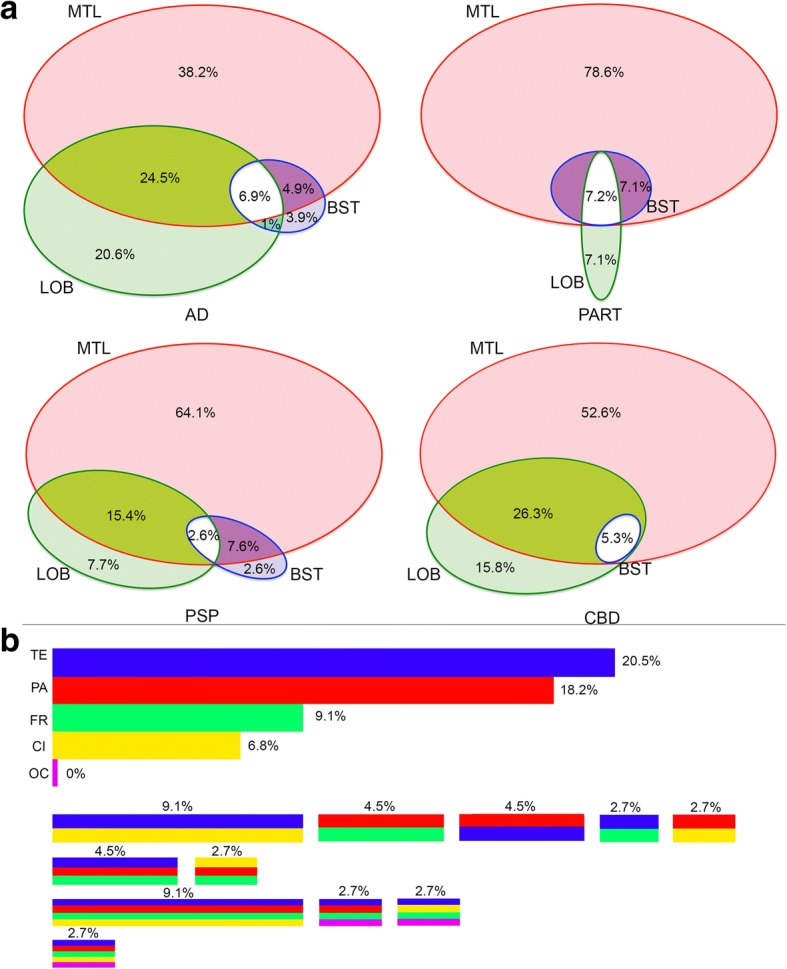
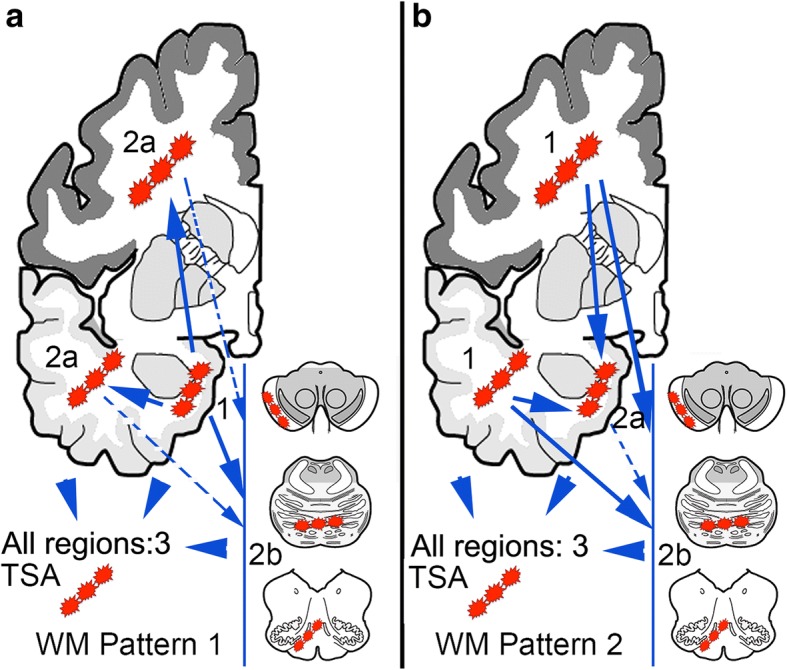
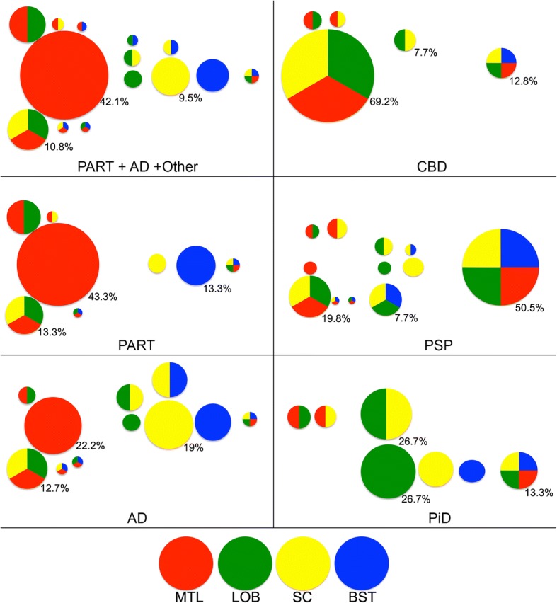
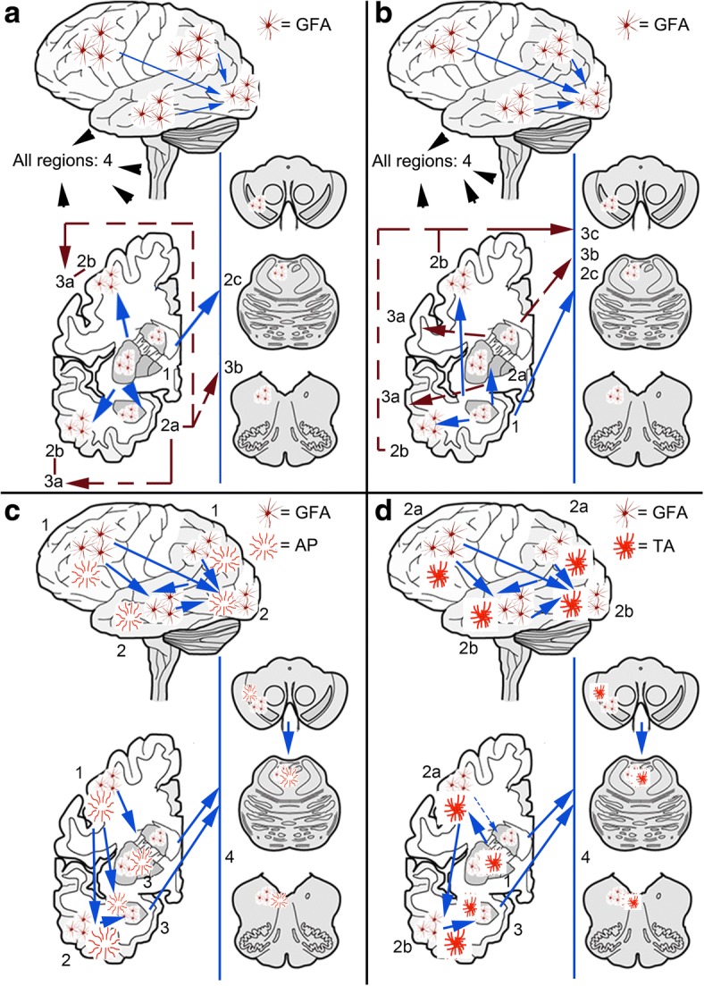
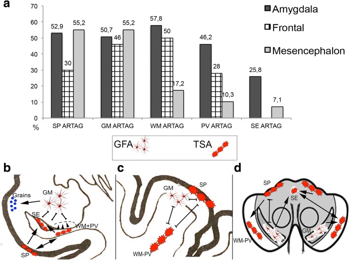
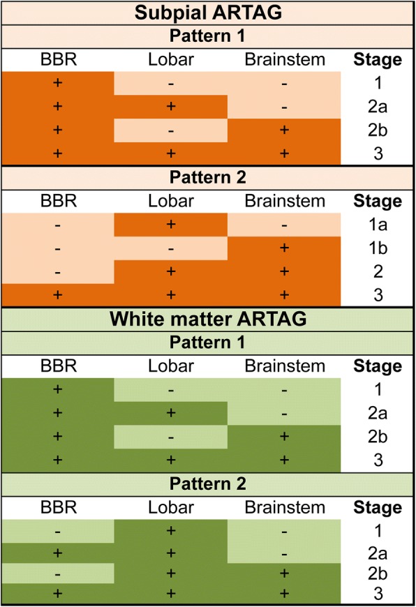
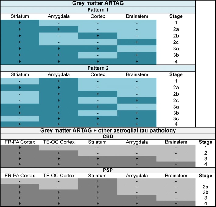
Aging-related tau astrogliopathy (ARTAG) describes tau pathology in astrocytes in different locations and anatomical regions. In the present study we addressed the question of whether sequential distribution patterns can be recognized for ARTAG or astroglial tau pathologies in both primary FTLD-tauopathies and non-FTLD-tauopathy cases. By evaluating 687 postmortem brains with diverse disorders we identified ARTAG in 455. We evaluated frequencies and hierarchical clustering of anatomical involvement and used conditional probability and logistic regression to model the sequential distribution of ARTAG and astroglial tau pathologies across different brain regions. For subpial and white matter ARTAG we recognize three and two patterns, respectively, each with three stages initiated or ending in the amygdala. Subependymal ARTAG does not show a clear sequential pattern. For grey matter (GM) ARTAG we recognize four stages including a striatal pathway of spreading towards the cortex and/or amygdala, and the brainstem, and an amygdala pathway, which precedes the involvement of the striatum and/or cortex and proceeds towards the brainstem. GM ARTAG and astrocytic plaque pathology in corticobasal degeneration follows a predominantly frontal-parietal cortical to temporal-occipital cortical, to subcortical, to brainstem pathway (four stages). GM ARTAG and tufted astrocyte pathology in progressive supranuclear palsy shows a striatum to frontal-parietal cortical to temporal to occipital, to amygdala, and to brainstem sequence (four stages). In Pick's disease cases with astroglial tau pathology an overlapping pattern with PSP can be appreciated. We conclude that tau-astrogliopathy type-specific sequential patterns cannot be simplified as neuron-based staging systems. The proposed cytopathological and hierarchical stages provide a conceptual approach to identify the initial steps of the pathogenesis of tau pathologies in ARTAG and primary FTLD-tauopathies.

Keywords: ARTAG; Aging-related tau astrogliopathy; Astrocytic plaque; Brain barrier; Hierarchical involvement; Ramified astrocyte; Spreading; Tau; Tauopathy; Tufted astrocyte.


Conflict of interest statement

Ethics approval and consent to participate

Informed consent was obtained from next of kin in accordance with institutional review board guidelines of the University of Pennsylvania.

Competing interests

The authors declare that they have no competing interests.

Publisher’s Note

Springer Nature remains neutral with regard to jurisdictional claims in published maps and institutional affiliations.

Figures

Fig. 1
Fig. 1
Tau immunoreactive astrocytes in progressive supranuclear palsy (PSP), corticobasal degeneration (CBD), and Pick’s disease (PiD). Note the variety of morphologies where fine granular deposits evolve into more coarse ones and then typical tufted astrocytes (PSP), astrocytic plaques (CBD), and ramified astrocytes (PiD) reminiscent of a maturation process (from left to right) of tau immunoreactive deposits. Bar represents 25 μm for all images
Fig. 2
Fig. 2
Frequencies and distribution patterns of subpial ARTAG. Frequency of subpial ARTAG in different regions (basal brain regions, BBR; lobar regions, LOB; and brainstem regions, BST) in a pooled cohort of non-FTLD-tauopathies (PART+AD+other), PSP, and CBD (a). Note the differences in concomitant involvement of regions. The sequential stages of subpial (SP) ARTAG in the pooled cohort of non-FTLD-tauopathies comprise pattern 1 (b) when basal brain regions show subpial ARTAG first (stage 1) followed by a bidirectional sequence rostrally (lobar) and caudally (brainstem), which two are affected rarely separately (stages 2a or b) and more frequently together (stage 3); pattern 2 (c) when subpial ARTAG in lobar regions or in brainstem appear first (stage 1a or b; two-headed dashed arrows indicate that we do not know which precedes the other); when affected together is stage 2 and finally when additionally basal brain regions are involved is stage 3; and pattern 3 (d) as exemplified by CBD, where subpial tau immunoreactivity of astrocytic feet is the predominant pathology independently of subpial ARTAG in basal brain regions (together representing stage 1) and both are followed by the involvement of the brainstem, representing stage 2. The pathogenesis of subpial astrocyte feet tau immunoreactivity in CBD is most likely different from subpial lobar ARTAG (thus indicated with an asterisk), therefore this sequence could be termed as “masked” bidirectional. This means that the typical subpial TSAs in CBD follow the subpial ARTAG in the basal brain regions (indicated by dashed arrows) are masked by the predominant end-feet tau immunoreactivity
Fig. 3
Fig. 3
Heatmap of severity scores of subpial (a), white matter (b) and grey matter (c) ARTAG in the cohort of non-FTLD tauopathies. The more dark colours reflect higher severity scores
Fig. 4
Fig. 4
Frequency of white matter ARTAG in three major regions (a; medial temporal lobe, MTL; lobar regions, LOB; and brainstem regions, BST) in AD, PART, PSP, and CBD and its combinations in five lobar areas (b) in AD. Note the differences in concomitant involvement of regions. TE: temporal, PA: parietal, FR: frontal, CI: cingular, OC: occipital
Fig. 5
Fig. 5
Sequential distribution patterns of white matter ARTAG in the pooled cohort of non-FTLD-tauopathies. Pattern 1 (a) is characterized by the appearance of white matter ARTAG in basal brain regions (stage 1) followed by lobar regions (stage 2a) or eventually brainstem (stage 2b) before involving all regions (stage 3). In Pattern 2 (b) lobar involvement (stage 1) is followed by the involvement of the basal brain regions or the brainstem (stages 2 a or b, respectively), before involving all regions (stage 3)
Fig. 6
Fig. 6
Frequency of combinations of grey matter ARTAG in different regions (medial temporal lobe, MTL; lobar regions, LOB; subcortical, SC; and brainstem regions, BST) in the pooled cohort of PART, AD and other non-FTLD tauopathies, AD, PART, PSP, Pick disease, and CBD. Note the differences and overlaps in concomitant involvement of regions. Only the three highest percentage values are shown in lower right corners for better overview. The size of the bubbles represent their frequency
Fig. 7
Fig. 7
Sequential distribution patterns of astroglial tau pathology in the grey matter. In non-FTLD-tauopathies Patterns 1 and 2 are recognized. The distribution of grey matter ARTAG (granular-fuzzy astrocytes, GFA) shows Pattern 1 (a) characterized by the early involvement of the striatum (stage 1) followed by the amygdala (stage 2a), or cortex (here occipital is the latest to be involved) (stage 2b), or the brainstem (stage 2c); then a further region (striatum + amygdala + cortex, stage 3a; or striatum + amygdala + brainstem, stage 3b) followed by the involvement of all regions (stage 4). In pattern 2 (b) the amygdala (stage 1) precedes the involvement of the striatum (stage 2a) or the cortex (stage 2b), or the brainstem (stage 2c); then a further region (striatum + amygdala + cortex, stage 3a; or striatum + amygdala + brainstem, stage 3b; or amygdala + cortex + brainstem, stage 3c) followed by the involvement of all regions (stage 4). In CBD (c) the distribution of astrocytic plaques (AP) and grey matter ARTAG begins in the frontal (including premotor) and parietal cortex (stage 1) followed by temporal and occipital cortex (stage 2), paralelly moving into subcortical areas including either or both the striatum and the amygdala (stage 3) followed by the brainstem (stage 4) including the substantia nigra followed by pons and medulla oblongata. Regarding tufted astrocytes (TA) and grey matter ARTAG in PSP (d), a striatum (stage 1) to cortical (frontal-parietal to temporal to occipital) areas (stage 2 and b, respectively) to amygdala (stage 3) and to brainstem (stage 4), including the substantia nigra followed by pons and medulla oblongata, sequence can be recognized
Fig. 8
Fig. 8
Frequency (a) and sequential distribution patterns of different ARTAG types in the amygdala region (b), frontal cortex (c) and mesencephalon (d) in pooled cases where any ARTAG type was seen. Arrows and small arrowheads indicate the proposed direction of the sequential distribution, two-headed arrows indicate concomitant, and lines the independent presence of different ARTAG types (SP: subpial, GM: grey matter, WM: white matter, PV: perivascular, SE: subependymal)
Fig. 9
Fig. 9
Staging scheme for subpial and white matter ARTAG
Fig. 10
Fig. 10
Staging scheme for grey matter ARTAG and for all astroglial tau pathologies in corticobasal degeneration (CBD) and progressive supranuclear palsy (PSP). FR: frontal, PA: parietal, TE: temporal, OC: occipital cortex

Similar articles

Cited by

References

    1. Abbott NJ, Pizzo ME, Preston JE, Janigro D, Thorne RG. The role of brain barriers in fluid movement in the CNS: is there a ‘glymphatic’ system? Acta Neuropathol. 2018;135:387–407. doi: 10.1007/s00401-018-1812-4. - DOI - PubMed
    1. Arnold SE, Toledo JB, Appleby DH, Xie SX, Wang LS, Baek Y, Wolk DA, Lee EB, Miller BL, al LVM. Comparative survey of the topographical distribution of signature molecular lesions in major neurodegenerative diseases. J Comp Neurol. 2013;521:4339–4355. doi: 10.1002/cne.23430. - DOI - PMC - PubMed
    1. Bancher C, Brunner C, Lassmann H, Budka H, Jellinger K, Wiche G, Seitelberger F, Grundke-Iqbal I, Iqbal K, Wisniewski HM. Accumulation of abnormally phosphorylated tau precedes the formation of neurofibrillary tangles in Alzheimer's disease. Brain Res. 1989;477:90–99. doi: 10.1016/0006-8993(89)91396-6. - DOI - PubMed
    1. Botez G, Probst A, Ipsen S, Tolnay M. Astrocytes expressing hyperphosphorylated tau protein without glial fibrillary tangles in argyrophilic grain disease. Acta Neuropathol. 1999;98:251–256. doi: 10.1007/s004010051077. - DOI - PubMed
    1. Braak H, Braak E. Neuropathological stageing of Alzheimer-related changes. Acta Neuropathol. 1991;82:239–259. doi: 10.1007/BF00308809. - DOI - PubMed

Publication types

MeSH terms