. 2018 Jun 11;18(1):178.
doi: 10.1186/s12906-018-2241-6.

Anti-cancer effects of Kaempferia parviflora on ovarian cancer SKOV3 cells

Affiliations

Anti-cancer effects of Kaempferia parviflora on ovarian cancer SKOV3 cells

Suthasinee Paramee et al. BMC Complement Altern Med. .

Abstract

Background: Kaempferia parviflora (KP) is an herb found in the north of Thailand and used as a folk medicine for improving vitality. Current reports have shown the anti-cancer activities of KP. However, the anti-cancer effects of KP on highly aggressive ovarian cancer have not been investigated. Therefore, we determined the effects of KP on cell proliferation, migration, and cell death in SKOV3 cells.

Methods: Ovarian cancer cell line, SKOV3 was used to investigate the anti-cancer effect of KP extract. Cell viability, cell proliferation, MMP activity, cell migration, and invasion were measured by MTT assay, cell counting, gelatin zymography, wound healing assay, and Transwell migration and invasion assays, respectively. Cell death was determined by trypan blue exclusion test, AnnexinV/PI with flow cytometry, and nuclear staining. The level of ERK and AKT phosphorylation, and caspase-3, caspase-7, caspase-9 was investigated by western blot analysis.

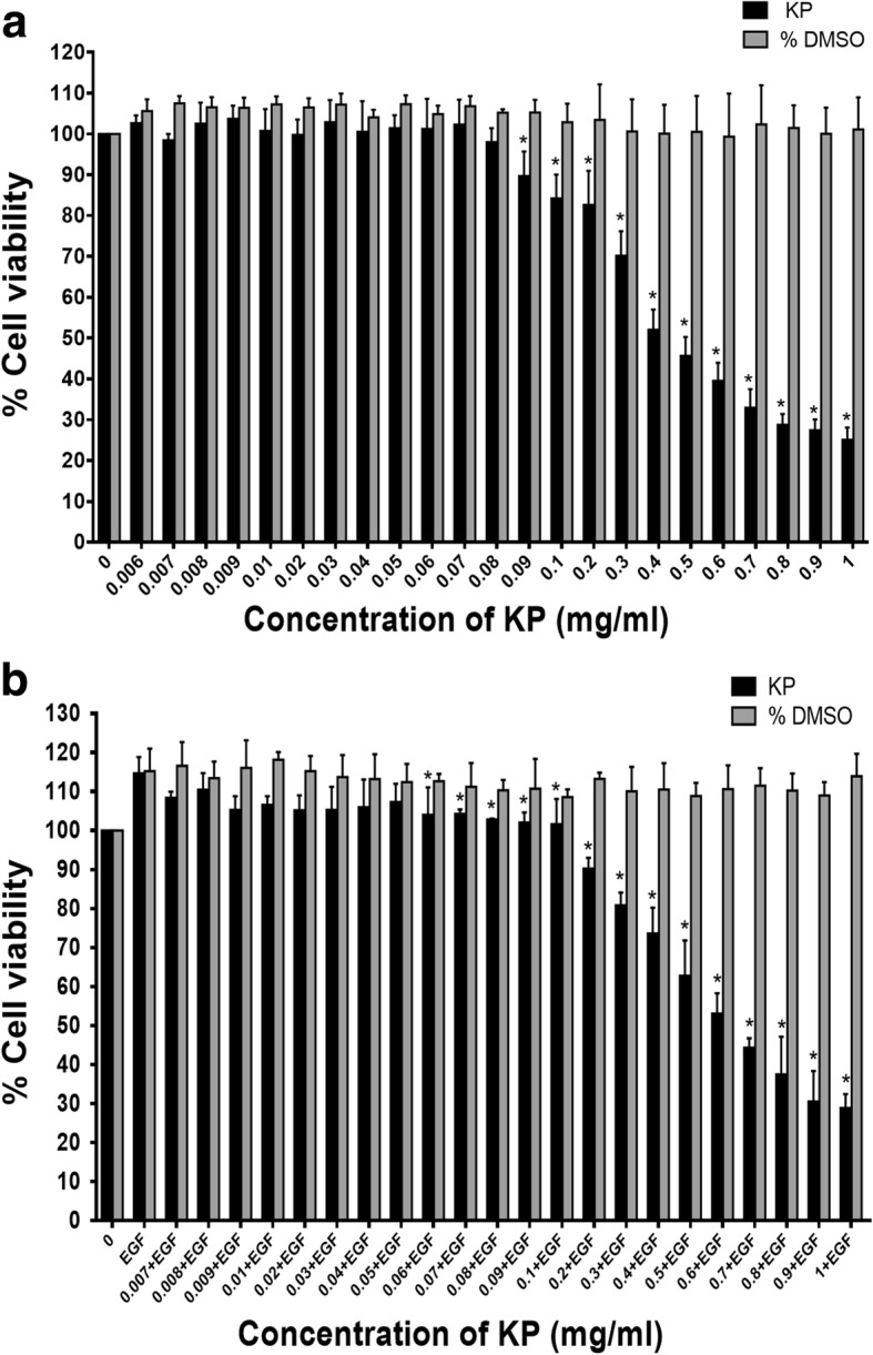
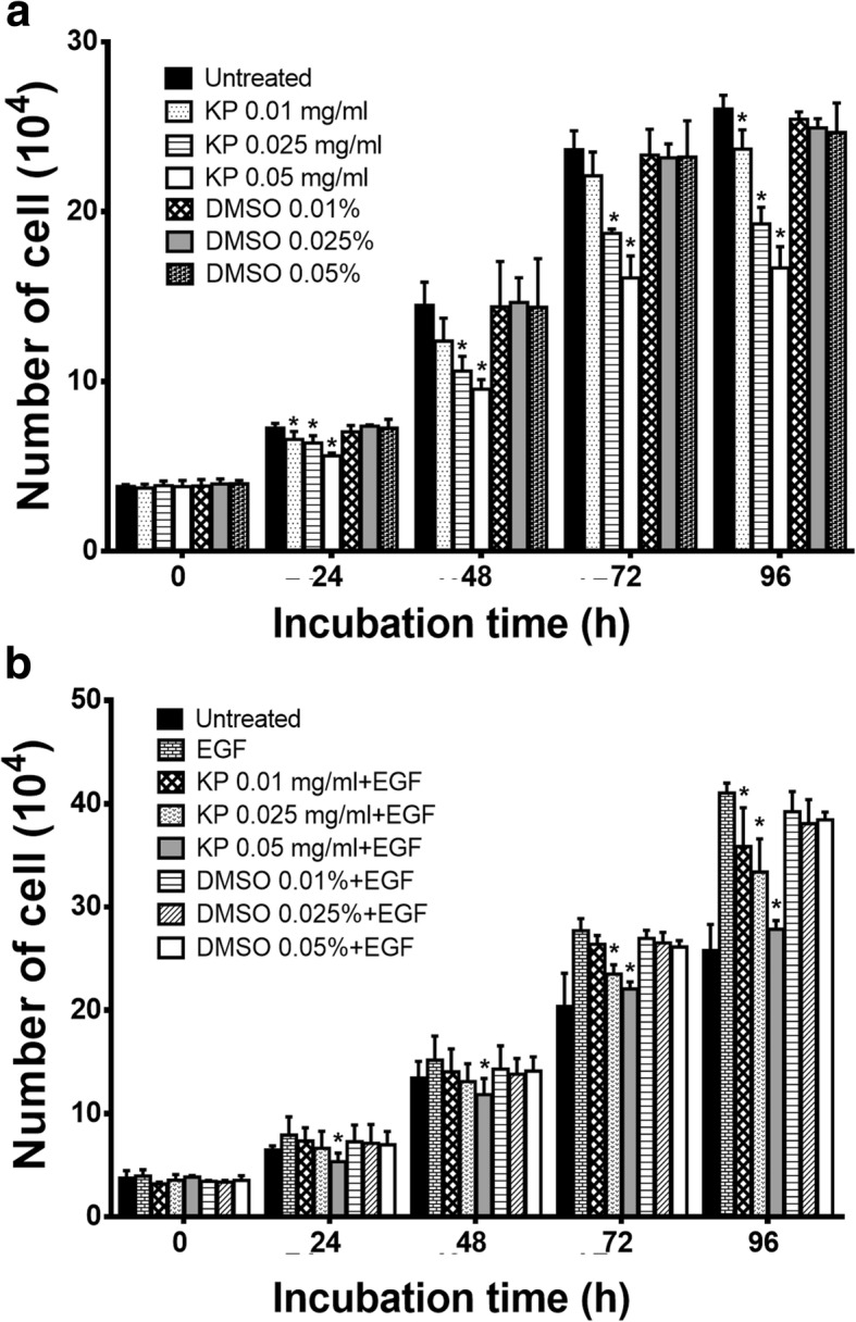
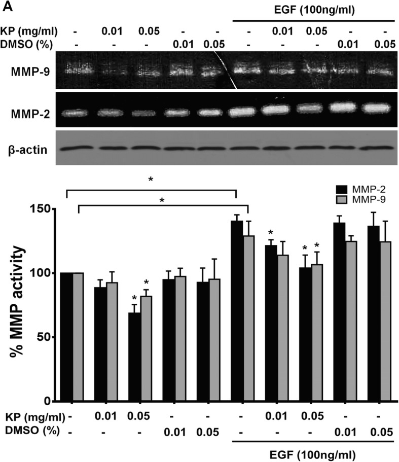
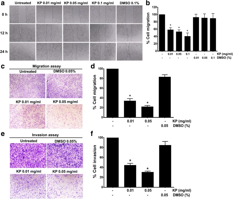
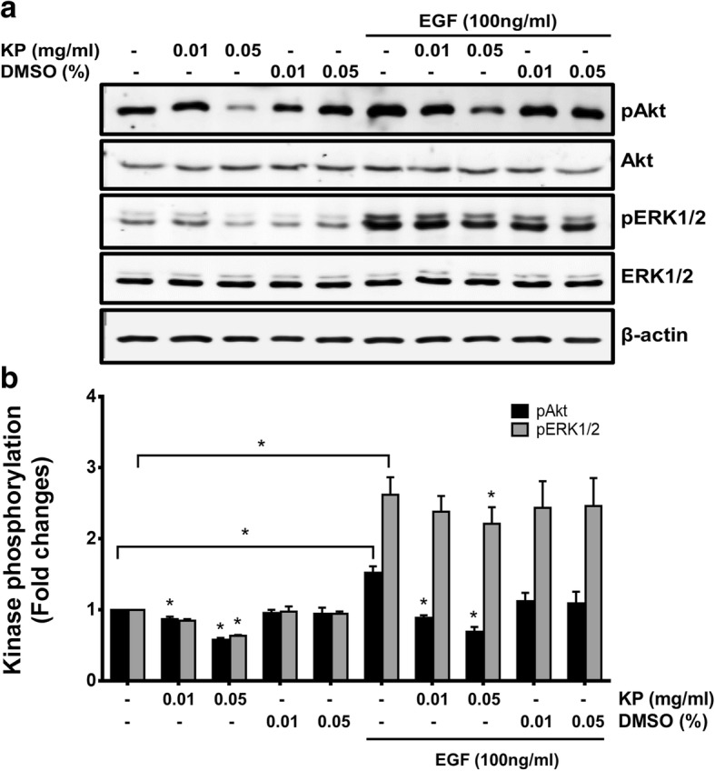
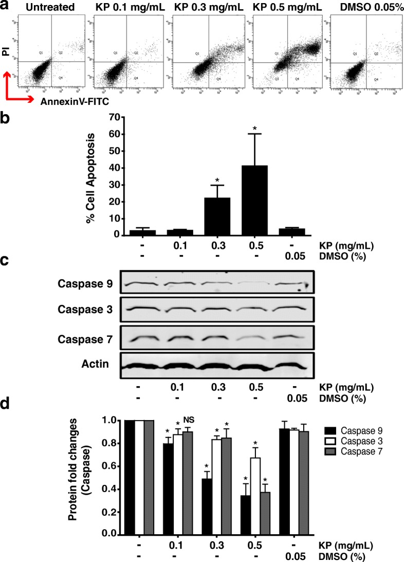
Results: KP extract was cytotoxic to SKOV3 cells when the concentration was increased, and this effect could still be observed even though EGF was present. Besides, the cell doubling time was significantly prolonged in the cells treated with KP. Moreover, KP strongly suppressed cell proliferation, cell migration and invasion. These consequences may be associated with the ability of KP in inhibiting the activity of MMP-2 and MMP-9 assayed by gelatin zymography. Moreover, KP at high concentrations could induce SKOV3 cell apoptosis demonstrated by AnnexinV/PI staining and flow cytometry. Consistently, nuclear labelling of cells treated with KP extract showed DNA fragmentation and deformity. The induction of caspase-3, caspase-7, and caspase-9 indicates that KP induces cell death through the intrinsic apoptotic pathway. The antitumor activities of KP might be regulated through PI3K/AKT and MAPK pathways since the phosphorylation of AKT and ERK1/2 was reduced.

Conclusions: The inhibitory effects of KP in cell proliferation, cell migration and invasion together with apoptotic cell death induction in SKOV3 cells suggest that KP has a potential to be a new candidate for ovarian cancer chemotherapeutic agent.

Keywords: Anti-cancer activity; Kaempferia parviflora; Ovarian cancer; Thai black ginger.


Conflict of interest statement

Ethics approval and consent to participate

Not applicable.

Competing interests

The authors declare that they have no competing interests.

Publisher’s Note

Springer Nature remains neutral with regard to jurisdictional claims in published maps and institutional affiliations.

Figures

Fig. 1
Fig. 1
The cytotoxicity effect of different concentrations of KP ethanol extract on SKOV3 cells without EGF (a) and with 100 ng/mL EGF (b). All data were from 3 independent experiments and reported as means ± SD of each quadruplicate *P < 0.05 compared to the control (untreated and EGF)
Fig. 2
Fig. 2
The number of SKOV3 cells treated with non-toxic concentrations of KP (0.01, 0.025, 0.05 mg/mL) without EGF (a) and with 100 ng/mL EGF (b) at 24, 48, 72 and 96 h. Data are expressed as mean ± SD (n = 3). *P < 0.05 as compared to untreated (a) or EGF (b)
Fig. 3
Fig. 3
The effect of KP ethanol extract on MMP-9 and MMP-2 activity. Gelatin zymogram showing MMP9 and MMP2 activities (a), Immunoreactive bands of β-actin was used as a loading control. Histogram of MMP-2 and MMP-9 activity is presented as percent of activity (b). All data were from 3 independent experiments. *P < 0.05
Fig. 4
Fig. 4
The effect of KP on SKOV3 cell migration. Wound-healing assay of SKOV3 cells treated with KP ethanol extract at 0, 12, and 24 h after performing the scratch (a). Histogram represents the percentage of cell migration (b). Transwell migration assay and represented histogram are shown in c and d. Invasion assay was shown in e and f. All data were from 3 independent experiments and reported as means ± SD for measurements in quadruplicate *P < 0.05 as compared to the control
Fig. 5
Fig. 5
The effect of KP on the PI3K/AKT and EKR1/2 MAPK signal transduction in SKOV3 cells. The immunoreactive bands of pAKT, AKT, pERK1/2 and ERK1/2 (a). Histogram of phosphorylation level of AKT and ERK1/2 (b). β-actin was used as a loading control. Data expressed as mean ± SD (n = 3). *P < 0.05
Fig. 6
Fig. 6
The effect of KP on cell death in SKOV3 cells by trypan blue exclusion assay. Percent of cell death of cells treated with KP without EGF (a) and with 100 ng/mL EGF (b). Data presented as means ± SD, n = 3, *P < 0.05 compared to DMSO control. The DNA staining (Hoechst 33342) of KP treated cells shows nuclear deformity (c). The condensation of the nucleus was observed in KP ethanol extract treatments compared to the vehicle control DMSO. Original magnification, 400X
Fig. 7
Fig. 7
The effect of KP ethanol extract on apoptosis and caspases in SKOV3 cells. Annexin V-FITC and PI labeling in KP treated cells was measured by flow cytometer (a) and histogram of percent of apoptotic cells is shown in b. The level of caspase-3, - 9, and  -7 by western blotting are shown in c. Histogram of relative intensity of full-length of caspase-3, − -9, and -7. β-actin is used as a protein loading control (d). These data are represented as mean ± SD of three replicates. *P < 0.05 indicates significant difference compared to control

Similar articles

Cited by

References

    1. Waldmann A, Eisemann N, Katalinic A. Epidemiology of Malignant Cervical, Corpus Uteri and Ovarian Tumours - Current Data and Epidemiological Trends. Geburtshilfe Frauenheilkd. Thieme Medical Publishers; 2013 [cited 2017 Nov 14];73:123–9. - PMC - PubMed
    1. Siegel RL, Miller KD, Jemal A. Cancer statistics, 2017. CA Cancer J Clin. 2017;67:7–30. doi: 10.3322/caac.21387. - DOI - PubMed
    1. Jelovac D, Armstrong DK. Recent progress in the diagnosis and treatment of ovarian cancer. CA Cancer J Clin. 2011;61:183–203. doi: 10.3322/caac.20113. - DOI - PMC - PubMed
    1. Vang R, Shih I-M, Kurman RJ. Ovarian low-grade and high-grade serous carcinoma: pathogenesis, clinicopathologic and molecular biologic features, and diagnostic problems. Adv Anat Pathol. 2009;16:267–282. doi: 10.1097/PAP.0b013e3181b4fffa. - DOI - PMC - PubMed
    1. Kurman RJ. Origin and molecular pathogenesis of ovarian high-grade serous carcinoma. Ann Oncol. 2013;24:x16–x21. doi: 10.1093/annonc/mdt463. - DOI - PubMed

MeSH terms