. 2018 Sep;17(9):1917-1926.
doi: 10.1158/1535-7163.MCT-17-1194. Epub 2018 Jun 11.

STAT3 Cyclic Decoy Demonstrates Robust Antitumor Effects in Non-Small Cell Lung Cancer

Affiliations

STAT3 Cyclic Decoy Demonstrates Robust Antitumor Effects in Non-Small Cell Lung Cancer

Christian Njatcha et al. Mol Cancer Ther. 2018 Sep.

Abstract

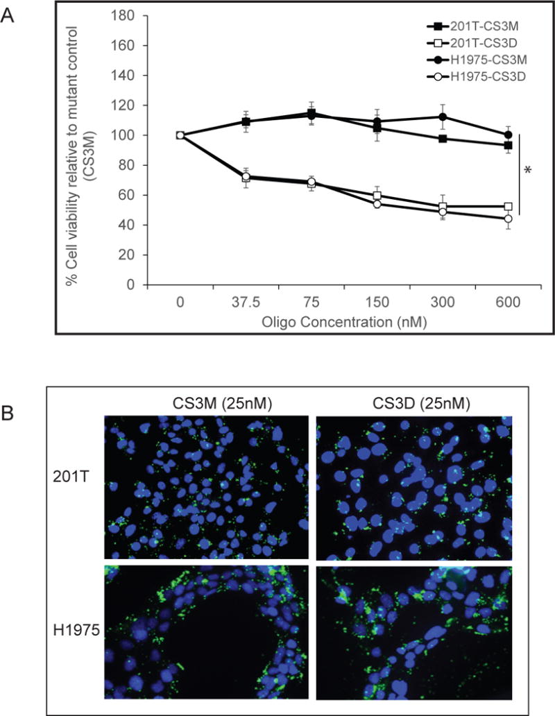
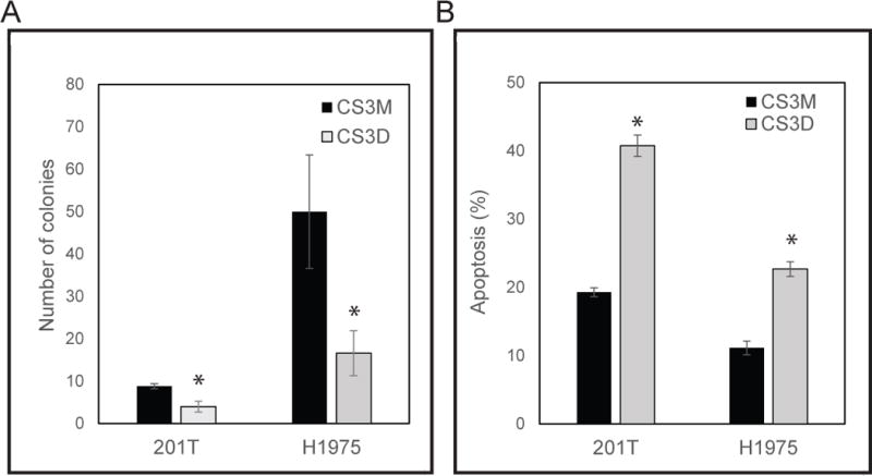
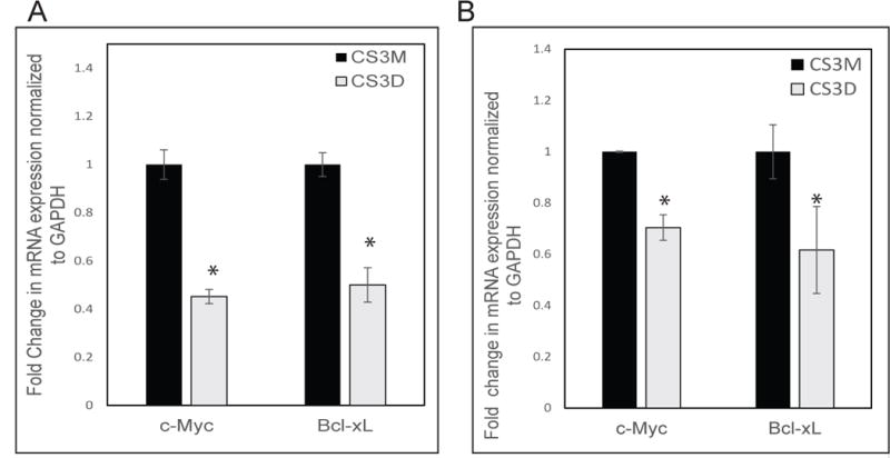
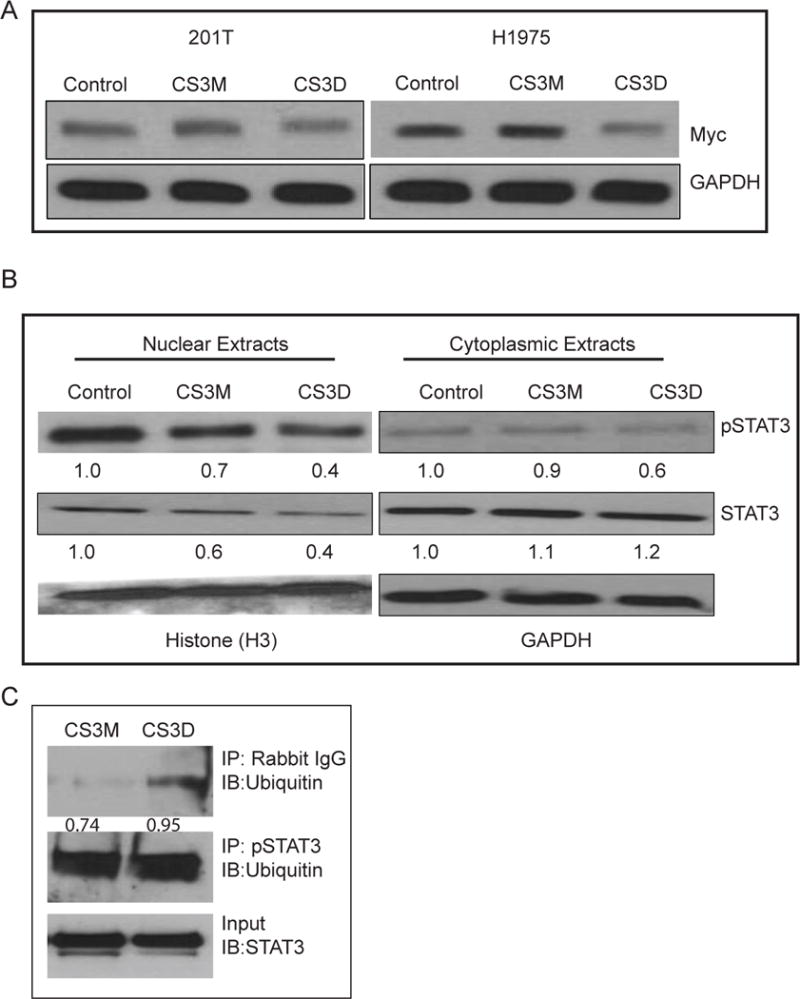
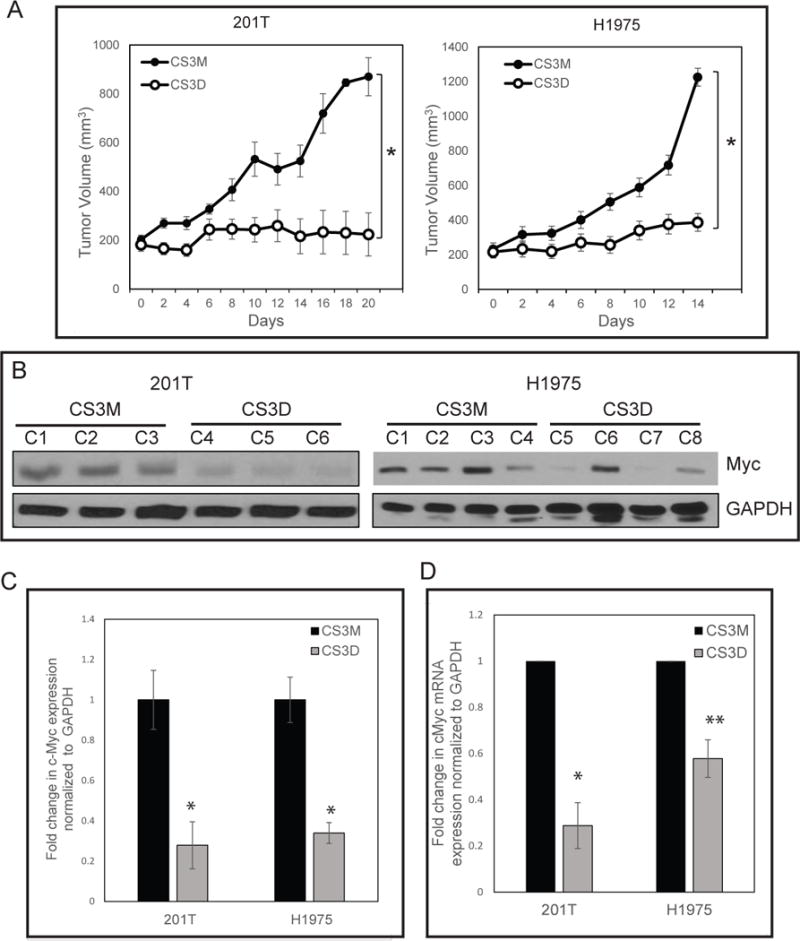
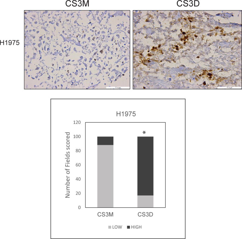
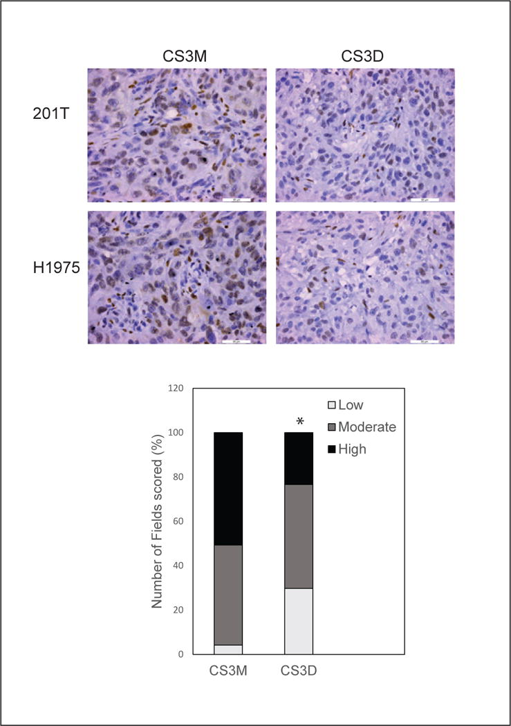
Constitutively activated STAT3 plays a critical role in non-small cell lung carcinoma (NSCLC) progression by mediating proliferation and survival. STAT3 activation in normal cells is transient, making it an attractive target for NSCLC therapy. The therapeutic potential of blocking STAT3 in NSCLC was assessed utilizing a decoy approach by ligating a double-stranded 15-mer oligonucleotide that corresponds to the STAT3 response element of STAT3-target genes, to produce a cyclic STAT3 decoy (CS3D). The decoy was evaluated using NSCLC cells containing either wild-type EGFR (201T) or mutant EGFR with an additional EGFRi resistance mutation (H1975). These cells are resistant to EGFR inhibitors and require an alternate therapeutic approach. CS3D activity was compared with an inactive cyclic control oligonucleotide (CS3M) that differs by a single base pair, rendering it unable to bind to STAT3 protein. Transfection of 0.3 μmol/L of CS3D caused a 50% inhibition in proliferation in 201T and H1975 cells, relative to CS3M, and a 2-fold increase in apoptotic cells. Toxicity was minimal in normal cells. CS3D treatment caused a significant reduction of mRNA and protein expression of the STAT3 target gene c-Myc and inhibited colony formation by 70%. The active decoy decreased the nuclear pool of STAT3 compared with the mutant. In a xenograft model, treatments with CS3D (5 mg/kg) caused a potent 96.5% and 81.7% reduction in tumor growth in 201T (P < 0.007) and H1975 models (P < 0.0001), respectively, and reduced c-Myc and p-STAT3 proteins. Targeting STAT3 with the cyclic decoy could be an effective therapeutic strategy for NSCLC. Mol Cancer Ther; 17(9); 1917-26. ©2018 AACR.


Conflict of interest statement

The authors declare no potential conflicts of interest.

Figures

Figure 1
Figure 1. Effects of the cyclic STAT3 decoy in vitro
(A) Effect on cell viability. NSCLC cell lines (201T and H1975) were transfected with CS3D or CS3M at concentrations ranging from 0 nM to 600 nM. Using MTS assays, cell viabilities was assessed 72 hours later. Three independent experiments were performed, using 24-well plates and 4 wells/concentration. (B) Uptake of fluorescein-labeled CS3D by NSCLC in vitro. 24 hours post-transfection, confocal imaging shows intracellular localization of CS3M or CS3D in 201T and H1975 cells.
Figure 2
Figure 2. CS3D inhibits anchorage independent growth and promotes apoptosis
(A) NSCLC cell growth was assessed in colony forming assays in the presence of either 300 nM CS3D or CS3M. CS3D significantly blocks the ability of NSCLC to grow in an anchorage independent manner in soft-agar post-transfection. (B) Detection of annexin V and propidium iodide (PI)-positive cells by flow cytometry: H1975 and 201T cells transfected with 100nM of CS3M or CS3D for 24hr were stained with apoptosis marker (annexin V) and cell viability dye. Statistical significance was determined as *P<0.05, n=3.
Figure 3
Figure 3. Down-regulation of c-Myc and bcl-xL mRNA expression by CS3D
24 hours post-transfection, cells were treated with EGF (10 ng/ml) for 1.5 hr and mRNA was harvested from wild-type (A) and mutant EGFR T790M (B) NSCLC cell lines, and mRNA expression was assessed by RT-qPCR. Relative mRNA expression was normalized to GAPDH mRNA levels as an internal control. Statistical significance was determined as *P<0.05.
Figure 4
Figure 4. Change in c-Myc and STAT3 protein by CS3D
(A) Down-regulation of c-Myc protein by CS3D. Cells were treated as in Fig. 3, and 3 hr after EGF treatment, lysates were collected from wild-type (left) and mutant EGFR T797M (right) NSCLC cell lines. Protein expression was assessed by Western blot. Relative c-Myc expression was normalized to GAPDH protein levels as an internal control. (B) CS3D alters STAT3 levels. 24 hours post-transfection with either CS3M, CS3D, or lipofectamine alone H1975 were stimulated with IL-6 (50ng/mL) for 1 hr to strongly activate STAT3. After cell lysis, nuclear and cytoplasmic extracts were obtained by subcellular fractionation using centrifugation, and Western blotting was used to assessed to protein expression. GAPDH was used as a marker of the cytoplasmic fraction and histone H3 was used as a marker of the nuclear fraction. Densitometric quantification of the c-Myc bands are as indicated. (C) CS3D increases the proportion of pSTAT3-ubiquitin bound complexes. 24 hours post-transfection, P-STAT3 was immunoprecipitated and the presence of ubiquitin was assessed by immunoblotting. Greater ratio of pSTAT3-ubiquitin complexes was detected with CS3D treatment. The increase in pSTAT3-unbiquitin complex correlated with decrease in total STAT3 present. Densitometric analysis revealed a greater ratio of pSTAT3-ubiquitin complex to STAT3 in response to CS3D (0.95) as compared to CS3M (0.74).
Figure 5
Figure 5. CS3D suppresses NSCLC xenograft tumor growth
(A) Intravenous delivery of CS3D inhibits tumor growth in both wild-type (201T) and mutant EGFR T790M (H1975) xenografts. Approximately 1x10⁶ cells were inoculated subcutaneously in the flanks of nude mice. Following the development of palpable tumors (about 200mm3), mice were randomized and given daily injections of either CS3D or CS3M (5mg/kg/day; 10 tumors/group). Experiments were done twice as biological replicates. Tumor volumes were recorded every other day while animal weights were also monitored during the course of the treatment. The difference in tumor volume between the CS3D- and CS3M-treated groups was significant for both 201T (P<0.007) and H1975 (P<0.0001). (B) c-Myc is suppressed after CS3D treatment in vivo. At the end of the treatments, tumors were harvested, whole cell lysates were prepared and RNA extracted for target gene analysis by Western blotting and RT-qPCR. GAPDH protein was used as internal loading control for immunoblotting. CS3D treated tumors (C4, C5, and C6) from separate animals show a decrease in c-Myc expression level relative to the CS3M-treated group (C1, C2, and C3) in 201T. Immunoblotting analysis of H1975 residual tumors treated with CS3D (C8-C8) also showed reduction in c-Myc expression levels relative to CS3M (C1-C4). The expression of c-Myc was significantly reduced in response to CS3D, relative to CS3M, in both wild-type (201T, (P<0.042)) and mutant EGFR (H1975, (P<0.05)) derived tumors (*P<0.05). Data are shown as mean +/− SEM. (C) Densitometry quantification of c-Myc protein expression levels in 201T and H1975 from tumor-derived xenografts. The expression of c-Myc was significantly reduced in response to CS3D, relative to CS3M, in both wild-type (201T, (P<0.042)) and mutant EGFR (H1975, (P<0.05)) derived tumors. Data shown are means +/− SEM between two groups (CS3M and CS3D). (D) c-Myc mRNA expression levels in 201T and H1975: CS3D suppresses c-Myc gene expression levels in 201T and H1975-derived tumors. Data are presented as mean +/− SEM. *P<0.05 compared with the mutant control group (CS3M).
Figure 6
Figure 6. STAT3 decoy induces caspase-3 cleavage
Representative sections of H1975 tumors stained for expression of cleaved caspase-3. H1975-derived xenografts administered daily intravenous injections of either CS3D or CS3M were harvested at the end of treatment (day 14), and expression of cleaved caspase-3 was used as a marker for apoptotic cells. CS3D significantly induces apoptosis relative to CS3M. Similar results was also observed in the 201T derived xenografts.
Figure 7
Figure 7. Short-term CS3D-treatment suppresses p-STAT3 levels
Tumors derived from H1975 xenografts were analyzed by immunohistochemical staining for p-STAT3 after 5 daily iv injections of either STAT3 decoy or mutant decoy. Representative sections of H1975 tumors show a decrease in p-STAT3 levels. Bar graph quantitates the frequency of low-, medium-, and high-grade staining in multiple images; P < 0.001, decoy compared to mutant.Bright field images captured at 40X magnfication

Comment in

References

    1. Siegel RL, Miller KD, Jemal A. Cancer Statistics: 2017. CA Cancer J Clin. 2017;67:7–30. - PubMed
    1. Jemal A, Ward EM, Johnson CJ, Cronin KA, Ma J, Ryerson B, Mariotto A, Lake AJ, et al. Annual Report to the Nation on the Status of Cancer: 1975-2014. Featuring Survival J Natl Cancer Inst. 2017;109(9) - PMC - PubMed
    1. Torre LA, Siegel RL, Ward EM, Jemal A. Global Cancer Incidence and Mortality Rates and Trends: An Update. Cancer Epidemiol Biomarkers Prev. 2016;25:16–27. - PubMed
    1. Chen Z, Fillmore CM, Hammerman PS, Kim CF, Wong KK. Non-small-cell lung cancers: a heterogeneous set of diseases. Nat Rev Cancer. 2014;14:535–46. - PMC - PubMed
    1. Gainor JF, Shaw AT. Emerging paradigms in the development of resistance to tyrosine kinase inhibitors in lung cancer. J Clin Oncol. 2013;31:3987–96. - PMC - PubMed

Publication types

MeSH terms