Review
. 2019 Mar 1;11(3):a033001.
doi: 10.1101/cshperspect.a033001.

Translational Control in Virus-Infected Cells

Affiliations
Review

Translational Control in Virus-Infected Cells

Noam Stern-Ginossar et al. Cold Spring Harb Perspect Biol. .

Abstract

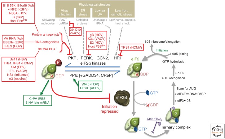
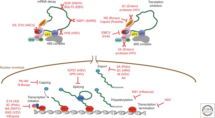
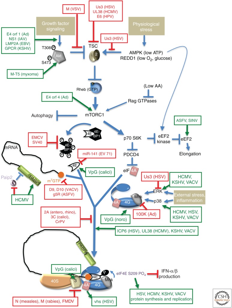
As obligate intracellular parasites, virus reproduction requires host cell functions. Despite variations in genome size and configuration, nucleic acid composition, and their repertoire of encoded functions, all viruses remain unconditionally dependent on the protein synthesis machinery resident within their cellular hosts to translate viral messenger RNAs (mRNAs). A complex signaling network responsive to physiological stress, including infection, regulates host translation factors and ribosome availability. Furthermore, access to the translation apparatus is patrolled by powerful host immune defenses programmed to restrict viral invaders. Here, we review the tactics and mechanisms used by viruses to appropriate control over host ribosomes, subvert host defenses, and dominate the infected cell translational landscape. These not only define aspects of infection biology paramount for virus reproduction, but continue to drive fundamental discoveries into how cellular protein synthesis is controlled in health and disease.


Figures

Figure 1.
Figure 1.
Targeting stress-responsive host defenses by viral functions controls eukaryotic initiation factor (eIF)2α phosphorylation and translation initiation. Composed of three subunits (α,β,γ), eIF2 is a GTP-binding translation initiation factor that loads eIF3-bound 40S ribosomal subunits with methionyl-transfer RNA (Met-tRNAi) (right panel). Subsequent mRNA recruitment, AUG recognition, and eIF5-stimulated GTP hydrolysis is followed by 60S subunit joining and translation initiation by the 80S ribosome. Recycling inactive eIF2•GDP to the active GTP-bound form requires the GEF eIF2B. Phosphorylation of eIF2α on S51 blocks initiation by binding to and inhibiting eIF2B, preventing GDP-GTP exchange. Four eIF2 kinases, each of which is activated by specific molecules that accumulate in response to a discrete physiological stress, and the protein phosphatase 1 catalytic subunit (PP1c), partnered with an inducible (GADD34) or constitutively active (CreP) regulatory subunit, control eIF2α phosphorylation. Viral functions that activate (green), respond to (green), or repress (red) the indicated host effectors are shown. aa, amino acid.
Figure 2.
Figure 2.
Viruses use multiple mechanisms to block host messenger RNA (mRNA) translation. Cellular mRNA biogenesis starts in the nucleus where RNA polymerase II generates primary transcripts that undergo several processing steps including 5′ capping, splicing, and polyadenylation before functional mRNAs are produced. Mature mRNAs are exported to the cytoplasm where they are translated. Initiation of cap-dependent mRNA translation typically requires recruitment of the multisubunit eukaryotic translation initiation factor (eIF)4F, which consists of the cap-binding protein eIF4E, the RNA helicase eIF4A, and the adaptor protein eIF4G. Binding of eIF4G to poly(A)-binding protein (PABP) mediates communication between 5′ and 3′ ends. Virus-encoded factors that degrade (scissors) cellular proteins and mRNAs or repress cellular functions are shown.
Figure 3.
Figure 3.
Subverting mechanistic target of rapamycin complex 1 (mTORC1) signaling to control translation in virus-infected cells. Occupying a nexus of intersecting signaling networks, the cellular ser/thr kinase mTORC1 plays a critical role stimulating anabolic programs, like protein synthesis, and repressing catabolic outcomes like autophagy. Briefly, growth factor–induced Akt phosphorylation (T308, S473) and activation represses TSC1/2, which, in turn, allows Rheb-GTP to activate mTORC1. Nutrient, energy, amino acid (aa), or oxygen insufficiency (physiological stress) all repress mTORC1 through discrete effectors. Signaling through mTORC1 allows swift changes in translational output in response to differing environmental and physiological inputs by controlling initiation and elongation. Initiation is stimulated by phosphorylating and inactivating 4E-BP translational repressor family members (e.g., 4E-BP1), which bind the cap-binding protein eIF4E to prevent its interaction with eIF4G. Through its substrate p70S6K, mTORC1 controls the DEAD-box-containing RNA helicase eIF4A, which together with eukaryotic initiation factor (eIF)4E and eIF4G comprises the multisubunit initiation factor eIF4F. Regulated eIF4F assembly controls cap-dependent mRNA translation as eIF4F recruits 40S subunits to the mRNA capped 5′ end. By repressing eEF2 kinase, mTORC1 stimulates eEF2 and elongation. The impact of mTORC1 activation on translation initiation and elongation is shown, as are viral factors that stimulate (green) and repress (red) the indicated cellular effectors. SV40, simian virus 40; calici, calicivirus; noro, norovirus; entero, enterovirus; rhino, rhinovirus.
Figure 4.
Figure 4.
Ribosomal proteins are required for translation of viral RNAs. Locations of ribosomal proteins on 40S and 60S subunits that are important for viral messenger RNA (mRNA) translation are shown on the ribosome structure derived from PDB entry 4v88. (Figure based on data in Ben-Shem et al. 2011.) Both intrasubunit (left) and solvent surfaces (right) are shown. Ribsomal RNA (rRNA) (gray) and ribosomal proteins (tan) are depicted with the proteins implicated in viral mRNA translation in color: eS25 (red), RACK1 (turquoise), eL40 (green), uL30 (purple), eL18 (orange), and P1/P2 (dark blue/light blue).
Figure 5.
Figure 5.
Strategies to maximize viral genome coding capacity. Internal ribosome entry site (IRES)-driven polyprotein production, programmed ribosome frameshifting (PRF), reinitiation, stop-codon readthrough, leaky scanning, ribosome stalling, and translational bypass/ribosome hopping are shown in the cartoon. (See the text for a detailed description.) Stop codons (stop signs), proteolytic cleavage sites (scissors), 5′-cap structure (orange circle), open reading frames (green), ribosome subunits (brown and blue), and initiation factor loaded with transfer RNA (tRNA) are shown.

References

    1. Abbas YM, Laudenbach BT, Martinez-Montero S, Cencic R, Habjan M, Pichlmair A, Damha MJ, Pelletier J, Nagar B. 2017. Structure of human IFIT1 with capped RNA reveals adaptable mRNA binding and mechanisms for sensing N1 and N2 ribose 2′-O methylations. Proc Natl Acad Sci 114: E2106–E2115. - PMC - PubMed
    1. Abernathy E, Glaunsinger B. 2015. Emerging roles for RNA degradation in viral replication and antiviral defense. Virology 479–480: 600–608. - PMC - PubMed
    1. Abernathy E, Gilbertson S, Alla R, Glaunsinger B. 2015. Viral nucleases induce an mRNA degradation-transcription feedback loop in mammalian cells. Cell Host Microbe 18: 243–253. - PMC - PubMed
    1. Abrahão JS, Araújo R, Colson P, La Scola B. 2017. The analysis of translation-related gene set boosts debates around origin and evolution of mimiviruses. PLoS Genet 13: e1006532. - PMC - PubMed
    1. Albornoz A, Carletti T, Corazza G, Marcello A. 2014. The stress granule component TIA-1 binds tick-borne encephalitis virus RNA and is recruited to peri-nuclear sites of viral replication to inhibit viral translation. J Virol 88: 6611–6622. - PMC - PubMed