Review
. 2019 Jan 2;11(1):a032714.
doi: 10.1101/cshperspect.a032714.

Single-Molecule Fluorescence Applied to Translation

Affiliations
Review

Single-Molecule Fluorescence Applied to Translation

Arjun Prabhakar et al. Cold Spring Harb Perspect Biol. .

Abstract

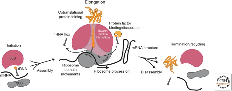
Single-molecule fluorescence methods have illuminated the dynamics of the translational machinery. Structural and bulk biochemical experiments have provided detailed atomic and global mechanistic views of translation, respectively. Single-molecule studies of translation have bridged these views by temporally connecting the conformational and compositional states defined from structural data within the mechanistic framework of translation produced from biochemical studies. Here, we discuss the context for applying different single-molecule fluorescence experiments, and present recent applications to studying prokaryotic and eukaryotic translation. We underscore the power of observing single translating ribosomes to delineate and sort complex mechanistic pathways during initiation and elongation, and discuss future applications of current and improved technologies.


Figures

Figure 1.
Figure 1.
Dynamic picture of translation. Translation is regulated through a complex network of interactions and movements of the ribosomal subunits, the translated messenger RNA (mRNA), the adaptor transfer RNAs (tRNAs), protein translation factors, and the growing nascent peptide chain. Key dynamic features of translation are highlighted.
Figure 2.
Figure 2.
Compositional studies of translation using single-molecule fluorescence. (A) Fluorescently labeled biomolecule (green) functionalized with a biotin is immobilized to the polyethylene glycol (PEG)-derivatized SiO2 surface through biotin–streptavidin interactions. Total internal reflectance (TIR) shown here produces an evanescent field, exciting only fluorophores within 100 nm from the surface. (B) Binding of a second labeled biomolecule (red) in solution to the immobilized biomolecule results in detection of red fluorescence burst, from which arrival and departure times can be measured. (C) Zero-mode waveguides (ZMWs) allow delivery of up to four different fluorescently labeled ligands at micromolar concentrations caused by its shorter excitation lengths with direct illumination by green (532 nm), red (642 nm), and cyan (488 nm) lasers. (D) 70S ribosomes with fMet-Cy3-tRNAfMet (green) were immobilized by the biotinylated mRNA and real-time transit of Phe-Cy5-tRNAPhe (red) and Lys-Cy2-tRNALys (cyan) ternary complexes (TCs) was monitored during translation (Uemura et al. 2010). (E) Tracking the binding events of Cy5-IF2 (red), fMet-Cy3-tRNAfMet (green), and Cy3.5-50S subunit (yellow) to the Alexa 488-30S-mRNA complex (cyan) (Tsai et al. 2012) revealed multiple pathways of translation initiation in bacteria (see text for details).
Figure 3.
Figure 3.
Conformational studies of translation using single-molecule Förster resonance energy transfer (FRET). (A) With single-laser excitation of the donor dye, fluorescence intensities of donor (green, Id) and acceptor (red, Ia) dyes attached to a surface-immobilized biomolecular complex are measured to calculate the FRET efficiency (EFRET), which reports distance between the fluorophores (R) used to define conformational states of a biological process. (B) Cy3 and Cy5 labeling of P-site and A-site transfer RNAs (tRNAs), respectively, was used to follow the tRNA conformations during tRNA selection stage of elongation (Blanchard et al. 2004a). (C) (Left) Helix44 of 30S subunit was labeled with Cy3 and Helix101 of 50S subunit was labeled with Cy5, bringing the dyes within FRET distance to report intersubunit conformational changes (Marshall et al. 2008b). (Right) With immobilization of Cy3-30S (green) as a preinitiation complex and delivery of Cy5-50S (red) along with Phe-tRNAPhe and elongation factors, subunit joining is signaled by the simultaneous burst of Cy5 fluorescence and dip in Cy3 fluorescence at the beginning, and anticorrelated changes in the Cy3 and Cy5 fluorescence intensities correspond to transitions between the nonrotated and rotated conformational states evolved from cycles of peptidyl transfer and translocation during elongation. (D) Replacing the acceptor dyes in tRNA and ribosome labels in B and C with nonfluorescent quenchers (QSY9, QSY21, respectively) allowed simultaneous detection of ribosome intersubunit and tRNA conformations with Cy3 labeling on Lys-tRNALys and Cy5 labeling on the 30S subunit (Choi and Puglisi 2017).
Figure 4.
Figure 4.
Recent work on bacterial translation involved use of intersubunit dye–quencher labeling (Cy3B-30S and BHQ-2-50S; see Fig. 3C for labeling positions) to track ribosome conformation (green signal). (A) Monitoring Lys-Cy5-tRNALys-binding dynamics (red) on ribosomes with Nm-modified mRNAs (Choi et al. 2018) shows multiple short Cy5 pulses representing tRNA rejection events, explaining the long ribosome stall in nonrotated state. (B) Tracking translation of gene 60 mRNA revealed that the subset of ribosomes that bypassed stalled at the take-off Gly-45 codon in the rotated state. (C) Simultaneously tracking the ribosome intersubunit conformation (green), Cy5-RF binding (red), and P-site tRNA occupancy (Phe-Cy5.5-tRNAPhe, violet) during translation termination and recycling (Prabhakar et al. 2017a) helped resolve the posttermination rotated state, a key intermediate preceding subunit splitting step (catalyzed by RRF and EF-G) and subsequent 30S complex disassembly (performed by IF3).
Figure 5.
Figure 5.
Recent work on eukaryotic translation. (A) Conformational dynamics of hepatitis C virus (HCV) internal ribosome entry site (IRES) bound to human 40S subunit was monitored using Cy3-Cy5 Förster resonance energy transfer (FRET) (Fuchs et al. 2015), detecting two different conformational states important in the pathway of HCV IRES initiation. (B) Tracking real-time assembly of yeast Cy3.5-40S (yellow), yeast Cy5-60S (red), and Phe-Cy3-tRNAPhe (green) on immobilized Cy5.5-CrPV IRES (violet) (Petrov et al. 2016) led to discovery of parallel pathways of CrPV IRES initiation. (C) Petrov et al. (2016) also found that ribosomes initiate on the CrPV IRES in both 0 and +1 reading frames, another pathway branchpoint in the mechanism of CrPV IRES initiation.

References

    1. Aitken CE, Puglisi JD. 2010. Following the intersubunit conformation of the ribosome during translation in real time. Nat Struct Mol Biol 17: 793–800. - PMC - PubMed
    1. Antoun A, Pavlov MY, Andersson K, Tenson T, Ehrenberg M. 2003. The roles of initiation factor 2 and guanosine triphosphate in initiation of protein synthesis. EMBO J 22: 5593–5601. - PMC - PubMed
    1. Ban N, Nissen P, Hansen J, Moore PB, Steitz TA. 2000. The complete atomic structure of the large ribosomal subunit at 2.4 Å resolution. Science 289: 905–920. - PubMed
    1. Barbet-Massin E, Huang CT, Daebel V, Hsu ST, Reif B. 2015. Site-specific solid-state NMR studies of “trigger factor” in complex with the large ribosomal subunit 50S. Angew Chem Int Ed Engl 54: 4367–4369. - PubMed
    1. Becker T, Franckenberg S, Wickles S, Shoemaker CJ, Anger AM, Armache JP, Sieber H, Ungewickell C, Berninghausen O, Daberkow I, et al. 2012. Structural basis of highly conserved ribosome recycling in eukaryotes and archaea. Nature 482: 501–506. - PMC - PubMed

Publication types