. 2018 Jun 26;115(26):6816-6821.
doi: 10.1073/pnas.1804262115. Epub 2018 Jun 11.

Potent laminin-inspired antioxidant regenerative dressing accelerates wound healing in diabetes

Affiliations

Potent laminin-inspired antioxidant regenerative dressing accelerates wound healing in diabetes

Yunxiao Zhu et al. Proc Natl Acad Sci U S A. .

Abstract

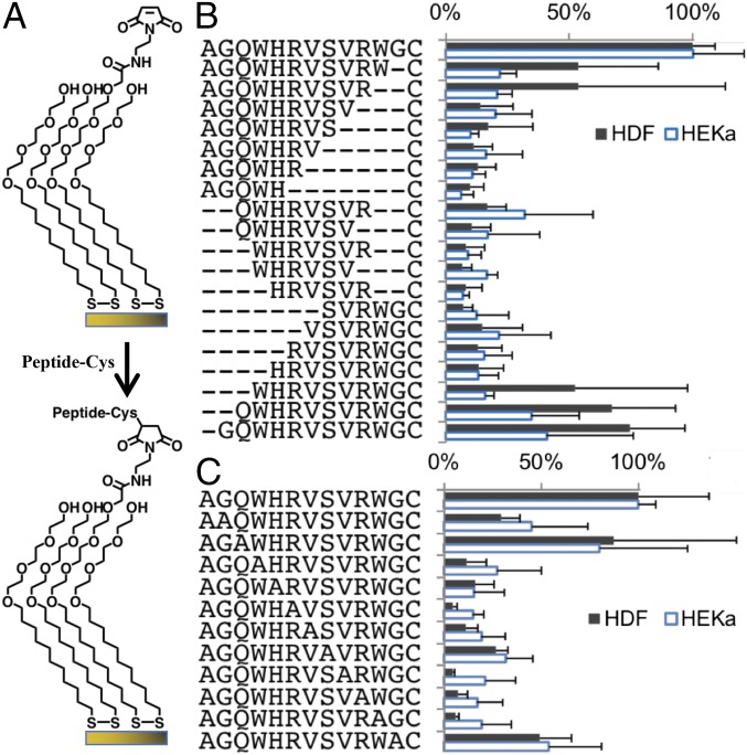
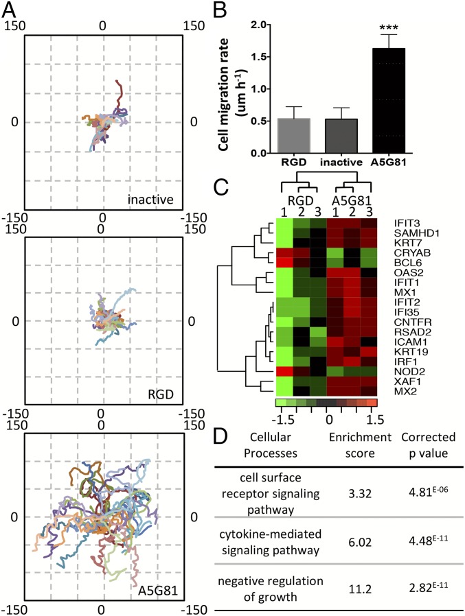
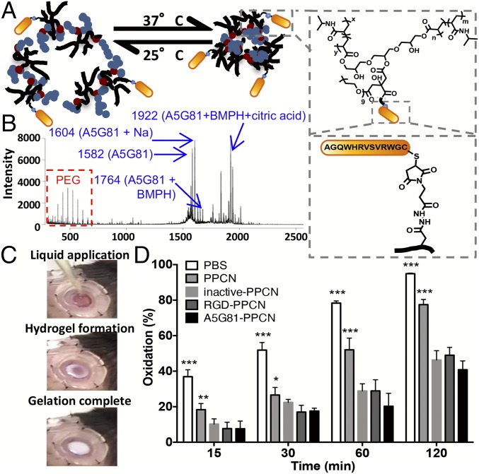
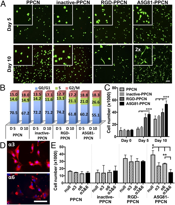
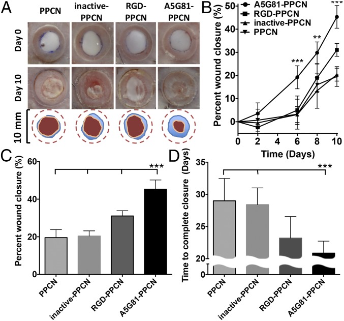
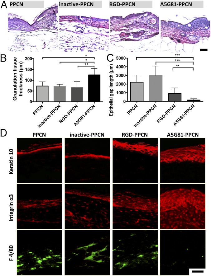
The successful treatment of chronic dermal wounds, such as diabetic foot ulcers (DFU), depends on the development of safe, effective, and affordable regenerative tools that the surgeon can rely on to promote wound closure. Although promising, strategies that involve cell-based therapies and the local release of exogenous growth factors are costly, require very long development times, and result in modest improvements in patient outcome. We describe the development of an antioxidant shape-conforming regenerative wound dressing that uses the laminin-derived dodecapeptide A5G81 as a potent tethered cell adhesion-, proliferation-, and haptokinesis-inducing ligand to locally promote wound closure. A5G81 immobilized within a thermoresponsive citrate-based hydrogel facilitates integrin-mediated spreading, migration, and proliferation of dermal and epidermal cells, resulting in faster tissue regeneration in diabetic wounds. This peptide-hydrogel system represents a paradigm shift in dermoconductive and dermoinductive strategies for treating DFU without the need for soluble biological or pharmacological factors.

Keywords: citric acid; diabetic foot ulcers; laminin; regenerative biomaterials; wound healing.


Conflict of interest statement

Conflict of interest statement: Y.Z., M.M., and G.A.A. are coinventors on a patent application pertaining to the hydrogel system disclosed in the manuscript.

Figures

Fig. 1.
Fig. 1.
All amino acid residues in the original sequence are required for A5G81 bioactivity. (A) HDF and HEKa were cultured on SAMs presenting a peptide (1 mol%) against a background of tri(ethylene glycol). Monolayers were prepared by immobilizing cysteine-terminated peptides onto maleimide-terminated dioctadecyl disulfide. (B) Adhesion of HDF and HEKa on SAMs presenting truncated versions of A5G81. (C) Adhesion of HDF and HEKa on SAMs presenting alanine-substituted versions of A5G81 (n = 3; all data are presented as mean ± SD).
Fig. 2.
Fig. 2.
A5G81 immobilized on 2D SAM surfaces promotes HEKa migration via haptokinesis. (A) The trajectories of 30 cells on inactive-, RGD-, and A5G81-presenting surfaces (all at 1 mol% peptide) were plotted in one field, oriented so that the cell’s original location is x(0), y(0). The axes denote the migration distance in microns. (B) Mean rate of cell migration. All data are presented as mean ± SD; statistical significance was performed using one-way ANOVA with a Turkey’s multiple comparison test (n = 30; ***P < 0.001). (C) Transcriptional profiling of HEKa cultured on A5G81 and RGD SAM surfaces (same molar concentration). The heat map of the top differentially expressed genes at the end of a 12-h culture period is shown. (D) The clusters of GO processes impacted by culturing the cells on the peptide-presenting SAM surfaces for 12 h.
Fig. 3.
Fig. 3.
Click chemistry enables the facile synthesis of thermoresponsive, antioxidant peptide-PPCN. (A) Schematic illustrating A5G81-PPCN self-assembly due to temperature change (Left). A5G81 peptide conjugation to PPCN via the cross-linker BMPH (Right). The same amount of peptide was conjugated across different peptide groups. (B) MALDI-TOF spectrum of A5G81-PPCN confirming the presence of the PPCN, peptide, peptide-linker, and peptide-linker-citric acid constructs. (C) A5G81-PPCN is applied as a liquid that covers the wound bed and conforms to the wound edges before gelation, due to body temperature, within seconds. (D) The β-carotene lipid peroxidation inhibition assay shows improved antioxidant activity of the peptide-modified PPCN (n = 9) (*P < 0.05; **P < 0.01; ***P < 0.001).
Fig. 4.
Fig. 4.
A5G81-PPCN promotes cell spreading and α3- and α6-mediated proliferation of entrapped HDF. (A) HDF entrapped within hydrogels and stained with Calcein-AM (live cells, green) and ethidium homodimer 1 (dead cells, red) at 5 and 10 d. (Scale bar: 100 μm.) Insets show a 2× magnified view of cell spreading. (B) Cell cycle analysis of the HDF in the hydrogels. (C) Cell proliferation within the hydrogel. (D) Staining of the integrin subunits α3 and α6 in HDF. (Scale bar: 100 μm.) (E) HDF proliferation response due to blocking integrin subunits α3 and/or α6. All data are presented as mean ± SD (n ≥ 5; *P < 0.05; **P < 0.01; ***P < 0.001).
Fig. 5.
Fig. 5.
A5G81-PPCN promotes accelerated regenerative wound closure of excisional splinted wounds in diabetic mice. (A) Representative images of the wound area immediately after wounding and at day 10 postwounding (Top, Middle). The wound-closure boundary at day 0 and day 10 postwounding is overlaid on the image (Bottom). (B) Wound healing 10 d postwounding. (C) Quantification of wound closure at day 10 postwounding for all four groups. (D) Summary of the complete wound-closure times. All data are presented as mean ± SD (n ≥ 5; ***P < 0.01).
Fig. 6.
Fig. 6.
A5G81-PPCN accelerates tissue regeneration in vivo. (A) H&E staining of tissue sections shows complete resorption of the hydrogel in all four groups 30 d postwounding. (B) Thicker granulation tissue was found in the A5G81-PPCN−treated wounds. (C) Quantification of the epithelial gap demonstrates that wounds treated with A5G81-PPCN exhibited the smallest epithelial gap and maximum wound closure. (D) Immunofluorescence staining of the wound, demonstrating enhanced expression of keratin-10, integrin α3, and reduced positive staining for macrophages (F4/80) in the A5G81-PPCN−treated wounds. (Scale bar: 100 μm.) All data are presented as mean ± SD (n ≥ 5; *P < 0.05; **P < 0.01; ***P < 0.001).

Comment in

References

    1. Centers for Disease Control and Prevention 2014. National Diabetes Statistics Report: Estimates of diabetes and its burden in the United States (Centers Dis Control Prevention, Atlanta)
    1. World Health Organization . Global Report on Diabetes. World Health Org; Geneva: 2016.
    1. Falanga V. Wound healing and its impairment in the diabetic foot. Lancet. 2005;366:1736–1743. - PubMed
    1. Smiell JM, et al. Efficacy and safety of becaplermin (recombinant human platelet-derived growth factor-BB) in patients with nonhealing, lower extremity diabetic ulcers: A combined analysis of four randomized studies. Wound Repair Regen. 1999;7:335–346. - PubMed
    1. Hu S, Kirsner RS, Falanga V, Phillips T, Eaglstein WH. Evaluation of Apligraf persistence and basement membrane restoration in donor site wounds: A pilot study. Wound Repair Regen. 2006;14:427–433. - PubMed

LinkOut - more resources