. 2018 Jun 26;115(26):6822-6827.
doi: 10.1073/pnas.1806308115. Epub 2018 Jun 11.

Differentiated human airway organoids to assess infectivity of emerging influenza virus

Affiliations

Differentiated human airway organoids to assess infectivity of emerging influenza virus

Jie Zhou et al. Proc Natl Acad Sci U S A. .

Abstract

Novel reassortant avian influenza H7N9 virus and pandemic 2009 H1N1 (H1N1pdm) virus cause human infections, while avian H7N2 and swine H1N1 virus mainly infect birds and pigs, respectively. There is no robust in vitro model for assessing the infectivity of emerging viruses in humans. Based on a recently established method, we generated long-term expanding 3D human airway organoids which accommodate four types of airway epithelial cells: ciliated, goblet, club, and basal cells. We report differentiation conditions which increase ciliated cell numbers to a nearly physiological level with synchronously beating cilia readily discernible in every organoid. In addition, the differentiation conditions induce elevated levels of serine proteases, which are essential for productive infection of human influenza viruses and low-pathogenic avian influenza viruses. We also established improved 2D monolayer culture conditions for the differentiated airway organoids. To demonstrate the ability of differentiated airway organoids to identify human-infective virus, 3D and 2D differentiated airway organoids are applied to evaluate two pairs of viruses with known distinct infectivity in humans, H7N9/Ah versus H7N2 and H1N1pdm versus an H1N1 strain isolated from swine (H1N1sw). The human-infective H7N9/Ah virus replicated more robustly than the poorly human-infective H7N2 virus; the highly human-infective H1N1pdm virus replicated to a higher titer than the counterpart H1N1sw. Collectively, we developed differentiated human airway organoids which can morphologically and functionally simulate human airway epithelium. These differentiated airway organoids can be applied for rapid assessment of the infectivity of emerging respiratory viruses to human.

Keywords: airway organoid; infectivity; influenza virus; proximal differentiation.


Conflict of interest statement

The authors declare no conflict of interest.

Figures
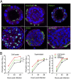
Fig. 1.
Fig. 1.
Demonstration of four types of airway epithelial cells and replication of influenza viruses in human AOs. (A) Four lineages of airway epithelial cells are identified in the human AOs, i.e., ACCUB+ and FOXJ1+ ciliated cells, P63+ basal cells, CC10+ club cells, and MUC5AC+ goblet cells. Nuclei and cellular actin filaments were counterstained with DAPI (blue) and Phalloidin-647 (purple), respectively. (Scale bars, 10 µm.) (B) The AOs (n = 3) were inoculated with H1N1pdm, H5N1, and H7N9/Ah at an MOI of 0.01. The infected organoids (cell lysate) (Left) and supernatants (Center) were harvested at the indicated times to detect the viral loads. Supernatant samples were used for viral titration (Right). Data are presented as mean ± SD. The experiment was performed three times independently. **P < 0.01; ***P < 0.005.
Fig. 2.
Fig. 2.
PD of human AOs. (A) The AOs were cultured in PD medium or the original human AO medium in parallel for 16 d. Bright-field images of organoids at the indicated days are shown. (Magnification, 20×.) (B) Images of organoids cultured in PD medium and AO medium were used to measure the diameters of individual organoids (n = 300) using ImageJ. NS, not significant. ***P < 0.005. (C) Cilia in the PD AOs are captured by video. (Scale bar, 10 µm.) (D) Confocal images of P63+ basal cells and ACCTUB+ ciliated cells in the organoids cultured in PD and AO medium. (Scale bar, 10 μm.)
Fig. 3.
Fig. 3.
Characterization of the differentiation status of AOs. (A) Fold changes in expression levels of cell-type markers (Left) and serine proteases (Right) in the organoids cultured in PD medium versus those cultured in AO medium at the indicated days. Data show the mean and SD of two lines of organoids. *P < 0.05; **P < 0.01. (B) The percentages of individual cell types in the organoids cultured in PD medium and AO medium. The representative histograms of one organoid line are shown. (C) Fold change in positive cell percentages in organoids cultured in PD medium versus those in AO medium. Data are presented as the mean and SD of four independent experiments. *P < 0.05.
Fig. 4.
Fig. 4.
Influenza virus infection in the 3D PD AOs. The 3D PD AOs (n = 3) were inoculated with H7N9/Ah and H7N2 virus at an MOI of 0.01. The infected organoids (cell lysate) and supernatants were harvested at the indicated times to detect the viral loads (Left and Middle). Supernatant samples were used for viral titration (Right). The experiment was performed three times independently. ***P < 0.005.
Fig. 5.
Fig. 5.
Establishment of 2D differentiated AOs and replication capacity of influenza viruses. (A) Confocal images of the abundant ACCTUB+ ciliated cells (Left and Center) taken in LSM 800 and 780 microscopes, respectively, and H7N9/Ah-infected (NP+) cells (Right) in the 2D PD AOs. Nuclei and cellular actin filaments were counterstained with DAPI (blue) and Phalloidin-647 (purple). (Scale bar, 10 μm.) (B) The 2D PD AOs (n = 2) were inoculated with H7N9/Ah and H7N2 (Left) and H1N1pdm and H1N1sw (Right) at an MOI of 0.001. The cell-free media were harvested from the apical and basolateral chambers at the indicated times for viral titration. The experiment was performed three times independently.

Similar articles

Cited by

References

    1. Klenk HD. Influenza viruses en route from birds to man. Cell Host Microbe. 2014;15:653–654. - PubMed
    1. To KK, Chan JF, Chen H, Li L, Yuen KY. The emergence of influenza A H7N9 in human beings 16 years after influenza A H5N1: A tale of two cities. Lancet Infect Dis. 2013;13:809–821. - PMC - PubMed
    1. Chen Y, et al. Human infections with the emerging avian influenza A H7N9 virus from wet market poultry: Clinical analysis and characterisation of viral genome. Lancet. 2013;381:1916–1925. - PMC - PubMed
    1. World Health Organization 2017 Human infection with avian influenza A(H7N9) virus China. Available at www.who.int/csr/don/26-october-2017-ah7n9-china/en/WHO. Accessed May 30, 2018.
    1. Dawood FS, et al. Novel Swine-Origin Influenza A (H1N1) Virus Investigation Team Emergence of a novel swine-origin influenza A (H1N1) virus in humans. N Engl J Med. 2009;360:2605–2615. - PubMed

Publication types

MeSH terms

Associated data

LinkOut - more resources