. 2018 Jun 26;115(26):6590-6595.
doi: 10.1073/pnas.1806153115. Epub 2018 Jun 11.

Bright quantum dots emitting at ∼1,600 nm in the NIR-IIb window for deep tissue fluorescence imaging

Affiliations

Bright quantum dots emitting at ∼1,600 nm in the NIR-IIb window for deep tissue fluorescence imaging

Mingxi Zhang et al. Proc Natl Acad Sci U S A. .

Abstract

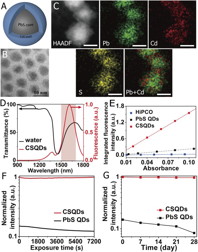
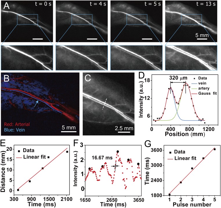
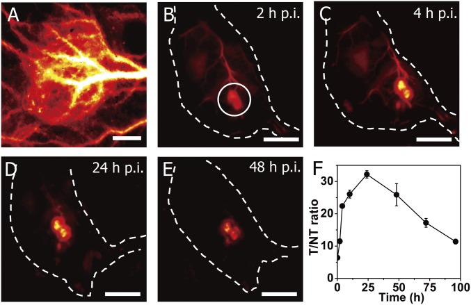
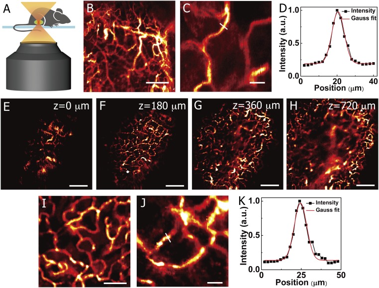
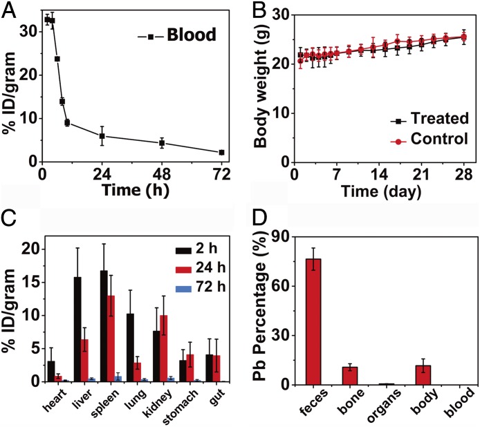
With suppressed photon scattering and diminished autofluorescence, in vivo fluorescence imaging in the 1,500- to 1,700-nm range of the near-IR (NIR) spectrum (NIR-IIb window) can afford high clarity and deep tissue penetration. However, there has been a lack of NIR-IIb fluorescent probes with sufficient brightness and aqueous stability. Here, we present a bright fluorescent probe emitting at ∼1,600 nm based on core/shell lead sulfide/cadmium sulfide (CdS) quantum dots (CSQDs) synthesized in organic phase. The CdS shell plays a critical role of protecting the lead sulfide (PbS) core from oxidation and retaining its bright fluorescence through the process of amphiphilic polymer coating and transferring to water needed for imparting aqueous stability and compatibility. The resulting CSQDs with a branched PEG outer layer exhibited a long blood circulation half-life of 7 hours and enabled through-skin, real-time imaging of blood flows in mouse vasculatures at an unprecedented 60 frames per second (fps) speed by detecting ∼1,600-nm fluorescence under 808-nm excitation. It also allowed through-skin in vivo confocal 3D imaging of tumor vasculatures in mice with an imaging depth of ∼1.2 mm. The PEG-CSQDs accumulated in tumor effectively through the enhanced permeation and retention effect, affording a high tumor-to-normal tissue ratio up to ∼32 owing to the bright ∼1,600-nm emission and nearly zero autofluorescence background resulting from a large ∼800-nm Stoke's shift. The aqueous-compatible CSQDs are excreted through the biliary pathway without causing obvious toxicity effects, suggesting a useful class of ∼1,600-nm emitting probes for biomedical research.

Keywords: NIR-IIb window; deep tissue; fluorescence imaging; in vivo; quantum dots.


Conflict of interest statement

The authors declare no conflict of interest.

Figures

Fig. 1.
Fig. 1.
Characterization of NIR-IIb–emitting CSQDs. (A) Schematic design of CSQDs. (B) TEM image and high-resolution TEM image of as-prepared PbS/CdS QDs. (Scale bar: 10 nm.) (C) HAADF-STEM images of the PbS/CdS core/shell nanostructure. The energy-dispersive X-ray spectroscopy elemental maps show the distribution of Pb (green), Cd (red), and S (yellow). (Scale bar: 5 nm.) (D) Fluorescence emission spectrum of PEGylated CSQDs (right axis) plotted together with water transmittance spectrum (left axis). The pink region highlights the NIR-IIb window. (E) Plot of the integrated fluorescence spectra of PEG-CSQDs, PEGylated PbS QDs, and HiPCO-SWNTs at five different absorbance values of 808 nm or concentrations. Linear fits were used to calculate quantum yield by comparing the slopes with reference HiPCO-SWNTs (QY ∼ 0.04–0.4%). (F) Photostability of PEGylated PbS QDs and PbS/CdS CSQDs in PBS under continuous 808-nm laser exposure for 2 h. The y axis shows the fluorescence intensity of the samples normalized to CSQD. (G) The long-time stability of PEGylated PbS QDs and PbS/CdS CSQDs in PBS at 4 °C over the course of 4 wk. The y axis shows the fluorescence intensity of the samples normalized to CSQD.
Fig. 2.
Fig. 2.
Fast NIR-IIb imaging of blood flow in the NIR-IIb window at 60 fps. (A) A time course of ∼1,600-nm fluorescence images recorded at 808-nm excitation of a mouse hind limb after i.v. injection of PEG-CSQDs showing the blood flow returning to the femoral vein after filling the femoral artery; t = 0 is defined as the time point when NIR-IIb signal started to show up in the femoral vein. The frame rate of imaging is 60 fps (808-nm laser, 2.5× objective, 1,500-nm long-pass filter, 5-ms exposure time using a Ninox 640 InGaAs camera, laser power density ∼70 mW/cm2). (Scale bar: 5 mm.) (B) PCA for differentiation of arterial and venous components. (C) A zoomed in image of a subregion in the hind limb. (D) Cross-sectional fluorescence intensity profile of femoral vein and artery marked in C. (E) Blood speed analysis in femoral vein. The calculated venous speed is 11.7 mm/s. (F) Periodic intensity oscillations of hind limb vessel. (G) Cardiac cycle analysis based on periodic variation over pulses. The calculated cardiac cycle is 455.7 mm per pulse.
Fig. 3.
Fig. 3.
In vivo fluorescence imaging of tumor in the NIR-IIb window with a T/NT tissue ratio >30. (A) High-magnification (10× objective), wide-field fluorescence imaging (∼1,600-nm emission, 808-nm excitation) of an s.c. xenograft MC38 tumor on a mouse after tail vein i.v. injection of PEG-CSQDs. (Scale bar: 1 mm.) (BE) Wide-field fluorescence imaging (1× objective) at different time points after injection. (Scale bar: 10 mm.) (F) Time course curve of T/NT tissue ratios over the course of 96 h p.i. Wide-field imaging experiment parameters: 808-nm laser, 1,500-nm long-pass filter, laser power density ∼60 mW/cm2.
Fig. 4.
Fig. 4.
In vivo noninvasive ∼1,600-nm fluorescence confocal imaging in the NIR-IIb window. (A) A schematic drawing illustrating in vivo confocal imaging of the mouse through the skin. (B and C) In vivo fluorescence confocal imaging of mouse hind limb vessels at a depth of ∼270 μm after i.v. injection of PEG-CSQDs. (B) 2,000 × 2,000 μm. (Scale bar: 500 μm.) (C) 300 μm × 300 μm. (Scale bar: 50 μm.) (D) Cross-sectional fluorescence intensity profile of the hind limb vessel marked in C with the FWHM of ∼7.9 μm and S/B ratio of 6.3. (EH) In vivo layer-by-layer fluorescence confocal imaging of tumor vessels over an area (2,500 × 2,500 μm) after an i.v. injection of PEG-CSQDs; z = 0 is defined as the position when NIR-IIb signal started to show up in tumor. (Scale bar: 500 μm.) (I and J) High-resolution fluorescence confocal imaging of tumor vessels at a depth of 180 μm. (I) 800 × 800 μm. (Scale bar: 200 μm.) (J) 300 × 300 μm. (Scale bar: 50 μm.) (K) Cross-sectional fluorescence intensity profile of the tumor vessel marked in J with the FWHM of ∼9.2 and S/B ratio of 8.8. Confocal imaging experiment parameters: 785-nm laser, 1,500-nm long-pass filter, laser power ∼40 mW, photomultiplier tube voltage of 600 V.
Fig. 5.
Fig. 5.
In vivo pharmacokinetics and biodistribution of PEG-CSQDs. (A) Time course of CSQDs concentration in the blood of CSQDs treated mice over 72 h p.i., with a blood circulation half-life time of ∼7 h. (B) Body weight of CSQDs-treated mice over a period time of 28 d. (C) Biodistribution of CSQDs in main organs of CSQDs-treated mice at 2, 24, and 72 h p.i. (D) Percentage of Pb in different parts of CSQDs-treated mice after 28 d p.i. All of feces during 28 d were collected. Organs: heart, liver, spleen, lungs, kidneys, stomach, and gut. Body: other parts of mouse, including skin, muscle, brain, et al.

References

    1. Ellenbroek SIJ, van Rheenen J. Imaging hallmarks of cancer in living mice. Nat Rev Cancer. 2014;14:406–418. - PubMed
    1. Yang W, Yuste R. In vivo imaging of neural activity. Nat Methods. 2017;14:349–359. - PMC - PubMed
    1. Hong G, Diao S, Antaris AL, Dai H. Carbon nanomaterials for biological imaging and nanomedicinal therapy. Chem Rev. 2015;115:10816–10906. - PubMed
    1. Hong G, Antaris AL, Dai H. Near-infrared fluorophores for biomedical imaging. Nat Biomed Eng. 2017;1:0010.
    1. O’Connell MJ, et al. Band gap fluorescence from individual single-walled carbon nanotubes. Science. 2002;297:593–596. - PubMed

Publication types

MeSH terms

LinkOut - more resources